[Preprint]. 2024 Oct 29:2024.10.26.620318.
doi: 10.1101/2024.10.26.620318.

DNMT3AR882H Is Not Required for Disease Maintenance in Primary Human AML, but Is Associated With Increased Leukemia Stem Cell Frequency

Affiliations

DNMT3AR882H Is Not Required for Disease Maintenance in Primary Human AML, but Is Associated With Increased Leukemia Stem Cell Frequency

Thomas Köhnke et al. bioRxiv. .

Update in

Abstract

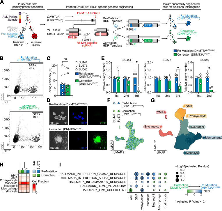
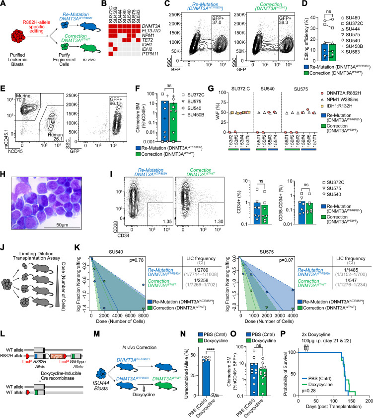
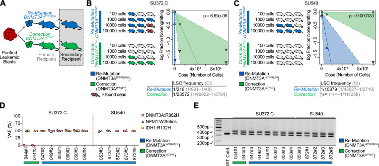
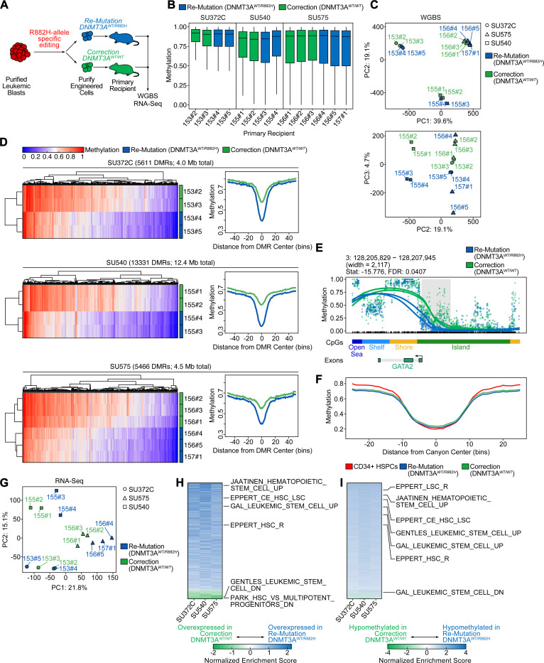
Genetic mutations are being thoroughly mapped in human cancers, yet a fundamental question in cancer biology is whether such mutations are functionally required for cancer initiation, maintenance of established cancer, or both. Here, we study this question in the context of human acute myeloid leukemia (AML), where DNMT3A R882 missense mutations often arise early, in pre-leukemic clonal hematopoiesis, and corrupt the DNA methylation landscape to initiate leukemia. We developed CRISPR-based methods to directly correct DNMT3A R882 mutations in leukemic cells obtained from patients. Surprisingly, DNMT3A R882 mutations were largely dispensable for disease maintenance. Replacing DNMT3A R882 mutants with wild-type DNMT3A did not impair the ability of AML cells to engraft in vivo, and minimally altered DNA methylation. Taken together, DNMT3A R882 mutations are initially necessary for AML initiation, but are largely dispensable for disease maintenance. The notion that initiating oncogenes differ from those that maintain cancer has important implications for cancer evolution and therapy.


Conflict of interest statement

Declaration of interests: R.M. is on the Advisory Boards of Kodikaz Therapeutic Solutions, Orbital Therapeutics, Pheast Therapeutics, 858 Therapeutics, Prelude Therapeutics, Mubadala Capital, and Aculeus Therapeutics. R.M. is a co-founder and equity holder of Pheast Therapeutics, MyeloGene, and Orbital Therapeutics.

Figures

Fig. 1:
Fig. 1:. Correction of DNMT3AR882H in pre-leukemic HSCs results in normalization of aberrant self-renewal.
A: Schematic of DNMT3AR882H correction approach. Cell populations of interest (either residual HSPCs containing pre-leukemic HSPCs or leukemic blasts) are purified by flow cytometry. Subsequently, pre-complexed Cas9 and sgRNA targeting the mutant, R882H allele, are electroporated into the target cells and cells are transduced with rAAV6 containing either the codon-optimized mutant (R882H) or wildtype (R882R) sequence, and a selectable fluorescent marker (BFP or GFP). Successfully engineered cells are then isolated for functional experiments. B: Isolation by FACS of successfully edited residual HSPCs (from patient SU575) for either re-mutation (top panel) or correction (bottom panel) of DNMT3AR882H. C: Editing efficiency across all experiments in residual HSPCs as percentage of fluorescent protein positive cells (n=3 independent patient specimens). D: Representative hematopoietic colonies 14 days after plating into semisolid media (Methocult). E: Serial replating assay of gene-edited residual HSPCs derived from three independent patient specimens (n=3–4 technical replicates per patient), * P < 0.05. F: UMAP embedding of single-cell RNA-Seq from gene-edited residual HSPCs differentiated in vitro for 14 days after editing colored by genotype and donor. G: UMAP embedding as in F colored by assigned cell type. H: Relative abundance of cell types within each sample. Sample identities and DNMT3A genotypes are indicated at the top. I: Gene set enrichment analysis of Hallmark gene sets between re-mutation (DNMT3AWT/R882H, blue) and correction (DNMT3AWT/WT, green) across cell types. * P < 0.1.
Fig. 2:
Fig. 2:. Correction of DNMT3AR882H in AML blasts has no effect on leukemogenicity or leukemia disease maintenance.
A: Schematic of leukemic blast editing and xenotransplantation. B: Oncoprint of samples included in this study. C: Isolation of successfully edited leukemic blasts for either re-mutation (left panel) or correction (right panel) of DNMT3AR882H from patient SU480. D: Editing efficiency across patient specimens quantified as percentage of fluorescent protein positive cells 3 days after gene-editing. E: Representative flow cytometry analysis of bone marrow aspirate 12 weeks after injection of human, gene corrected (DNMT3AWT/WT) leukemic blasts showing human engraftment with retention of the GFP fluorescent protein. F: Human chimerism in bone marrow aspirates at 12 weeks after injection of the indicated patient sample with either DNMT3A re-mutation or correction. G: Variant allele frequency (VAF) of somatic mutations found in the indicated patient specimens in human genomic DNA extracted from mice 4 months after engraftment indicating clonal retention of all somatic variants, except for DNMT3A in DNMT3A-corrected specimens. H: Representative Giemsa staining of cytospin of human leukemic cells isolated 4 months after DNMT3AR882H correction. I: Representative flow cytometry plots (left) and quantification (right) of CD34+ and CD34+CD38− leukemic subpopulations 4 months after re-mutation or correction of DNMT3AR882H. J: Schematic of limiting dilution transplantation assay. K: Quantification of leukemia initiating cell (LIC) frequency for patient specimens SU540 (left) and SU575 (right) after re-mutation or correction of DNMT3AR882H. Estimated frequency of LIC and test statistics are indicated. L: Schematic of doxycycline-inducible Cre recombinase driven correction of the endogenous DNMT3AR882H allele in iPSC-derived AML blasts (iSU444). M: Schematic of in vivo correction experiment. Mice are engrafted with mutant (DNMT3AWT/R882H) leukemic blasts, and administration of doxycycline 3 weeks after engraftment leads to correction of DNMT3AWT/R882H to DNMT3AWT/WT in vivo. N: Quantification of successful in vivo recombination of DNMT3AWT/R882H, P < 0.0001. O: Human Chimerism 5 weeks after in vivo DNMT3AR882H correction. P: Kaplan-Meier survival curve of mice with or without in vivo DNMT3AR882H correction.
Fig. 3:
Fig. 3:. Secondary transplantation reveals a reduction of leukemic stem frequency upon DNMT3AR882H correction.
A: Schematic of secondary transplantation experiments. Human leukemic cells are harvested from primary recipients 4 months after gene-editing and then transplanted in limiting dilution into secondary host animals. B: Quantification of secondary limiting dilution assay results for patient specimen SU372.C. Estimated LSC frequencies and test statistics are indicated. C: Quantification of secondary limiting dilution assay results for patient specimen SU540. None of the mice receiving corrected (DNMT3AWT/WT) leukemic cells showed engraftment at the doses indicated. Estimated LSC frequencies and test statistics are shown. D: VAF in human genomic DNA isolated from secondary recipients for recurrent somatic mutations found in patient samples utilized for secondary transplantation studies. E: FLT3-ITD PCR from human genomic DNA isolated from secondary recipients.
Fig. 4:
Fig. 4:. Epigenetic and transcriptional identity is highly patient-specific with only minimal changes upon correction of DNMT3AR882H.
A: Schematic of sample generation for whole-genome bisulfite (WGBS) and RNA-Sequencing (RNA-Seq). Human cells were harvested from recipient mice 4 months after gene-editing. B: Average CpG methylation across the human genome for three patient specimens with either correction (green) or re-mutation (blue). Each bar represents an individual sample harvested from a primary recipient mouse (mouse ID indicated on x-axis). C: Principal component analysis of global CpG methylation from engrafted mice. Patient ID is indicated by shape and DNMT3A genotype is indicated by color. D: Methylation at differentially methylated loci (heatmaps, left) for all DMRs identified within each patient sample as well as a summary meta-region plot of methylation at DMRs normalized for width (right). DMRs in meta-region plots are extended by 2 kb for visualization of flanking regions. E: Methylation at a representative DMR (adjacent to GATA2) for patient sample SU372.C. Individual lines indicate cells harvested from separate mice, color indicates the DNMT3A genotype. F: Summary meta-region plot of methylation at methylation canyon loci from Jeong et al. for re-mutated (blue) or corrected (green) patient samples, as well as healthy CD34+ HSPC control (red). G: Principal component analysis of global RNA-Seq patterns from engrafted mice. Patient ID is indicated by shape and DNMT3A genotype is indicated by color. H: Gene set enrichment analysis of differential gene expression within each patient plotted as a heatmap of normalized enrichment scores (NES) sorted by descending average. Gene sets enriched in re-mutated (DNMT3AWT/R882H) are colored in blue, gene sets enriched in corrected (DNMT3AWT/WT) are colored in green. Representative gene sets relating to hematopoietic or leukemic stem cell function are highlighted. I: Gene set enrichment analysis of differential promoter methylation within each patient as a heatmap of NES sorted by descending average. Gene sets enriched in hypomethylated DMRs in re-mutated (DNMT3AWT/R882H) samples are colored in blue, and gene sets enriched in hypermethylated DMRs in re-mutated samples are colored in red. Representative gene sets relating to hematopoietic or leukemic stem cell function are highlighted.

References

    1. Kats L.M., Reschke M., Taulli R., Pozdnyakova O., Burgess K., Bhargava P., Straley K., Karnik R., Meissner A., Small D., et al. (2014). Proto-oncogenic role of mutant IDH2 in leukemia initiation and maintenance. Cell Stem Cell 14, 329–341. 10.1016/j.stem.2013.12.016. - DOI - PMC - PubMed
    1. Dunbar A.J., Bowman R.L., Park Y.C., O’Connor K., Izzo F., Myers R.M., Karzai A., Zaroogian Z., Kim W.J., Fernandez-Maestre I., et al. (2024). Jak2V617F Reversible Activation Shows Its Essential Requirement in Myeloproliferative Neoplasms. Cancer Discov 14, 737–751. 10.1158/2159-8290.CD-22-0952. - DOI - PMC - PubMed
    1. Dow L.E., O’Rourke K.P., Simon J., Tschaharganeh D.F., van Es J.H., Clevers H., and Lowe S.W. (2015). Apc Restoration Promotes Cellular Differentiation and Reestablishes Crypt Homeostasis in Colorectal Cancer. Cell 161, 1539–1552. 10.1016/j.cell.2015.05.033. - DOI - PMC - PubMed
    1. Ley T.J., Ding L., Walter M.J., McLellan M.D., Lamprecht T., Larson D.E., Kandoth C., Payton J.E., Baty J., Welch J., et al. (2010). DNMT3A mutations in acute myeloid leukemia. N Engl J Med 363, 2424–2433. 10.1056/NEJMoa1005143. - DOI - PMC - PubMed
    1. Network T.C.G.A.R. (2013). Genomic and Epigenomic Landscapes of Adult De Novo Acute Myeloid Leukemia. N. Engl. J. Med. 368, 2059–2074. 10.1056/nejmoa1301689. - DOI - PMC - PubMed

Publication types

LinkOut - more resources