[Preprint]. 2024 Nov 1:2024.11.01.621496.
doi: 10.1101/2024.11.01.621496.

Defective Neurogenesis in Lowe Syndrome is Caused by Mitochondria Loss and Cilia-related Sonic Hedgehog Defects

Affiliations

Defective Neurogenesis in Lowe Syndrome is Caused by Mitochondria Loss and Cilia-related Sonic Hedgehog Defects

Chien-Hui Lo et al. bioRxiv. .

Abstract

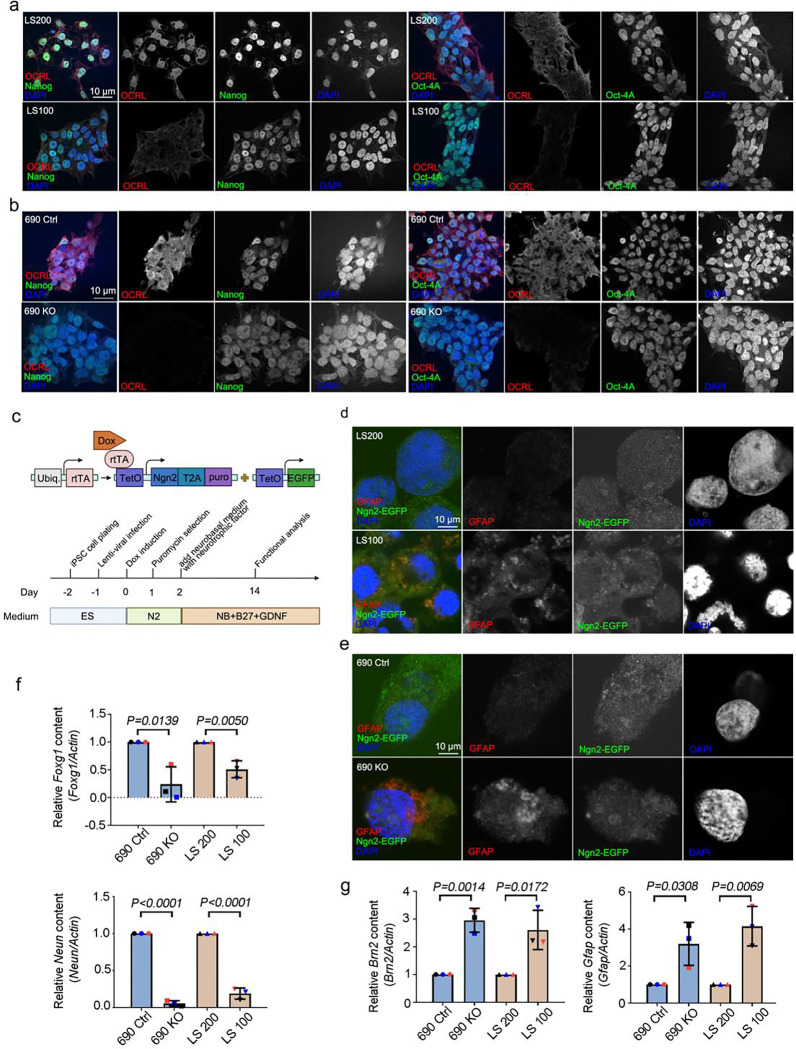
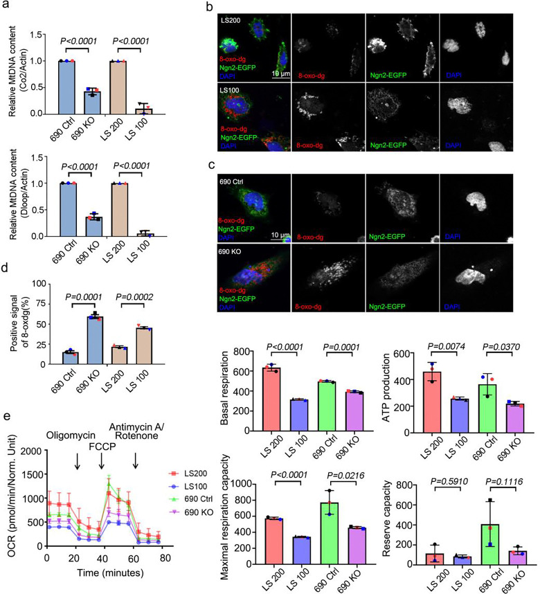
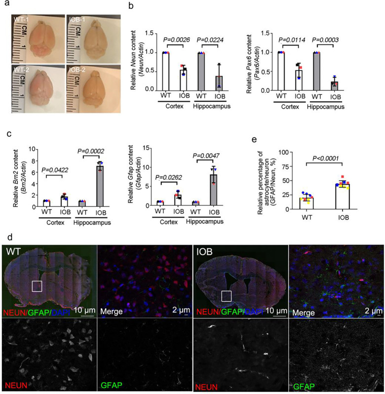
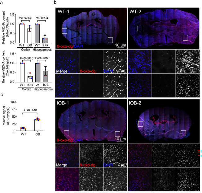
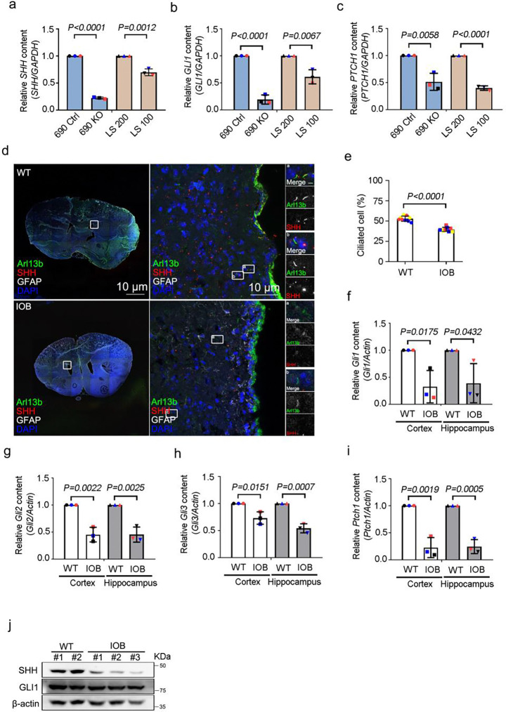
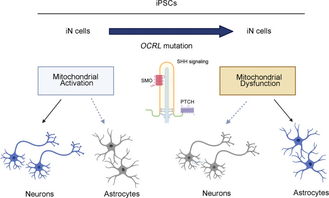
Human brain development is a complex process that requires intricate coordination of multiple cellular and developmental events. Dysfunction of lipid metabolism can lead to neurodevelopmental disorders. Lowe syndrome (LS) is a recessive X-linked disorder associated with proximal tubular renal disease, congenital cataracts and glaucoma, and central nervous system developmental delays. Mutations in OCRL, which encodes an inositol polyphosphate 5-phosphatase, lead to the development of LS. The cellular mechanism responsible for neuronal dysfunction in LS is unknown. Here we show depletion of mitochondrial DNA and decrease in mitochondrial activities result in neuronal differentiation defects. Increased astrocytes, which are secondary responders to neurodegeneration, are observed in neuronal (iN) cells differentiated from Lowe patient-derived iPSCs and an LS mouse model. Inactivation of cilia-related sonic hedgehog signaling, which organizes the pattern of cellular neuronal differentiation, is observed in an OCRL knockout, iN cells differentiated from Lowe patient-derived iPSCs, and an LS mouse model. Taken together, our findings indicate that mitochondrial dysfunction and impairment of the ciliary sonic hedgehog signaling pathway represent a novel pathogenic mechanism underlying the disrupted neuronal differentiation observed in LS.

Keywords: Cilia formation; Lowe Syndrome; Mitochondria; Neuronal differentiation; OCRL.


Conflict of interest statement

Conflict of Interest Statement The authors declare no competing interests.

Figures

Figure 1.
Figure 1.. Increased astrocyte production during neuronal differentiation in OCRL-deficient and Lowe syndrome iPSCs.
(a and b) Lowe syndrome iPSCs and OCRL knockout iPSCs stained with OCRL (red) and Nanong; OCT4 (green) antibodies. DNA stained with DAPI (blue). Scale bars are as indicated. (c) Design of lentiviral vectors to induce Ngn2-mediated conversion of iPSCs to neuronal (iN) cells. (d and e) Lowe syndrome iPSCs and OCRL knockout iPSCs stained with GFAP (red) antibodies and expressed Ngn2-EGAP. DNA stained with DAPI (blue). Scale bars are as indicated. (f) Quantitative real-time PCR (RT-PCR) validation of FOXG1 and NEUN in iN cells. RT-PCR was repeated three times with different batches. Gene expression values are normalized to GAPDH (g) Quantitative real-time PCR (RT-PCR) validation of BRN2 and GFAP in iN cells. RT-PCR was repeated three times with different batches. Gene expression values are normalized to GAPDH. The bars in each graph represent mean ± SD. Statistical significance was determined using Student’s t-test, with exact p-values reported.
Figure 2.
Figure 2.. Mitochondria defects in iN cells derived from OCRL-deficient iPSCs.
(a) Quantitative real-time PCR (RT-PCR) validation of mitochondrial DNA genes COX2 and DLOOP in iN cells. RT-PCR was repeated three times with different batches. Gene expression values are normalized to ACTIN. (b and c) iN cells derived from Lowe syndrome iPSCs and OCRL knockout iPSCs stained with 8-oxo-dg (red) antibodies and expressed Ngn2-EGAP. DNA stained with DAPI (blue). Scale bars are as indicated. (d) Quantification of the percentage of iPSCs-derived iN cells positive for 8-oxo-dg signal. > 100 cells analyzed for each independent experiment. (e) Oxygen consumption rate of Lowe syndrome iPSCs-derived iN cells and OCRL knockout iPSCs-derived iN cells measured by Seahorse Analyzer. The bars in each graph represent mean ± SD. Statistical significance was determined using Student’s t-test, with exact p-values reported.
Figure 3.
Figure 3.. Elevated astrocyte population during neuronal differentiation in Lowe syndrome mouse model.
(a) Images showing brain of wild-type and IOB mouse. (b) Quantitative real-time PCR (RT-PCR) validation of Neun and Pax6 in brain sections. RT-PCR was repeated three times with different batches. Gene expression values are normalized to actin. (c) Quantitative real-time PCR (RT-PCR) validation of Brn2 and Gfap in brain tissues. RT-PCR was repeated three times with different batches. Gene expression values are normalized to actin. (d) Wild-type and IOB mouse brain section stained with Neun (red) and GFAP (green) antibodies. DNA stained with DAPI (blue). Scale bars are as indicated. (e) Quantification of the ratio of Neun and GFAP signals in brain section(s) of wild-type and IOB mouse. > 100 cells analyzed for each independent experiment. The bars in each graph represent mean ± SD. Statistical significance was determined using Student’s t-test, with exact p-values reported.
Figure 4.
Figure 4.. Mitochondrial defects in the brain of Lowe syndrome mouse model.
(a) Quantitative real-time PCR (RT-PCR) validation of mitochondrial DNA genes Cox2 and Dloop in brain sections of wild-type and IOB mouse. RT-PCR was repeated three times with different batches. Gene expression values are normalized to Actin. (b) Brain sections of wild-type and IOB mouse stained with 8-oxo-dg (red) antibody. DNA stained with DAPI (blue). Scale bars are as indicated. (c) Quantification of the positive percentage of 8-oxo-dg signals in brain sections of wild-type and IOB mouse > 100 cells analyzed for each independent experiment. The bars in each graph represent mean ± SD. Statistical significance was determined using Student’s t-test, with exact p-values reported.
Figure 5.
Figure 5.. Involvement of cilia-mediated Shh signaling in neuronal differentiation of OCRL-deficient iN cells.
(a, b and c) Quantitative real-time PCR (RT-PCR) validation of Shh signaling genes SHH, GLI1 and PTCH1 in iN cells. RT-PCR was repeated three times with different batches. Gene expression values are normalized to GAPDH. (d) Brain section of wild-type and IOB mouse stained with SHH (red) and Arl13b (green) antibodies. DNA stained with DAPI (blue). Scale bars as indicated. (e) Quantification of the percentage of positive ciliated cells. > 100 cells analyzed for each independent experiment. (f, g, h and i) Quantitative real-time PCR (RT-PCR) validation of Shh signaling genes Gli1, Gli2, Gli3 and Ptch1 in brain sections of wild-type and IOB mouse. RT-PCR was repeated three times with different batches. Gene expression values are normalized to actin. (j) Western blot analysis using antibodies against SHH, GLI1 and β-actin in brain sections of wild-type and IOB mouse. The bars in each graph represent mean ± SD. Statistical significance was determined using Student’s t-test, with exact p-values reported.
Figure 6.
Figure 6.. Mitochondrial-mediated neuronal differentiation defects in OCRL-deficient Cells.
Neuronal cells induced (iN) from iPSCs derived from mutant OCRL and Lowe syndrome mouse models contain a high level of astrocytes. OCRL knockout, mutant iPSCs-derived iN cells, and Lowe syndrome mouse model also possess reduced cilia-related sonic hedgehog pathways (created by BioRender.com).

References

    1. Shah D.M., et al. , Lowe Oculocerebrorenal Syndrome Comparison of Anterior Segment Anatomy in Eyes with and without Glaucoma. Ophthalmol Glaucoma, 2024. 7(1): p. 101–102. - PMC - PubMed
    1. Murdock N. and Chou E., Oculocerebrorenal Syndrome, in StatPearls. 2024: Treasure Island (FL) ineligible companies. Disclosure: Eva Chou declares no relevant financial relationships with ineligible companies.
    1. Schaub J.A., et al. , Regional Retinal Ganglion Cell Axon Loss in a Murine Glaucoma Model. Invest Ophthalmol Vis Sci, 2017. 58(5): p. 2765–2773. - PMC - PubMed
    1. Nusbaum D.M., Wu S.M., and Frankfort B.J., Elevated intracranial pressure causes optic nerve and retinal ganglion cell degeneration in mice. Exp Eye Res, 2015. 136: p. 38–44. - PMC - PubMed
    1. Sharma S., Skowronek A., and Erdmann K.S., The role of the Lowe syndrome protein OCRL in the endocytic pathway. Biol Chem, 2015. 396(12): p. 1293–300. - PubMed

Publication types

LinkOut - more resources