[Preprint]. 2025 Feb 13:2024.09.17.613365.
doi: 10.1101/2024.09.17.613365.

Type-II kinase inhibitors that target Parkinson's Disease-associated LRRK2

Affiliations

Type-II kinase inhibitors that target Parkinson's Disease-associated LRRK2

Nicolai D Raig et al. bioRxiv. .

Update in

  • Type II kinase inhibitors that target Parkinson's disease-associated LRRK2.
    Raig ND, Surridge KJ, Sanz-Murillo M, Dederer V, Krämer A, Schwalm MP, Lattal NM, Elson L, Chatterjee D, Mathea S, Hanke T, Leschziner AE, Reck-Peterson SL, Knapp S. Raig ND, et al. Sci Adv. 2025 Jun 6;11(23):eadt2050. doi: 10.1126/sciadv.adt2050. Epub 2025 Jun 4. Sci Adv. 2025. PMID: 40465731 Free PMC article.

Abstract

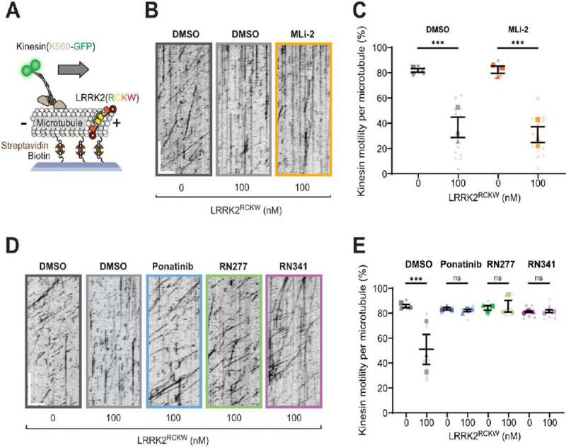
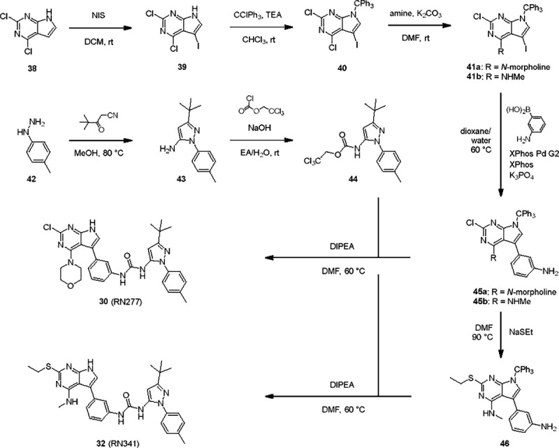
Aberrant increases in kinase activity of leucine-rich repeat kinase 2 (LRRK2) are associated with Parkinson's disease (PD). Numerous LRRK2-selective type-I kinase inhibitors have been developed and some have entered clinical trials. In this study, we present the first LRRK2-selective type-II kinase inhibitors. Targeting the inactive conformation of LRRK2 is functionally distinct from targeting the active-like conformation using type-I inhibitors. We designed these inhibitors using a combinatorial chemistry approach fusing selective LRRK2 type-I and promiscuous type-II inhibitors by iterative cycles of synthesis supported by structural biology and activity testing. Our current lead structures are selective and potent LRRK2 inhibitors. Through cellular assays, cryo-electron microscopy structural analysis, and in vitro motility assays, we show that our inhibitors stabilize the open, inactive kinase conformation. These new conformation-specific compounds will be invaluable as tools to study LRRK2's function and regulation, and expand the potential therapeutic options for PD.


Conflict of interest statement

Competing interest statement Authors declare that they have no competing interests.

Figures

Fig. 1.
Fig. 1.. LRRK2 type-II inhibitor design strategy.
(A) Schematic domain structure of LRRK2. The three constructs used in this study are indicated: full-length LRRK2, LRRK2RCKW and LRRK2KW; (B) and (C) Close up of the inhibitor binding pocket from cryo-EM maps and models of LRRK2RCKW bound to the type-I inhibitor MLi-2 (PDB: 8TXZ) (B) and type-II inhibitor GZD-824 (PDB: 8TZE) (C). Key residues and features are labelled. Both structures are shown in the same view, aligned through the C-lobe of the kinase. Dark orange: C-lobe; light orange: N-lobe; black: DYG motif; grey: G-loop; green: activation loop. (D) Scheme depicting our hybrid design strategy to develop potent and selective type-II inhibitors for LRRK2.
Fig. 2.
Fig. 2.. Co-crystal structure of RN129 bound to CLK3 and cryo-EM structure of RN277 bound to LRRK2RCKW.
(A) The co-crystal structure of RN129 (28) with CLK3 highlighting the type-II binding mode and interactions between the protein and inhibitor (PDB: 9EZ3); (B) Ribbon diagram of the atomic model of LRRK2RCKW:RN277:E11 DARPin complex (PDB: 9DMI); (C) and (D) Close ups of the active sites of the cryo-EM structures of LRRK2RCKW:RN277 (C) and LRRK2RCKW:GZD824 (PDB: 8TZE) (D).
Fig. 3.
Fig. 3.. Kinome selectivity of RN341.
(A) Kinome phylogenetic tree, with 96 kinases screened in the DSF assay against Rebastinib highlighted in blue. The 18.5 K ΔTm shift of LRRK2KW is highlighted in red. For all screened kinases, the bubble size correlates with the degree of ΔTm shift, as indicated in the legend; (B) Kinome phylogenetic tree, with 103 kinases screened in the DSF assay against RN341 highlighted in blue. The 20 K ΔTm shift of LRRK2KW is highlighted in red. The bubble size for each kinase correlates with the ΔTm shifts, as indicated in the legend (as in A). Kinases with ΔTm > 6 K are labeled; (C) Waterfall plots of the ReactionBiology 33PanQinase screen of RN341 at 1 μM and 10 μM against 350 wild-type kinases. Kinases with decreased activity in the presence of RN341 to < 22 % of the control value are labeled; (D) Off-target validation from both screens via in cellulo nanoBRET assay in 2 biological replicates, error bars ± sd, EC50 (JNK2) = 2.7 μM, EC50 (STK10) = 1.5 μM, EC50 (MAPK14) = 1.7 μM, EC50 (TTK) = 3.2 μM, EC50 (CDKL1) = 17 μM, EC50 (CLK1) = 6.0 μM, EC50 (JNK3) = 15 μM, EC50 (DYRK2) = >20 μM, EC50 (SLK) >20 μM, EC50 (DDR2) >20 μM, EC50 (STK17B) = >20 μM.
Fig. 4.
Fig. 4.. Inhibition of LRRK2’s phosphorylation of Rab8a in vitro and in cellulo.
(A) and (B) Dose response curve of RN277 (30) and RN341 (32) inhibiting LRRK2RCKW-mediated phosphorylation of Rab8a. Activity was calculated as the percentage (%) of phosphorylated Rab8a vs. non-phosphorylated Rab8a detected in the presence of different concentrations of RN277/RN341; (C) Western blots from 293T cells transiently co-transfected with LRRK2 (full-length) and GFP-Rab8a for 48h prior to treatment with a dilution series of RN277 (30) and RN341 (32) for 4h. DMSO and MLi-2 (500 nM) treatment for 4h were used as negative and positive controls, respectively. Lysed cells were immunoblotted for LRRK2, total GFP-Rab8a, phospho-Rab8a (pT72) and GAPDH as a loading control; (D) Quantification from four independent western blots showing the ratio of GFP-pRab8a to total GFP-Rab8a upon treatment with RN277 (30) and RN341 (32) at the indicated concentrations. Statistical analysis was performed using one-way ANOVA analysis with Tukey’s multiple comparison of means. ****p<0.0001, error bars ± s.e.m. (E) Western blots from 293T cells transiently co-transfected with LRRK2 (full-length) and GFP-Rab8a, treated with DMSO (control), 500 nM MLi-2, 5 μM RN277 or 5 μM RN341 for 4h, 48h post- transfection. Lysed cells were immunoblotted for LRRK2, phospho-LRRK2 (pS935), total GFP-Rab8a, phospho-Rab8a (pT72) and GAPDH as a loading control; (F) Quantification from four independent western blots showing the ratio of GFP-pRab8a to total GFP-Rab8a upon treatment with the indicated inhibitors (as in D). Statistical analysis was performed using a one-way ANOVA analysis with Tukey’s multiple comparison of means. ****p<0.0001, error bars ± s.e.m.; (G) Quantification from four independent western blots showing the ratio of pLRRK2 to total LRRK2 upon treatment with the indicated inhibitors. Statistical analysis was performed using a one-way ANOVA analysis with Tukey’s multiple comparison of means. *p 0.0469, error bars ± s.e.m. Note that data from one replicate shown in Fig. 4D forms part of the dataset presented in F/G.
Fig. 5.
Fig. 5.. LRRK2-specific type-II inhibitors RN277 and RN341 rescue kinesin motility in the presence of LRRK2RCKW.
(A) Schematic of the single-molecule in vitro motility assay; (B) Example kymographs from single-molecule motility assays showing kinesin motility with DMSO or the type-I inhibitor MLi-2 (5 μM) in the presence or absence of LRRK2RCKW. Scale bars 5 μm (x) and 30 s (y); (C) Quantification of the percentage (mean ± s.e.m) of motile events per microtubule as a function of LRRK2RCKW concentration in the absence (DMSO) or presence of MLi-2 (5 μM). Three technical replicates were collected per condition, with data points represented as circles, triangles and squares corresponding to single data points (microtubules) within each replicate. Statistical analysis was performed using the mean of each technical replicate; DMSO condition ***p 0.0007, MLi-2 condition ***p 0.0003, One-way ANOVA with Sidaks multiple comparison test within drug only; (D) Example kymographs from single-molecule motility assays showing kinesin motility with DMSO or the type-II inhibitors Ponatinib, RN277 and RN341 (5 μM) in the presence or absence of LRRK2RCKW. Scale bars 5 μm (x) and 30 s (y); (E) Quantification of the percentage (mean ± s.e.m) of motile events per microtubule as a function of LRRK2RCKW concentration in the absence (DMSO) or presence of type-II inhibitors Ponatinib, RN277 and RN341 (5 μM). Three technical replicates were collected per condition, with data points represented as circles, triangles and squares corresponding to single data points (microtubules) within each replicate. Statistical analysis was performed using the mean of each technical replicate; ***p 0.0003, One-way ANOVA with Sidaks multiple comparison test within drug only.
Scheme 1.
Scheme 1.. Synthesis of RN277 and RN341.
The convergent synthesis route of compound 30 (RN277) and 32 (RN341). The detailed procedures and analytics are shown in the supplementary information. rt: room temperature, NIS: N-Iodosuccinimide, DCM: dichloromethane, TEA: tri-ethylamine, DMF: dimethylformamide, XPhos: dicyclohexyl[2′,4′,6′-tris(propan-2-yl)[1,1′-biphenyl]-2-yl]phosphane, EA: ethyl acetate, DI-PEA: N,N-diisopropylethylamine.

Similar articles

References

    1. Willis A. W., Roberts E., Beck J. C., Fiske B., Ross W., Savica R., Van Den Eeden S. K., Tanner C. M., Marras C., on behalf of the Parkinson’s Foundation P4 Group, Alcalay R., Schwarzschild M., Racette B., Chen H., Church T., Wilson B., Doria J. M., Incidence of Parkinson disease in North America. npj Parkinsons Dis. 8, 170 (2022). - PMC - PubMed
    1. Paisán-Ruíz C., Jain S., Evans E. W., Gilks W. P., Simón J., Van Der Brug M., De Munain A. L., Aparicio S., Gil A. M., Khan N., Johnson J., Martinez J. R., Nicholl D., Carrera I. M., Peňa A. S., De Silva R., Lees A., Martí-Massó J. F., Pérez-Tur J., Wood N. W., Singleton A. B., Cloning of the Gene Containing Mutations that Cause PARK8-Linked Parkinson’s Disease. Neuron 44, 595–600 (2004). - PubMed
    1. Zimprich A., Biskup S., Leitner P., Lichtner P., Farrer M., Lincoln S., Kachergus J., Hulihan M., Uitti R. J., Calne D. B., Stoessl A. J., Pfeiffer R. F., Patenge N., Carbajal I. C., Vieregge P., Asmus F., Müller-Myhsok B., Dickson D. W., Meitinger T., Strom T. M., Wszolek Z. K., Gasser T., Mutations in LRRK2 Cause Autosomal-Dominant Parkinsonism with Pleomorphic Pathology. Neuron 44, 601–607 (2004). - PubMed
    1. Satake W., Nakabayashi Y., Mizuta I., Hirota Y., Ito C., Kubo M., Kawaguchi T., Tsunoda T., Watanabe M., Takeda A., Tomiyama H., Nakashima K., Hasegawa K., Obata F., Yoshikawa T., Kawakami H., Sakoda S., Yamamoto M., Hattori N., Murata M., Nakamura Y., Toda T., Genome-wide association study identifies common variants at four loci as genetic risk factors for Parkinson’s disease. Nat Genet 41, 1303–1307 (2009). - PubMed
    1. Simón-Sánchez J., Schulte C., Bras J. M., Sharma M., Gibbs J. R., Berg D., Paisan-Ruiz C., Lichtner P., Scholz S. W., Hernandez D. G., Krüger R., Federoff M., Klein C., Goate A., Perlmutter J., Bonin M., Nalls M. A., Illig T., Gieger C., Houlden H., Steffens M., Okun M. S., Racette B. A., Cookson M. R., Foote K. D., Fernandez H. H., Traynor B. J., Schreiber S., Arepalli S., Zonozi R., Gwinn K., Van Der Brug M., Lopez G., Chanock S. J., Schatzkin A., Park Y., Hollenbeck A., Gao J., Huang X., Wood N. W., Lorenz D., Deuschl G., Chen H., Riess O., Hardy J. A., Singleton A. B., Gasser T., Genome-wide association study reveals genetic risk underlying Parkinson’s disease. Nat Genet 41, 1308–1312 (2009). - PMC - PubMed

Publication types

LinkOut - more resources