[Preprint]. 2024 Oct 30:2024.10.25.620326.
doi: 10.1101/2024.10.25.620326.

Phosphorylation of HP1/Swi6 relieves competition with Suv39/Clr4 on nucleosomes and enables H3K9 trimethyl spreading

Affiliations

Phosphorylation of HP1/Swi6 relieves competition with Suv39/Clr4 on nucleosomes and enables H3K9 trimethyl spreading

Dana R Kennedy et al. bioRxiv. .

Abstract

Heterochromatin formation in Schizosaccharomyces pombe requires the spreading of histone 3 (H3) Lysine 9 (K9) methylation (me) from nucleation centers by the H3K9 methylase, Suv39/Clr4, and the reader protein, HP1/Swi6. To accomplish this, Suv39/Clr4 and HP1/Swi6 have to associate with nucleosomes both nonspecifically, binding DNA and octamer surfaces and specifically, via recognition of methylated H3K9 by their respective chromodomains. However, how both proteins avoid competition for the same nucleosomes in this process is unclear. Here, we show that phosphorylation tunes the nucleosome affinity of HP1/Swi6 such that it preferentially partitions onto Suv39/Clr4's trimethyl product rather than its unmethylated substrates. Preferential partitioning enables efficient conversion from di-to trimethylation on nucleosomes in vitro and H3K9me3 spreading in vivo. Together, our data suggests that phosphorylation of HP1/Swi6 creates a regime that relieves competition with the "read-write" mechanism of Suv39/Clr4 for productive heterochromatin spreading.


Figures

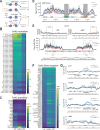
Figure 1:
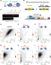
Figure 1:. S18 and S24 in Swi6 are required for spreading, but not nucleation of heterochromatin silencing.
A. Overview of the Swi6 protein domain architecture and previously identified (Shimada et al.) in vivo phosphorylation sites (green residue numbers). NTE: N-terminal extension; CD: chromodomain (H3K9me binding); HINGE: unstructured hinge region; CSD: chromo-shadow domain (dimerization and effector recruitment). B. Strategy for production of swi6 S-A mutants in the MAT ΔREIII HSS reporter background. C. Swi6 levels are not affected by S-A mutations. Total extracts of swi6 wild-type or indicated mutants were probed with an anti-Swi6 polyclonal antibody. In vitro purified Swi6 that was either phosphorylated (pSwi6) or not (unpSwi6) is run as size controls. Note, not all mutant Swi6 proteins display a band shift even if they retain phosphosites D.-I. 2-D Density hexbin plots examining silencing at nucleation “green” and spreading “orange” reporter in Δswi6, wild-type, and indicated S-A mutants. The yellow box indicates a “green” and “orange” regime consistent with silencing loss, and the magenta box indicates a regime consistent with loss of spreading, but not nucleation. The dashed line indicates the threshold for orange ON and the numbers the fraction of cells above the line.
Figure 2:
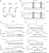
Figure 2:. Conversion from H3K9me2 to H3K9me3 is compromised outside nucleation centers in S18 and S24 Swi6 mutants.
A. Overview of the ChIP-seq experiments. B-D. ChIP-seq signal visualization plots. The solid ChIP/input line for each genotype represents the mean of three repeats, while the shading represents the 95% confidence interval. B. Plots of H3K9me2 (TOP) and H3K9me3 (BOTTOM) ChIP signal over input at the MAT ΔREIII HSS mating type locus for wild-type (black), swi6S18/24A (blue), and Δswi6 (gold). Signal over “green” and “orange” reporters are greyed out, as reads from these reporters map to multiple locations within the reference sequence, as all reporters contain control elements derived from the ura4 and ade6 genes. C. H3K9me2 (TOP) and H3K9me3 (BOTTOM) plots as in A. for subtelomere IIR for wild-type and swi6S18/24A. The red bar on the H3K9me2 plots indicates the distance from tlh2 to where H3K9me2 levels drop in swi6S18/24A relative to wild-type. Inset: a zoomed-in view proximal to tlh2 is shown for H3K9me2 and me3. The red arrows in the insets indicate the point of separation of the 95% confidence intervals, which is significantly further telomere proximal for H3K9me3. D. H3K9me2 (TOP) and H3K9me3 (BOTTOM) plots as in A. for centromere II for wild-type and swi6S18/24A. Inset: the left side of the pericentromere.
Figure 3:
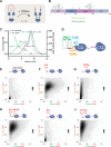
Figure 3:. Swi6 phosphorylation increases oligomerization and decreases nucleosome binding, without affecting specificity.
A. Production of phosphorylated Swi6 (pSwi6) in E. coli. Casein Kinase II (CKII) is co-expressed with Swi6. After lysis and purification, the 6X His tag is removed from the pSwi6 or unpSwi6 protein. B. Mass Spectrometry on pSwi6. Shown is a domain diagram of Swi6. Phosphorylation sites identified in pSwi6 by 2D-ETD-MS are indicated and grouped by detection prevalence in the sample. C. Size Exclusion Chromatography followed by Multi-Angle Light Scattering (SEC-MALS) on EDC/NHS cross-linked unpSwi6 (black) and pSwi6 (green). Relative refractive index signals (solid lines, left y-axis) and derived molar masses (lines over particular species, right y-axis) are shown as a function of the elution volume. [Swi6] was 100μM. D. Fluorescence polarization (FP) with fluorescein (star)- labeled H3 tail peptides (1–20) and pSwi6 (green) or unpSwi6 (black) for H3K9me0 (open circles) and H3K9me3 (filled circles) is shown. Error bars represent standard deviation. Binding was too low to be fit for unpSwi6 and H3K9me0 peptides. E. FP with H3K9me0 (open circles) or H3Kc9me3 (MLA, filled circles) mononucleosomes. Fluorescein (green star) is attached by a flexible linker at one end of the 147 bp DNA template. For D.&E., the average of three independent fluorescent polarization experiments for each substrate is shown. Error bars represent standard deviation. F. Summary table of affinities and specificities for D. and E. G. Representative maximum projection live microscopy images of indicated Swi6-GFP / Sad1-mKO2 strains. H. Analysis of signal intensity in Swi6-GFP foci in indicated strains. Wt Swi6, n=242; Swi6S18/24A, n=251; Swi6S18/24/117−220A, n=145; Swi6S46/52/117−220A, n=192. n, number of foci analyzed.
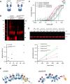
Figure 4:
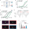
Figure 4:. Swi6 phosphorylation mitigates inhibition of the Clr4-mediated conversion of H3K9me2 to H3K9me3
A. Most Swi6 molecules in the cell are phosphorylated at S18 and S24. Quantitative western blots against total Swi6 and phosphorylated Swi6 at S18/S24. A standard curve of pSwi6 isolated as in Figure 3 is included in both blots. Total protein lysates from wild-type swi6 and swi6S18/24A strains were probed with a polyclonal anti-Swi6 antibody (α-Swi6) or an antibody raised against a phosphorylated S18/S24 peptide (α-S18P-S24P). α-tubulin was used as a loading control. One of two independent experiments is shown. L; ladder. B. Experimental scheme to probe the impact of Swi6 on H3K9 trimethylation. C. Quantitative western blots on the time-dependent formation of H3K9me3 from H3K9me2 mononucleosomes in the presence of pSwi6 or unpSwi6. The same blots were probed with α-H3K9me3 and α-H4 antibodies as a loading and normalization control. D. Single exponential fits of production of H3K9me3 tails over time for indicated concentrations of unpSwi6 or pSwi6. E. plot of kobs vs. [Swi6] (μM). F. Fluorescence polarization with H3K9me0 (open circles) or H3Kc9me3 (MLA, filled circles) mononucleosomes as in Figure 3E., with pSwi6 (green) or pSwi6S18/24A (magenta). Relative Kd values in Table 1. Error bars represent standard deviation. G. Model of the impact of pSwi6 on Clr4 activity. Top: pSwi6 does not engage with K3K9me0 nucleosomes, clearing the substrate for Clr4, and has reduced interactions with the nucleosome core. Bottom: Swi6 binds H3K9me3 and me0 nucleosomes, occluding Clr4 access.

Similar articles

References

    1. Grewal S. I. S. The molecular basis of heterochromatin assembly and epigenetic inheritance. Mol. Cell 83, 1767–1785 (2023). - PMC - PubMed
    1. Hamali B., Amine A. A. A. & Al-Sady B. Regulation of the heterochromatin spreading reaction by trans-acting factors. Open Biol. 13, 230271 (2023). - PMC - PubMed
    1. Elgin S. C. & Reuter G. Position-effect variegation, heterochromatin formation, and gene silencing in Drosophila. Cold Spring Harb Perspect Biol 5, a017780 (2013). - PMC - PubMed
    1. Zhang K., Mosch K., Fischle W. & Grewal S. I. S. Roles of the Clr4 methyltransferase complex in nucleation, spreading and maintenance of heterochromatin. Nat Struct Mol Biol 15, 381–8 (2008). - PubMed
    1. Muller M. M., Fierz B., Bittova L., Liszczak G. & Muir T. W. A two-state activation mechanism controls the histone methyltransferase Suv39h1. Nat Chem Biol 12, 188–93 (2016). - PMC - PubMed

Publication types

LinkOut - more resources