[Preprint]. 2024 Nov 2:2024.11.01.621145.
doi: 10.1101/2024.11.01.621145.

Collagen binding adhesin restricts Staphylococcus aureus skin infection

Affiliations

Collagen binding adhesin restricts Staphylococcus aureus skin infection

Mohini Bhattacharya et al. bioRxiv. .

Abstract

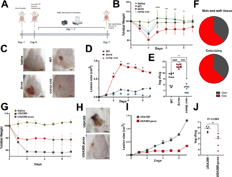
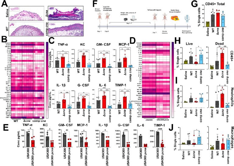
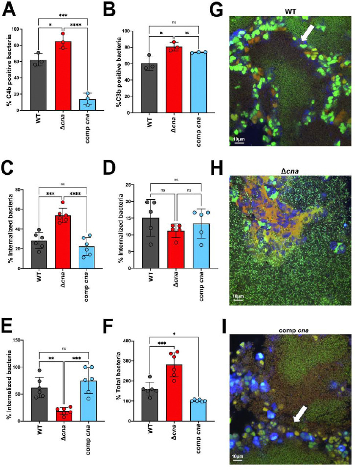
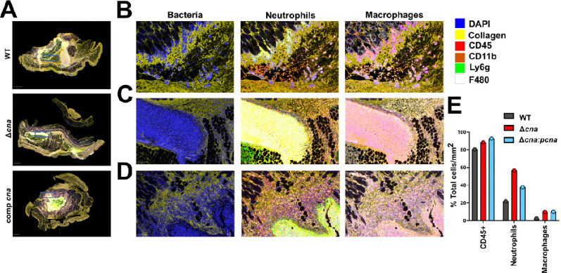
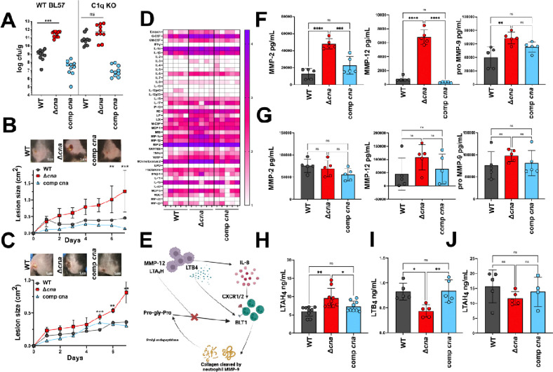
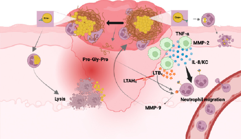
Staphylococcus aureus causes approximately 80% of skin and soft tissue infections (SSTIs). Collagen is the most abundant human extracellular matrix protein with critical roles in wound healing, and S. aureus encodes a collagen binding adhesin (Cna). The role of this protein during skin infections is unknown. Here we report that inability to bind collagen results in worsened pathology of intradermal Δcna S. aureus infection. WT/Cna+ S. aureus showed reduced infection severity, aggregate formation, and significantly improved clearance of bacteria. Cna binds to the collagen-like domain of serum C1q protein to reduce its opsonophagocytic functions. We demonstrate that infection of C1qKO mice with WT bacteria show results similar to the Δcna group. Conversely, inability to bind collagen resulted in an amplified inflammatory response caused in part by macrophage and neutrophil small molecule mediators released at the infection site (MMP-9, MMP-12, LTB4), resulting in increased immune cell infiltration and death.

Keywords: C1q; Collagen; MRSA; inflammation; skin.


Conflict of interest statement

Declaration of interests. The authors declare no competing interests.

Figures

Figure 1.
Figure 1.. Collagen binding adhesin reduces severity of S. aureus skin infection.
Method used for intradermal S. aureus infection and abscess formation (A). Weight loss measured over the 7-day infection period in mice (n=10 per bacterial strain) infected with WT MW2, isogenic Δcna or comp cna bacteria and calculated as a percentage of values at day 1(B). Images of abscess lesions taken at day 7, representative of mice infected with strains mentioned in B (C). Measurement of lesion sizes from mice infected with strains as above over a period of 7 days. Measurements were made using Image J (D). Bacterial burdens enumerated per gram of homogenized tissue excised at day 7 post infection with strains as described above (E). Percent distribution of the cna gene as detected in S. aureus clones associated with human skin and soft tissue infections (n=20) and colonizing healthy anterior nares (n=30) (F) Abscess model of skin infection performed as described in A, to compare pathology caused by USA300 (Cna−) and USA300:pcna (Cna+) bacteria (n=5 per group). Weight loss was measured over 7 days and calculated as a percentage of values at day 0 (G). Representative images of lesions formed 7 days post infection with strains mentioned in G (H). Lesion sizes measured over 7 days post infection with strains as described in G. Measurements were made using Image J (I). Colony forming units (CFU) per gram of homogenized tissue enumerated from abscesses biopsied from mice infected with strains as mentioned in G, at day 7 post inoculation (J). Results are representative of 3 (MW2) and 2 (USA300) independent analyses. Statistical analyses were performed with a one-way ANOVA (E) a two tailed Students t-test (I) or a two-way ANOVA with Bonferroni post test. Error was calculated based on SEM. Groups with significant differences are denoted. *P<0.05, **P<0.01, ****P<0.0001.
Figure 2.
Figure 2.. Expression of collagen binding adhesin is sufficient to restrict host inflammatory response.
Hematoxylin Eosin staining of tissue sections biopsied from mice infected with WT, isogenic Δcna or comp cna bacteria at day 7 post infection. Tissue section of mouse injected with saline is shown as a negative control (A). Cytokine array performed on mouse tissue collected from animals infected as described for A. The multiplexing analysis was performed to measure the concentration of 44 cytokines, using the Luminex 200 system by Eve Technologies Corp. Each column represents results from a single mouse (n=5 per group) (B). Individual graphs to show differences in cytokine measurements as made in B, for 8 cytokines of interest generated from mice in response to respective bacterial strains (C). Quantification of cytokine concentrations similar to B, made from mice infected with USA300 or the isogenic USA300:pcna strain (n=5 per group) (D). Individual graphs shown for 8 cytokines similar to C, measured from abscesses infected with strains as mentioned in D, 7 days post bacterial inoculation (E). Summary of methods used to perform flow cytometry quantification of immune cells from abscesses infected with bacteria as described for A (F). Quantification of the total number of single immune cells using an antibody specific to CD45, from tissue samples collected as described in F, for mice infected with bacterial strains described in A (n=7) (G). Differentiation of cells enumerated in G, based on exclusion of Am Cyan viability dye(live) from observably dead populations (H). Sub populations of total CD45 cells classified as neutrophils (I) or macrophages (J) based on staining with cell specific antibodies as described in Figure S2A, B. Results are representative of 3 independent analyses. Statistical analyses were performed with a one-way ANOVA with a Bonferroni post-test. Error was calculated based on SEM. *P<0.05, **P<0.01, ****P<0.0001
Figure 3.
Figure 3.. Cna binds to collagen motifs to alter the neutrophil response to bacteria.
Flow cytometry of WT, Δcna or comp cna bacteria opsonized with 10% serum and stained with antibodies targeting serum complement proteins C4b (A) and C3b (B). Uptake of bacterial strains mentioned above by primary human neutrophils, following opsonization with 10% pooled human serum. CFU was enumerated 10 minutes post incubation, following which samples were treated with lysostaphin to exclude extracellular populations (C). Experiment similar to C performed with bacteria opsonized with 10% C1q-depleted, pooled human serum (D). Experiments similar to C, with intracellular (E) and total (F) bacterial survival calculated at 30 minutes post incubation. Confocal microscopy performed on respective bacterial strains (Green=Syto-9/live) opsonized as mentioned above in the presence of type 1 collagen and incubated with Cell Tracker Blue-labelled primary human neutrophils for 20 minutes, following which samples were stained with ethidium homodimer-1 (red) to visualize dead/dying cells. Arrows indicate likely location of collagen boundary (G- I). Statistical analyses were performed with a one-way ANOVA and Bonferroni post-test. Error was calculated based on SEM. *P<0.05, **P<0.01, ****P<0.0001
Figure 4.
Figure 4.. S. aureus- collagen aggregates trigger increased neutrophilic response in skin abscess infection.
Multispectral quantitative pathology of skin abscess tissue excised from mice infected with WT, Δcna or comp cna bacteria 3 days post inoculation and stained with antibodies targeting 6 host proteins as described in the legend. Images depict staining of entire tissue section as used for quantitative analysis (A). Images digitally zoomed in (X5.9) from sections shown in A. Regions of interest are depicted in A as blue boxes. Images demonstrate staining from tissues infected with WT (B) Δcna (C) or comp cna (D) and stained with antibodies targeting 6 host proteins as shown in legend. Images are separated according to channels that demonstrate spatial distribution of bacteria with collagen (DAPI, Collagen), neutrophils (CD45, CD11b) and macrophages (CD45, F480). Quantification of staining demonstrated in A represented as total cells per millimeter of tissue sections infected with strains as described above (E).
Figure 5.
Figure 5.. C1q-Cna binding controls matrix metalloprotease activity and inflammation in skin abscess.
CFU per gram of homogenized tissue enumerated from either WT BL57 or C1qKO mice, 7 days post infection with WT, Δcna or comp cna bacteria (A). Representative images of lesions formed at day 7 post infection of WT BL57 (B) or C1qKO (C) mice with strains described in A (top). Lesion sizes measured over the course of the infection for each strain (n=10 per group, bottom). Comprehensive view for the concentrations of 44 inflammatory cytokines measured from abscess tissue 7 days after infection with respective bacteria, in the C1qKO mouse background. Concentrations are presented in logarithmic scale of picogram per mL homogenized tissue. Each column represents results from a single mouse (n=5 per group)(D). Graphical depiction of the known mechanisms by which matrix metalloproteases 9 and 12 can instigate a cascade of neutrophil and macrophage mediated inflammation of skin infection sites (E). Concentrations of MMP-2, pro-9 and 12 measured from abscess tissue excised from WT BL57 (F) or C1qKO (G) mice infected with respective strains. Concentrations of leukotriene A-4 hydrolase (LTAH4) measured using an enzyme linked immunosorbent assay, from WT BL57 mice infected with bacterial strains as described in G (H). Assay similar to H measuring the concentrations of leukotriene B4 from homogenized tissue samples (I). Assay similar to H, quantifying the concentrations of LTAH4 and performed with tissue samples from C1qKO mice infected with bacterial strains as described in H (J). Results are representative of 2 independent analyses. Statistical analyses were performed with a two-way ANOVA (B, C) or a one-way ANOVA (A, F-J) with Bonferroni posttest. Error was calculated based on SEM. *P<0.05, **P<0.01
Figure 6.
Figure 6.. Summary of results.
Expression of Cna allows S. aureus to bind to collagen and restrict bacterial contact with immune cells such as neutrophils. The N-terminal collagen like domain of C1q binds to Cna and reduces complement deposition and opsonophagocytosis by neutrophils. Bacteria that are taken up are eliminated by neutrophils (A). Bacteria lacking the ability to express Cna cannot directly bind to collagen. This allows direct contact between neutrophils and bacteria. C1q is not sequestered in the absence of Cna, causing increased bacterial uptake by neutrophils. This leads to neutrophil lysis and inflammation (B). Macrophages in the infection bed will release proinflammatory mediators including the matrix metalloprotease MMP-2, MMP-12 and neutrophil chemokine, IL-8. Macrophages also express leukotriene A4 hydrolase (LTAH4) in response to infection, which activates the soluble effector, leukotriene B 4 (LTB4). Neutrophils release MMP-9 which breaks down collagen to inflammatory Pro-Gly-Pro which assists in further neutrophil recruitment (C). Image created using Biorender.com

References

    1. Ricard-Blum S. (2011). The Collagen Family. Cold Spring Harb Perspect Biol 3, 1–19. 10.1101/CSHPERSPECT.A004978. - DOI - PMC - PubMed
    1. Mathew-Steiner S.S., Roy S., and Sen C.K. (2021). Collagen in Wound Healing. - PMC - PubMed
    1. Valotteau C., Prystopiuk V., Pietrocola G., Rindi S., Peterle D., De Filippis V., Foster T.J., Speziale P., and Dufrêne Y.F. (2017). Single-Cell and Single-Molecule Analysis Unravels the Multifunctionality of the Staphylococcus aureus Collagen-Binding Protein Cna. ACS Nano 11, 2160–2170. 10.1021/ACSNANO.6B08404. - DOI - PubMed
    1. Del Giudice P. (2020). Skin Infections Caused by Staphylococcus aureus. Acta Derm Venereol 100, 208–215. 10.2340/00015555-3466. - DOI - PMC - PubMed
    1. Esposito S., Noviello S., and Leone S. (2016). Epidemiology and microbiology of skin and soft tissue infections. Curr Opin Infect Dis 29, 109–115. 10.1097/QCO.0000000000000239. - DOI - PubMed

Publication types

LinkOut - more resources