[Preprint]. 2024 Nov 3:2024.10.31.621317.
doi: 10.1101/2024.10.31.621317.

An alternative route for β-hydroxybutyrate metabolism supports fatty acid synthesis in cancer cells

Affiliations

An alternative route for β-hydroxybutyrate metabolism supports fatty acid synthesis in cancer cells

Faith C Kaluba et al. bioRxiv. .

Update in

Abstract

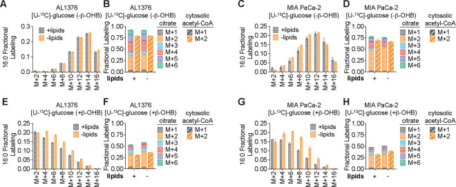
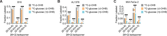
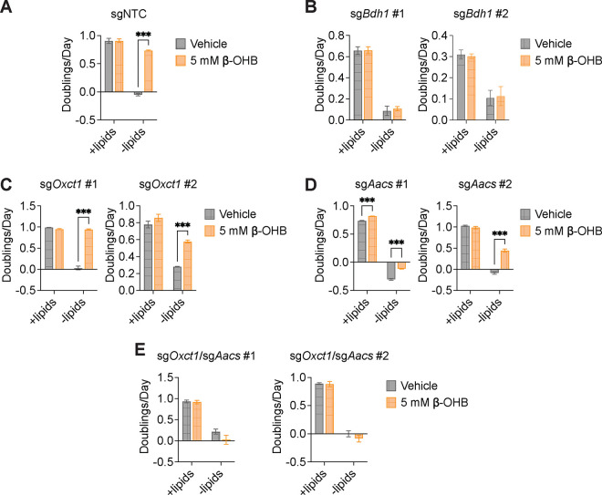
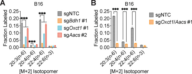
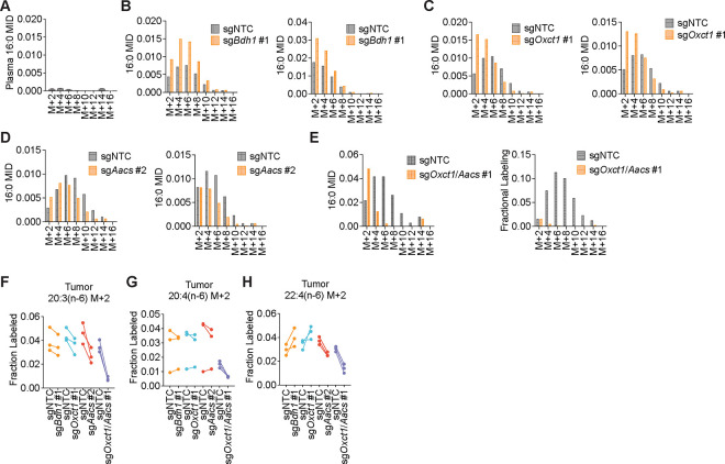
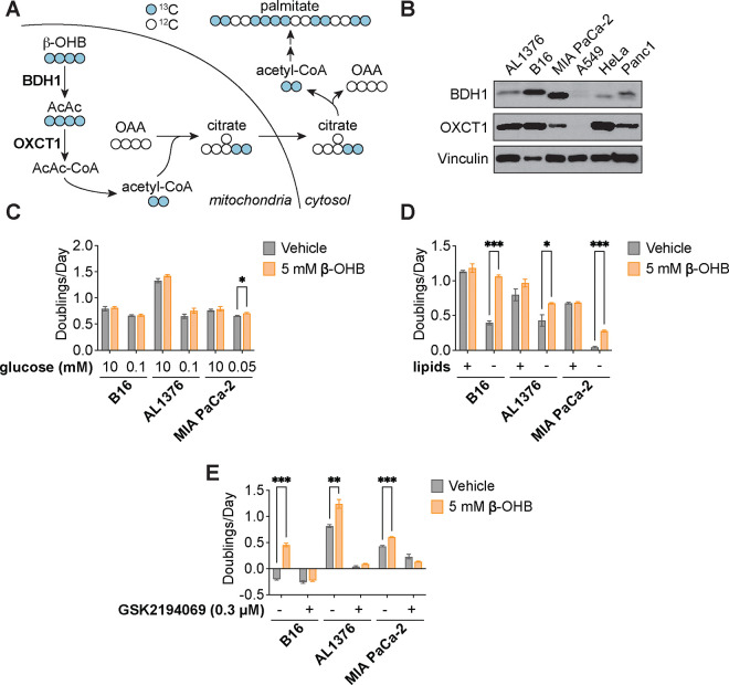
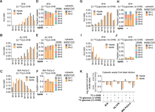
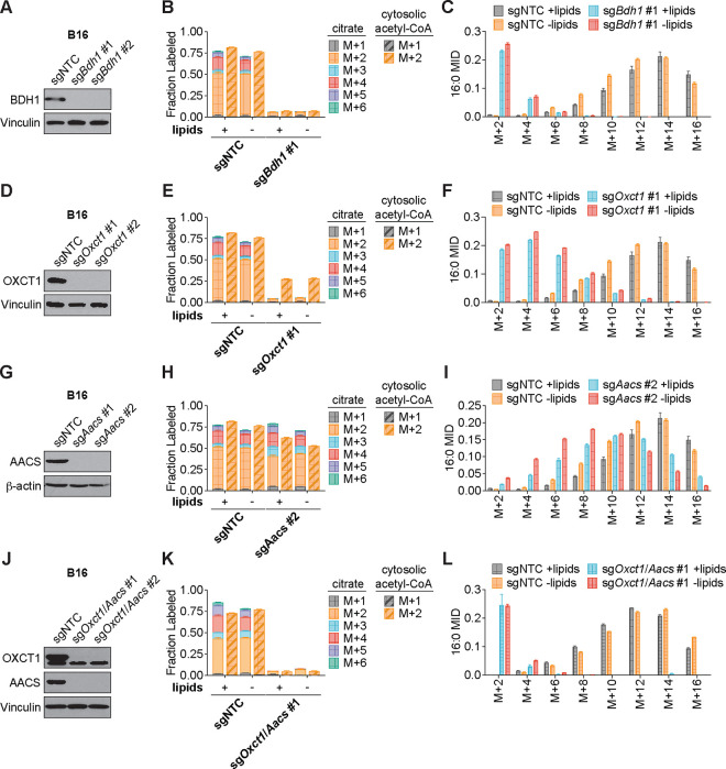
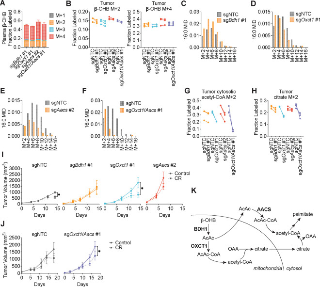
Cancer cells are exposed to diverse metabolites in the tumor microenvironment that are used to support the synthesis of nucleotides, amino acids, and lipids needed for rapid cell proliferation1-3. Recent work has shown that ketone bodies such as β-hydroxybutyrate (β-OHB), which are elevated in circulation under fasting conditions or low glycemic diets, can serve as an alternative fuel that is metabolized in the mitochondria to provide acetyl-CoA for the tricarboxylic acid (TCA) cycle in some tumors4-7. Here, we discover a non-canonical route for β-OHB metabolism, in which β-OHB can bypass the TCA cycle to generate cytosolic acetyl-CoA for de novo fatty acid synthesis in cancer cells. We show that β-OHB-derived acetoacetate in the mitochondria can be shunted into the cytosol, where acetoacetyl-CoA synthetase (AACS) and thiolase convert it into acetyl-CoA for fatty acid synthesis. This alternative metabolic routing of β-OHB allows it to avoid oxidation in the mitochondria and net contribute to anabolic biosynthetic processes. In cancer cells, β-OHB is used for fatty acid synthesis to support cell proliferation under lipid-limited conditions in vitro and contributes to tumor growth under lipid-limited conditions induced by a calorie-restricted diet in vivo. Together, these data demonstrate that β-OHB is preferentially used for fatty acid synthesis in cancer cells to support tumor growth.


Conflict of interest statement

Competing interests: R.G.J. is a scientific advisor to Servier Pharmaceuticals and is a member of the Scientific Advisory Board of Immunomet Therapeutics. All other authors declare no competing interests.

Figures

Extended Data Figure 1.
Extended Data Figure 1.. β-OHB is preferentially used over glucose for fatty acid synthesis, Related to Figure 2.
A-D, 16:0 MID (A, C) and citrate (solid bars) and cytosolic acetyl-CoA (dashed bars) MID (B, D) for AL1376 (A, B) and MIA PaCa-2 (C, D) cells labeled with [U-13C]-glucose for 48 h in lipid-replete versus lipid-depleted culture media without unlabeled β-OHB. E-H, 16:0 MID (E, G) and citrate (solid bars) and cytosolic acetyl-CoA (dashed bars) MID (F, H) for AL1376 (E, F) and MIA PaCa-2 (G, H) cells labeled with [U-13C]-glucose for 48 h in lipid-replete versus lipid-depleted culture media with 5 mM unlabeled β-OHB. Data are presented as mean ± s.e.m; n = 3 biologically independent replicates.
Extended Data Figure 2.
Extended Data Figure 2.. β-OHB is preferentially used over glucose for PUFA elongation, Related to Figure 2.
A-C, [M+2] fractional labeling of 20:3(n-6), 20:4(n-6), 22:4(n-6), and 22:6(n-3) in B16 (A), AL1376 (B), and MIA PaCa-2 (C) cells labeled with [U-13C]-β-OHB, [U-13C]-glucose without unlabeled β-OHB, or [U-13C]-glucose with 5 mM unlabeled β-OHB for 48 h in lipid-replete media. Data are presented as mean ± s.e.m; n = 3 biologically independent replicates. Comparisons were made using a two-tailed Student’s t test (A-C). ***P<0.001.
Extended Data Figure 3.
Extended Data Figure 3.. β-OHB can contribute to fatty acid synthesis through a citrate-independent route that requires AACS, Related to Figure 3.
A-E, Proliferation rates of the indicated B16 sgNTC (A), sgBdh1 (B), sgOxct1 (C), sgAacs (D), and sgOxct1/Aacs (E) cells grown in lipid-replete versus lipid-depleted culture media, with or without 5 mM β-OHB. Data are presented as mean ± s.e.m; n = 3 biologically independent replicates. Comparisons were made using a two-way ANOVA. ***P<0.001.
Extended Data Figure 4.
Extended Data Figure 4.. β-OHB contributes to PUFA elongation through both OXCT1 and AACS, Related to Figure 3.
A, [M+2] fractional labeling of 20:3(n-6), 20:4(n-6), 22:4(n-6), and 22:6(n-3) in B16 sgNTC, sgBdh1 #1, sgOxct1 #1, and sgAacs #2 cells labeled with [U-13C]-β-OHB for 48 h in lipid-replete media. B, [M+2] fractional labeling of 20:3(n-6), 20:4(n-6), 22:4(n-6), and 22:6(n-3) in B16 sgNTC and sgOxct1/Aacs #1 cells labeled with [U-13C]-β-OHB for 48 h in lipid-replete media. Data are presented as mean ± s.e.m; n = 3 biologically independent replicates. Comparisons were made using a two-way ANOVA. ***P<0.001.
Extended Data Figure 5.
Extended Data Figure 5.. β-OHB contributes to fatty acid synthesis through both OXCT1 and AACS in tumors in vivo, Related to Figure 4.
C57BL/6J mice bearing a B16 sgNTC tumor on one flank and a knockout tumor on the other flank were infused with [U-13C]-β-OHB for 6.5 h. A, Plasma 16:0 MID in all mice. B-E, 16:0 MIDs from each mouse bearing an sgNTC tumor versus a sgBdh1 #1 tumor (B), sgOxct1 #1 tumor (C), sgAacs #2 tumor (D), and sgOxct1/Aacs #1 tumor (E). F-H, [M+2] fractional labeling of 20:3(n-6) (F), 20:4(n-6) (G), and 22:4(n-6) (H) in the indicated sgNTC versus knockout tumors. Data are paired between sgNTC and knockout tumors from the same mouse. n = 3 biological replicates for each knockout tumor. Data are presented as mean ± s.e.m.
Figure 1.
Figure 1.. β-OHB promotes the proliferation of lipid-starved cancer cells.
A, Schematic of β-OHB metabolism and 13C labeling derived from [U-13C]-β-OHB. AcAc, acetoacetate; BDH1, β-OHB dehydrogenase 1; OXCT1, 3-oxoacid CoA-transferase 1; OAA, oxaloacetate. B, Immunoblot for BDH1, OXCT1, and vinculin in the indicated cancer cell lines. C, Proliferation rates of the indicated cancer cell lines grown in high or low glucose conditions, with or without 5 mM β-OHB. D, Proliferation rates of the indicated cancer cell lines grown in lipid-replete versus lipid-depleted culture media, with or without 5 mM β-OHB. E, Proliferation rates of the indicated cancer cell lines grown in lipid-depleted media, with or without 5 mM β-OHB and 0.3 μM of the FASN inhibitor GSK2194069. Data are presented as mean ± s.e.m; n = 3 biologically independent replicates. Comparisons were made using a two-tailed Student’s t test (C-E). *P<0.05, **P<0.01, ***P<0.001.
Figure 2.
Figure 2.. β-OHB is preferentially used over glucose for fatty acid synthesis.
A-C, 16:0 mass isotopomer distribution (MID) for B16 (A), AL1376 (B), and MIA PaCa-2 (C) cells labeled with [U-13C]-β-OHB for 48 h in lipid-replete versus lipid-depleted culture media. D-F, Citrate MID (solid bars) and cytosolic acetyl-CoA MID (dashed bars) for B16 (D), AL1376 (E), and MIA PaCa-2 (F) cells labeled with [U-13C]-β-OHB for 48 h in lipid-replete versus lipid-depleted culture media. G-J, 16:0 MID (G, I) and citrate and cytosolic acetyl-CoA MID (H, J) for B16 cells labeled with [U-13C]-glucose for 48 h in lipid-replete versus lipid-depleted culture media, either without unlabeled β-OHB (G, H) or with 5 mM unlabeled β-OHB (I, J). K, Cytosolic acetyl-CoA label dilution from citrate, as calculated by the log2(fold change) of the total fraction of cytosolic acetyl-CoA labeled versus the total fraction of citrate labeled, in B16, AL1376, and MIA PaCa-2 cells under the indicated tracing conditions. Data are presented as mean ± s.e.m; n = 3 biologically independent replicates.
Figure 3.
Figure 3.. β-OHB can contribute to fatty acid synthesis through a citrate-independent route that requires AACS.
A, Immunoblot for BDH1 and vinculin in the indicated B16 knockout lines. NTC, non-targeting control. B, C, Citrate (solid bars) and cytosolic acetyl-CoA (dashed bars) MIDs (B), and 16:0 MID (C) in B16 sgNTC versus sgBdh1 #1 cells labeled with [U-13C]-β-OHB for 48 h in lipid-replete versus lipid-depleted culture media. D, Immunoblot for OXCT1 and vinculin in the indicated B16 knockout lines. E, F, Citrate (solid bars) and cytosolic acetyl-CoA (dashed bars) MIDs (E), and 16:0 MID (F) in B16 sgNTC versus sgOxct1 #1 cells labeled with [U-13C]-β-OHB for 48 h in lipid-replete versus lipid-depleted culture media. G, Immunoblot for AACS and β-actin in the indicated B16 knockout lines. H, I, Citrate (solid bars) and cytosolic acetyl-CoA (dashed bars) MIDs (H), and 16:0 MID (I) in B16 sgNTC versus sgAacs #2 cells labeled with [U-13C]-β-OHB for 48 h in lipid-replete versus lipid-depleted culture media. J, Immunoblot for OXCT1, AACS, and vinculin in the indicated B16 knockout lines. K, L, Citrate (solid bars) and cytosolic acetyl-CoA (dashed bars) MIDs (K), and 16:0 MID (L) in B16 sgNTC versus sgOxct1/Aacs #1 cells labeled with [U-13C]-β-OHB for 48 h in lipid-replete versus lipid-depleted culture media. Data are presented as mean ± s.e.m; n = 3 biologically independent replicates.
Figure 4.
Figure 4.. β-OHB contributes to fatty acid synthesis through both OXCT1 and AACS in tumors in vivo.
C57BL/6J mice bearing a B16 sgNTC tumor on one flank and a knockout tumor on the other flank were infused with [U-13C]-β-OHB for 6.5 h. A, Plasma β-OHB labeling in mice bearing the indicated knockout tumors. B, [M+2] and [M+4] fractional labeling of β-OHB in the indicated sgNTC versus knockout tumors. Data are paired between sgNTC and knockout tumors from the same mouse. n = 3 biological replicates for each knockout tumor. C-F, Representative 16:0 MIDs from n = 1 biological replicate of a mouse bearing an sgNTC tumor versus a sgBdh1 #1 tumor (C), sgOxct1 #1 tumor (D), sgAacs #2 tumor (E), and sgOxct1/Aacs #1 tumor (F). See Extended Data Fig. 5 for additional biological replicates. G, H, [M+2] fractional labeling of cytosolic acetyl-CoA (G) and citrate (H) in the indicated sgNTC versus knockout tumors. Data are paired between sgNTC and knockout tumors from the same mouse. n = 3 biological replicates for each knockout tumor. I, J, Tumor volumes of subcutaneous B16 sgNTC, sgBdh1 #1, sgOxct1 #1, sgAacs #2 (I), and sgOxct1/Aacs #1 (J) allografts in male mice exposed to a control or caloric restriction (CR) diet. (I) sgNTC Control n = 6 mice, CR n = 6 mice; sgBdh1 #1 Control n = 6 mice, CR n = 6 mice; sgOxct1 #1 Control n = 5 mice, CR n = 5 mice; sgAacs #2 Control n = 6 mice, CR n = 6 mice. (J) sgNTC Control n = 3 mice, CR n = 4 mice; sgOxct1/Aacs #1 Control n = 4 mice, CR n = 4 mice. K, Schematic of β-OHB contribution to fatty acid synthesis through both a mitochondrial citrate-dependent route via OXCT1 and a citrate-independent route via AACS. Data are presented as mean ± s.e.m. Comparisons were made using a two-way ANOVA (I, J). *P<0.05.

References

    1. Hanahan D. & Weinberg R. A. Hallmarks of cancer: the next generation. Cell 144, 646–74 (2011). - PubMed
    1. Vander Heiden M. G. & DeBerardinis R. J. Understanding the Intersections between Metabolism and Cancer Biology. Cell 168, 657–669 (2017). - PMC - PubMed
    1. Finley L. W. S. What is cancer metabolism? Cell 186, 1670–1688 (2023). - PMC - PubMed
    1. Lien E. C. et al. Low glycaemic diets alter lipid metabolism to influence tumour growth. Nature 599, 302–307 (2021). - PMC - PubMed
    1. Gouirand V. et al. Ketogenic HMG-CoA lyase and its product β-hydroxybutyrate promote pancreatic cancer progression. EMBO J 41, e110466 (2022). - PMC - PubMed

Publication types

LinkOut - more resources