[Preprint]. 2024 Nov 1:2024.11.01.621488.
doi: 10.1101/2024.11.01.621488.

Structural Model of Bacteriophage P22 Scaffolding Protein in a Procapsid by Magic-Angle Spinning NMR

Affiliations

Structural Model of Bacteriophage P22 Scaffolding Protein in a Procapsid by Magic-Angle Spinning NMR

Changmiao Guo et al. bioRxiv. .

Abstract

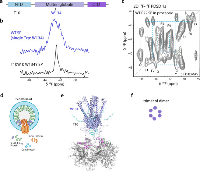
Icosahedral dsDNA viruses such as the tailed bacteriophages and herpesviruses have a conserved pathway to virion assembly that is initiated from a scaffolding protein driven procapsid formation. The dsDNA is actively packaged into procapsids, which undergo complex maturation reactions to form infectious virions. In bacteriophage P22, scaffolding protein (SP) directs the assembly of coat proteins into procapsids that have a T=7 icosahedral arrangement, en route to the formation of the mature P22 capsid. Other than the C-terminal helix-turn-helix involved in interaction with coat protein, the structure of the P22 303 amino acid scaffolding protein within the procapsid is not understood. Here, we present a structural model of P22 scaffolding protein encapsulated within the 23 MDa procapsid determined by magic angle spinning NMR spectroscopy. We took advantage of the 10-fold sensitivity gains afforded by the novel CPMAS CryoProbe to establish the secondary structure of P22 scaffolding protein and employed 19F MAS NMR experiments to probe its oligomeric state in the procapsid. Our results indicate that the scaffolding protein has both α-helical and disordered segments and forms a trimer of dimers when bound to the procapsid lattice. This work provides the first structural information for P22 SP beyond the C-terminal helix-turn-helix and demonstrates the power of MAS NMR to understand higher-order viral protein assemblies involving structural components that are inaccessible to other structural biology techniques.

Keywords: 19F NMR; Biological Sciences- Biophysics; Structural Biology; bacteriophage p22 procapsid; icosahedral dsDNA viruses; magic-angle spinning NMR; scaffolding protein.


Conflict of interest statement

DECLARATION OF INTERESTS The authors declare no conflict of interest.

Figures

Fig. 1.
Fig. 1.
a Schematic representation of bacteriophage P22 procapsid assembly. b 2D 13C-13C cross-polarization (CP)-based CORD MAS NMR spectrum of U-[13C,15N]-labeled P22 SP assembled in PC; the mixing time was 50 ms. c 2D direct-polarization-based DARR spectrum (gray) mixing overlaid with 2D CP-CORD spectrum (blue); the mixing time was 150 ms. The spectra were acquired at 20.0 T; the MAS frequency was 14 kHz. d Sequence and secondary structure of the P22 scaffolding protein. The N- and C-terminal domains of SP are colored in cyan. The cylinders represent α-helices. e AlphaFold 3 structural model of the P22 SP monomer.
Fig. 2.
Fig. 2.
a 2D dipolar-based 15N-1H HETCOR and scalar-based INEPT MAS NMR spectra of deuterated U-[13C,15N]-labeled P22 SP assembled in PC. The CP-based HETCOR spectrum with resolution enhancement processing is shown in purple. Rigid and highly dynamic residues in SP are mapped to the SP sequence and model, colored in purple and cyan, respectively. b,c 2D 13C-13C scalar-based INEPT-TOBSY and 2D dipolar-based 13C-1H HETCOR spectrum of U-[13C,15N]-SP PC assembly. d,e Backbone resonance assignments for the S285-V289 segment in P22 SP assembly (d), using the 3D 13C-detected NCACX and NCOCX spectra (e) acquired with the CPMAS CryoProbe.
Fig. 3.
Fig. 3.
a,b 2D CORD spectrum of P22 SP assembled in PC acquired with mixing times of 200 ms (blue) and 500 ms (gray). The aliphatic-aromatic and aromatic-aromatic regions are shown in a and b, respectively c Aromatic contacts in the 2D CORD spectrum with 50 ms. d Aromatic-based restraints mapped on the SP model.
Fig. 4.
Fig. 4.
a Domain organization of P22 SP. b 1D 19F MAS NMR spectrum of 5F-Trp WT SP (purple) and T10W/W134Y SP variant (black) assembled in PC. c 2D 19F-19F dipolar-based spin diffusion (SD) MAS NMR spectrum of 5F-Trp WT SP PC assembly. d Schematic illustration of the P22 PC assembly to show the structural variations of SP pairs near the portal vertex and interacting with the capsid lattice. e Structural model of the P22 SP oligomer in PC. ESMFold (ref) model of SP and cryo-EM density map of SP CTD and coat proteins (PDB 8I1V) were used. f Schematics of the possible arrangements for the trimer of dimers of P22 SP in the PC.

Similar articles

References

    1. Fuller M. & King J. Regulation of coat protein polymerization by the scaffolding protein of bacteriophage P22. Biophysical Journal 32, 381–401 (1980). - PMC - PubMed
    1. Newcomb W. W., Homa F. L., Thomsen D. R. & Brown J. C. In vitro assembly of the herpes simplex virus procapsid: formation of small procapsids at reduced scaffolding protein concentration. Journal of Structural Biology 133, 23–31 (2001). - PubMed
    1. Ostapchuk P., Almond M. & Hearing P. Characterization of empty adenovirus particles assembled in the absence of a functional adenovirus IVa2 protein. Journal of Virology 85, 5524–5531 (2011). - PMC - PubMed
    1. Casjens S. & King J. P22 morphogenesis I: Catalytic scaffolding protein in capsid assembly. Journal of Supramolecular Structure 2, 202–224 (1974). - PubMed
    1. Israel V. E proteins of bacteriophage P22. I. Identification and ejection from wild-type and defective particles. Journal of Virology 23, 91–97 (1977). - PMC - PubMed

Publication types