[Preprint]. 2024 Oct 28:2024.10.23.619897.
doi: 10.1101/2024.10.23.619897.

Coordinated translational control of multiple immune checkpoints by the integrated stress response pathway in lung cancer

Affiliations

Coordinated translational control of multiple immune checkpoints by the integrated stress response pathway in lung cancer

Shayna Thomas-Jardin et al. bioRxiv. .

Update in

Abstract

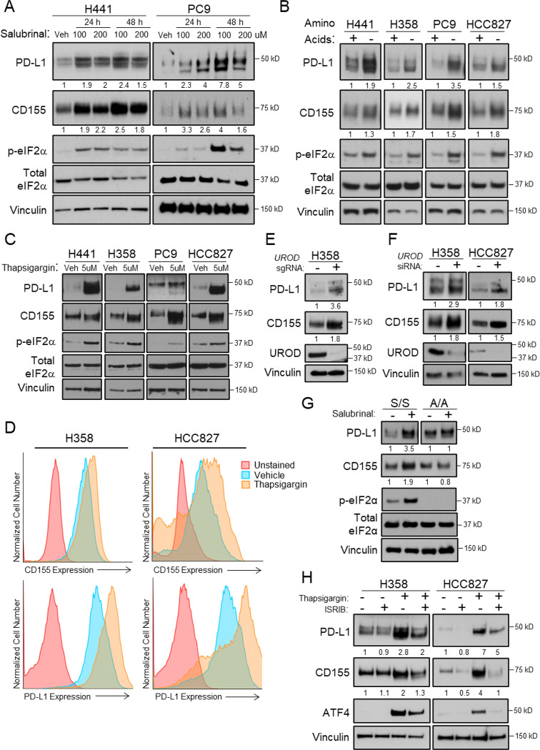
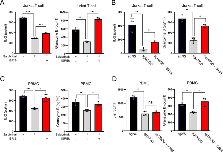
The integrated stress response (ISR) is an adaptive pathway hijacked by cancer cells to survive cellular stresses in the tumor microenvironment. ISR activation potently induces Programmed Death Ligand 1 (PD-L1), leading to suppression of anti-tumor immunity. Here we sought to uncover additional immune checkpoint proteins regulated by the ISR to elucidate mechanisms of tumor immune escape. We show that CD155 and PD-L1 are coordinately induced by the ISR, enhancing translation of both immune checkpoint proteins through bypass of inhibitory upstream open reading frames (uORFs) in their 5' UTRs. Analysis of primary human lung tumors identifies a significant correlation between PD-L1 and CD155 expression. ISR activation accelerates tumorigenesis and inhibits T cell function, effects that can be overcome by combining PD-1 blockade with the ISR inhibitor ISRIB. These studies uncover a novel mechanism by which two immune checkpoint proteins are coordinately regulated and suggest a new therapeutic strategy for lung cancer patients.

Statement of significance: This study uncovers a novel mechanism for the coordinated translational regulation of the PD-L1/PD1 and CD155/TIGIT immune checkpoint pathways and highlights the ISR as a therapeutic vulnerability for lung cancer. Inhibition of the ISR pathway bolsters PD-1 blockade, potentially unveiling a new therapeutic strategy for lung cancer patients.


Conflict of interest statement

The authors declare no potential conflicts of interest.

Figures

Figure 1.
Figure 1.. ISR pathway activation induces PD-L1 and CD155 protein.
(A) Western blot analysis of human KRAS mutant H441 or EGFR mutant PC9 cells treated for 24 or 48 hours with 100uM or 200uM Salubrinal or DMSO vehicle control. Vinculin served as a loading control for this and subsequent western blots. (B) Western blot analysis of human H441, H358, PC9, and HCC827 cells after 24 hours in RPMI 1640 media supplemented with or without amino acids. (C) Western blot analysis of human H441, H358, PC9, and HCC827 cells treated for 24 hours with 5uM thapsigargin (ER Ca+ ATPase pump inhibitor) to induce ER stress, or DMSO vehicle control. (D) Flow cytometric analysis of cell-surface CD155 and PD-L1 protein in DMSO vehicle control and thapsigargin-treated (5uM for 24 hours) H358 or HCC827 NSCLC cells. (E) Western blot analysis of H358 cells with control or UROD sgRNA. (F) Western blot analysis of H358 or HCC827 cells with control or UROD siRNA. (G) Western blot analysis in eIF2α wildtype (S/S) or mutant (Ser51Ala A/A) mouse embryonic fibroblasts (MEFs) treated with DMSO vehicle control or 100uM Salubrinal for 24 hours. (H) Western blot analysis of thapsigargin and ISRIB treated human NSCLC cells. Data from a single experiment are shown and are representative of at least 3 independent experiments.
Figure 2.
Figure 2.. ISR pathway activation enhances PD-L1 and CD155 translation. (
A) Polysome profiling of H1944 Vehicle or Salubrinal-treated cells (100uM for 24 hours). (B) Quantitative real-time PCR analysis of PD-L1(CD274) and CD155(PVR) mRNA (C) in ribosomal fractions from (A). qRT-PCR analysis for each gene shown was performed with 1 primer set spanning an exon-exon junction (Primer Set 1). Data for Primers Set 2 is available in Supplementary Figure 2. Fractions associated with <3 ribosomes were grouped to represent poorly translated mRNAs, fractions associated with >3 ribosomes were grouped as efficiently translated mRNAs. PD-L1 and CD155 mRNA expression in each fraction was normalized to Luciferase and mRNA abundance was calculated as the % of total in all fractions. Luciferase control mRNA was added to each fraction prior to RNA extraction to control for variability. Error bars represent standard deviation from the mean from three independent fractions (<3 or >3 ribosomes). (D) Diagram of the wildtype human CD155 5’ UTR with 6 CTGs, and mutant constructs with CTGs mutated to CTCs, cloned upstream of a luciferase reporter. (E) Dual luciferase assay of MEFs transfected with indicated CD155-5’ UTR-Firefly luciferase reporter constructs normalized to co-transfected control Renilla luciferase. Luciferase activity was monitored after 48h. Error bars represent standard deviation from the mean from n=3 biological replicates. Data from a single experiment are shown and representative of three independent experiments. (F) qRT-PCR analysis of mean luciferase mRNA normalized to actin in MEFs shown in (E). Error bars represent standard deviation from the mean from n=3 biological replicates. (G) Dual luciferase assay of the CD155 5’ UTR in MEFs, Vehicle or Salubrinal-treated (100uM for 24 hours). Error bars represent standard deviation from the mean from n=3 biological replicates. (H) qRT-PCR analysis of mean luciferase mRNA normalized to actin in MEFs shown in (G). n=3 biological replicates. A Student’s t-test was used to determine statistical significance (* p<0.05, ** p<0.005, *** p<0.0005).
Figure 3.
Figure 3.. ISR pathway activation diminishes immune cell function in vitro.
(A) ELISAs for IL-2 and Granzyme B of Jurkat T cells co-cultured with H358 cells. H358 cells were pre-treated for 24 hours with Salubrinal and 800nM ISRIB, then washed and co-cultured with Jurkat T cells for an additional 24 hours with α-CD3 and α-CD28 activating antibodies (4ug/ml each). Error bars represent standard deviation from the mean from n=3 biological replicates. Data from a single experiment are shown and representative of three independent experiments. (B) ELISAs for IL-2 and Granzyme B of Jurkat T cells co-cultured with H358 cells with control or UROD sgRNA and 800nM ISRIB. H358 cells were co-cultured with Jurkat T cells for 24 hours with α-CD3 and α-CD28 activating antibodies (4ug/ml each). Error bars represent standard deviation from the mean from n=3 biological replicates. Data from a single experiment are shown and representative of two independent experiments. (C) ELISAs for IL-2 and Granzyme B of primary human PBMCs co-cultured with H358 cells. H358 cells were pre-treated for 24 hours with Salubrinal and 800nM ISRIB, then washed and co-cultured with PBMCs for an additional 24 hours with α-CD3 and α-CD28 activating antibodies (1ug/ml each). Error bars represent standard deviation from the mean from n=3 biological replicates. Data from a single experiment are shown and representative of three independent experiments. (D) ELISAs for IL-2 and Granzyme B of PBMCs co-cultured with H358 cells with control or UROD sgRNA and 800nM ISRIB. H358 cells were co-cultured with PBMCs for 24 hours with α-CD3 and α-CD28 activating antibodies (1ug/ml each). Error bars represent standard deviation from the mean from n=3 biological replicates. Data from a single experiment are shown and representative of two independent experiments. A Student’s t-test was used to determine statistical significance (* p<0.05, ** p<0.005, *** p<0.0005).
Figure 4:
Figure 4:. ISR activation enhances tumorigenesis and reduces immune cell infiltration in vivo.
(A) Western blot analysis of KRAS mutant murine CMT167 cells treated for 24 or 48 hours with 100uM or 200uM Salubrinal or DMSO vehicle control. (B) Quantification of tumor volumes of CMT167 cells transplanted in C57BL/6J mice (n=11 mice per group for vehicle treated mice, n=10 mice per group for Salubrinal treated mice). Graph represents mean tumor volumes and error bars represent standard error of the mean. (C) End-point tumor volumes and (D) tumor mass of resected tumors shown in (B). Horizontal bars represent mean values. (E) Multiplexed IHC-F was performed on tumors from (B), 10X representative images shown, scale bar=200μm. (F,G,H) Quantification of CD3+, CD4+, and CD8+ tumor infiltrating lymphocytes (TILs, expressed as counts/mm3). 5 fields were quantified per mouse from 5 mice, graphs represent mean counts and error bars represent standard deviation from the mean. A Student’s t-test was used to determine statistical significance (* p<0.05, ** p<0.005, *** p<0.0005).
Figure 5.
Figure 5.. ISR pathway inhibition improves response to PD-1 blockade in a syngeneic mouse model.
(A) Western blot analysis of Kras mutant murine CMT167 cells expressing doxycycline-inducible control shRNA or two independent shRNAs targeting Urod. (B) Schematic illustration of CMT167 syngeneic experiment. (C) Quantification of tumor volumes of CMT167 cells expressing the indicated shRNA sequence transplanted in C57BL/6J mice (n=15 mice per group, scrambled shRNA data are shown in Supplementary Fig. 4). Graph represents mean tumor volumes and error bars represent standard error of the mean. (D) Uniform manifold approximation and projection (UMAP) analysis of tumor infiltrating lymphocytes (TILs) colored by cell types. (E) Quantification of TILs (expressed as a % of CD45+ cells) from flow mass cytometry (mass CyTOF). n=5 scrambled mice, n=5 Urod shRNA mice, n=5 Urod shRNA + ISRIB, n=4 Urod shRNA + αPD-1, n=5 Urod shRNA + ISRIB +αPD-1. Graphs represent mean values and error bars represent standard deviation from the mean. A Student’s t-test was used to determine statistical significance (* p<0.05, ** p<0.005, *** p<0.0005).
Figure 6:
Figure 6:. CD155 and PD-L1 are positively correlated in primary human lung adenocarcinoma tumors.
(A) Representative images of IHC for PD-L1 and CD155 of 33 primary human lung adenocarcinomas. 20X representative images shown, scale bar=100μm (B) Correlation of CD155 H-score and PD-L1 H-score of tissues in (A) (Pearson correlation r = 0.506, p = 0.0031). (C) Microphotographs of CD155 and PD-L1 immunohistochemistry analysis of primary lung adenocarcinomas displaying different combinations of expression. (CD155+/PDL1 +, CD155− /PD-L1 −, CD155+ /PD-L1−, CD155−/PD-L1 +). 20X representative images shown, scale bar=100μm. (D) Association of CD155 H-score expression in all primary lung adenocarcinoma with PD-L1 status (cut-off for positive PD-L1 expression: Tumor proportion score >=1%) (p= 0.0004). A Mann Whitney test was used to determine statistical significance. Graph represents the median; error bars represent the 95% confidence interval. (E) Association of CD155 H-score expression in all primary lung adenocarcinoma with pathological stage (p= 0.001). A Mann Whitney test was used to determine statistical significance. Graph represents the median; error bars represent the 95% confidence interval. (F) Correlation of CD155 H-score expression with tumor size (Spearman correlation r= 0.1946, p<0.0001).
Figure 7:
Figure 7:. Model of translational control of CD155 and PD-L1 in response to ISR activation.
Stresses commonly present in the tumor microenvironment activate the ISR pathway in tumor cells. This results in enhanced translation of CD155 and PD-L1, which suppresses T cell function by binding the inhibitory T cell receptors PD-1 and TIGIT respectively, thereby leading to enhanced tumorigenesis. Figure created with Biorender.com.

References

    1. Freeman GJ, Long AJ, Iwai Y, Bourque K, Chernova T, Nishimura H, et al. Engagement of the PD-1 immunoinhibitory receptor by a novel B7 family member leads to negative regulation of lymphocyte activation. J Exp Med. 2000;192(7):1027–34. - PMC - PubMed
    1. Dong H, Strome SE, Salomao DR, Tamura H, Hirano F, Flies DB, et al. Tumor-associated B7-H1 promotes T-cell apoptosis: a potential mechanism of immune evasion. Nat Med. 2002;8(8):793–800. - PubMed
    1. Barber DL, Wherry EJ, Masopust D, Zhu B, Allison JP, Sharpe AH, et al. Restoring function in exhausted CD8 T cells during chronic viral infection. Nature. 2006;439(7077):682–7. - PubMed
    1. Sharma P, Allison JP. Immune checkpoint targeting in cancer therapy: toward combination strategies with curative potential. Cell. 2015;161(2):205–14. - PMC - PubMed
    1. Meyers DE, Bryan PM, Banerji S, Morris DG. Targeting the PD-1/PD-L1 axis for the treatment of non-small-cell lung cancer. Current Oncology. 2018;25(4):e324–e34. - PMC - PubMed

Publication types