[Preprint]. 2024 Nov 1:2024.10.31.621372.
doi: 10.1101/2024.10.31.621372.

Elongation of the nascent avian foregut requires coordination of intrinsic and extrinsic cell behaviors

Affiliations

Elongation of the nascent avian foregut requires coordination of intrinsic and extrinsic cell behaviors

Olivia Powell et al. bioRxiv. .

Update in

Abstract

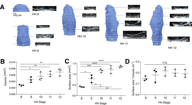
The foregut tube gives rise to the lungs and upper gastrointestinal tract, enabling vital functions of respiration and digestion. How the foregut tube forms during embryonic development has historically received considerable attention, but over the past few decades this question has primarily been addressed indirectly through studies on morphogenesis of the primitive heart tube, a closely related process. As a result, many aspects of foregut development remain unresolved. Here, we exploit the accessibility of the chick embryo to study the initial formation of the foregut tube, combining embryology with fate mapping, live imaging, and biomechanical analyses. The present study reveals that the foregut forms and elongates over a narrower time window than previously thought, and displays marked dorso-ventral and left-right asymmetries early in its development. Through tissue-specific ablation of endoderm along the anterior intestinal portal, we confirm its central biomechanical role in driving foregut morphogenesis, despite not directly contributing cells to the elongating tube. We further confirm the important role of this cell population in formation of the heart tube, with evidence that this role extends to later stages of cardiac looping as well. Together, these data reveal the need for an intricate balance between intrinsic cell behaviors and extrinsic forces for normal foregut elongation, and set the stage for future studies aimed at understanding the underlying molecular cues that coordinate this balance.


Figures

Figure 1:
Figure 1:. AIP displacement is poorly correlated with foregut elongation
(A, left) Representative depth-coded maximum intensity projections of DAPI-stained chick embryos from the initiation of foregut formation with the emergence of the head fold at HH stage 7 to onset of c-looping of the heart tube at HH stage 11. *indicates the position of the medial Anterior Intestinal Portal (AIP); scale = 200 μm. (A, right) Schematic sagittal section of the anterior chick embryo during foregut elongation. The forming foregut lumen is highlighted in blue, with the ventral foregut floor and dorsal foregut roof indicated. (B) AIP velocity between HH stages 8–9, 9–10, and 10–11 (n = 10 per group); *p<0.05, ***p<0.001 as determined by one-way ANOVA with Tukey’s multiple comparisons test. (C, left) HH stage 8 embryos following bisection (0 hours) to mechanically separate AIP from regressing node region and following 10 hours incubation ; scale = 200 μm. (C, right) Foregut length compared between bisected (n = 7) and intact (n = 8) control embryos; n.s. = not significant (p = 0.211 by Student’s t-test). (D, E) Foregut tube length normalized to antero-posterior embryo length (D) and foregut aspect ratio (E) across developmental stages HH stage 8- to 12 (n = 4–7 per stage). (F) Comparison of foregut elongation versus AIP displacement of individual embryos (n = 10 per stage) between HH stages 8–9 (left), 9–10 (middle), and 10–11 (right). Linear regression is indicated by a solid line, with shaded regions representing the 95% confidence interval calculated for the regression estimate, and dotted lines indicate y = x. Pearson correlation coefficient was used to measure the linear relationship between the two measures.
Figure 2:
Figure 2:. 3D volumetric imaging of the nascent foregut lumen
(A) At each HH stage between 8 and 12, 3-D reconstructions of the foregut lumen volume are shown at left, with transverse optical sections along the antero-posterior axis shown at right (lumen pseudocolored blue); scale bar = 100 μm. (B-D) Time course quantification of lumen volume (B), surface area (C) , and surface area-to-volume ratio (D) between HH stages 8 and 12; *p<0.05, **p<0.01, ***p<0.001 as determined by one-way ANOVA with Tukey’s multiple comparisons test (n = 3 per stage).
Figure 3:
Figure 3:. Fate maps reveal limited involution of the midline endoderm across the AIP
(A) Di labels injected onto AIP endoderm between HH stages 7–10 (left, 0 hours) and following 4 hours incubation (right); 4 hours spans the approximate time between HH stages in this range (HH7, n = 4; HH8, n = 7; HH9, n = 5; HH10, n = 3); scale = 200 μm. (B) DiI (red) and DiO (green) labels injected into midline presumptive roof endoderm at HH stage 8− move anteriorly toward the prechordal plate, but remain on the roof at HH stage 10+ (n = 4/4); scale = 200 μm. (C) Transverse sections through the foregut at HH stage 10 immunostained (red) for laminin (top) and E-cadherin (middle), and stained with phalloidin to visualize F-actin (bottom); counterstained with DAPI to visualize nuclei (cyan); R = foregut roof; F = foregut floor; scale = 50 μm (n = 3/3).
Figure 4:
Figure 4:. Role of endoderm contractility in AIP movement and FG formation
(A) mRNA electroporation 3nls-GFP and DeAct-GFP into AIP endoderm of HH stage 10 embryos followed by phalloidin staining to visualize effects on F-actin integrity. Phalloidin signal isolated in gray scale at right; scale = 20 μm. (B) Fast Green injection into the foregut lumen to visualize foregut length (quantified in C) immediately following electroporation with 3nl-GFP or DeAct-GFP mRNA at HH stage 8, and again after 10 hours incubation. Dashed white line indicates AIP lip; scale = 200 μm. (C) Percent elongation of foregut following mRNA electroporation of AIP endoderm with 3nls-GFP (n=9) or DeAct-GFP (n=10); ****p < 0.0001 by Student’s t-test. (D) Percentage of embryos displaying heart tube defects with 10 hours incubation following mRNA electroporation with 3nls-GFP (left) or DeAct-GFP mRNA (right). (E) Representative images of embryos electroporated at HH stage 8+ with 3nls-GFP, yielding normal control embryos (left), compared to DeAct-GFP electroporated embryos that displayed either heart tube malformations (middle) no heart tube at all (right). White dashed lines indicate heart tube boundary; scale = 200 μm.
Figure 5:
Figure 5:. Cell movements, morphology, and tissue deformations at the AIP during foregut elongation.
(A) Snapshots of a time lapse (Movie S1) dynamic changes in cell morphology of CAAX-EGFP electroporated endoderm cells at the AIP lip (dashed line) at HH stage 8-; red and yellow arrows indicate rapid polarization of cells; scale = 50 μm (B) Progressive time-projected cell tracks from time lapse (Movie S2) of a CAAX-GFP electroporated embryo spanning HH stages 8–11; white arrows indicate anterior cell flow in the presumptive foregut roof endoderm. Scale = 200 μm. (C) Whole-mount fluorescence imaging of tight junctions (ZO-1, green) and F-actin (Phalloidin, magenta), counterstained for nuclei (DAPI, blue), in the AIP endoderm (top, yellow box) and presumptive roof endoderm (bottom, orange box). White arrow indicates multicellular F-actin cables; scale =10 μm, brightfield image scale = 200 μm. (D) Velocity fields quantified from time lapse (Movie S2) of cell movements between HH stages 8+ and 10+. Direction of the velocity vector is indicated by the direction of arrows, and magnitude indicated by both the length of arrows and the underlying heat map (n = 3); scale = 200 μm. (E) Principal strain rates computed from velocity fields in (D), where major and minor cruciform axes represent maximum and minimum principal strain rates D1 and D2, respectively, and their orientations represent the associated principal directions. Negative/compressive principal strain rates are indicated in cyan, and positive/extensional strain rates indicated in red; strain rate scale (white) = 0.01%/s, spatial scale is as indicated in (D).

Similar articles

References

    1. Adelmann Howard B. 1922. “The Significance of the Prechordal Plate: An Interpretative Study.” American Journal of Anatomy 31 (1): 55–101. 10.1002/aja.1000310104. - DOI
    1. Aleksandrova Anastasiia, Andras Czirók Andras Szabó, Filla Michael B., Julius Hossain M., Whelan Paul F., Lansford Rusty, and Rongish Brenda J.. 2012. “Convective Tissue Movements Play a Major Role in Avian Endocardial Morphogenesis.” Developmental Biology 363 (2): 348–61. 10.1016/j.ydbio.2011.12.036. - DOI - PMC - PubMed
    1. Alsan Burak H., and Schultheiss Thomas M.. 2002. “Regulation of Avian Cardiogenesis by Fgf8 Signaling.” Development 129 (8): 1935–43. 10.1242/dev.129.8.1935. - DOI - PubMed
    1. Anderson Claire, Khan Mohsin A. F., Wong Frances, Solovieva Tatiana, Oliveira Nidia M. M., Baldock Richard A., Tickle Cheryll, Burt Dave W., and Stern Claudio D.. 2016. “A Strategy to Discover New Organizers Identifies a Putative Heart Organizer.” Nature Communications 7 (1): 12656. 10.1038/ncomms12656. - DOI - PMC - PubMed
    1. Angelo Jesse R., Guerrero-Zayas Mara-Isel, and Tremblay Kimberly D.. 2012. “A Fate Map of the Murine Pancreas Buds Reveals a Multipotent Ventral Foregut Organ Progenitor.” PLOS ONE 7 (7): e40707. 10.1371/journal.pone.0040707. - DOI - PMC - PubMed

Publication types