. 2024 Oct 30:9:100258.
doi: 10.1016/j.jtauto.2024.100258. eCollection 2024 Dec.

Homeostatic signals, including IL-7 and self-MHC recognition, induce the development of peripheral helper T cells, which are enriched in the joints of rheumatoid arthritis

Affiliations

Homeostatic signals, including IL-7 and self-MHC recognition, induce the development of peripheral helper T cells, which are enriched in the joints of rheumatoid arthritis

Ryosuke Tsurui et al. J Transl Autoimmun. .

Abstract

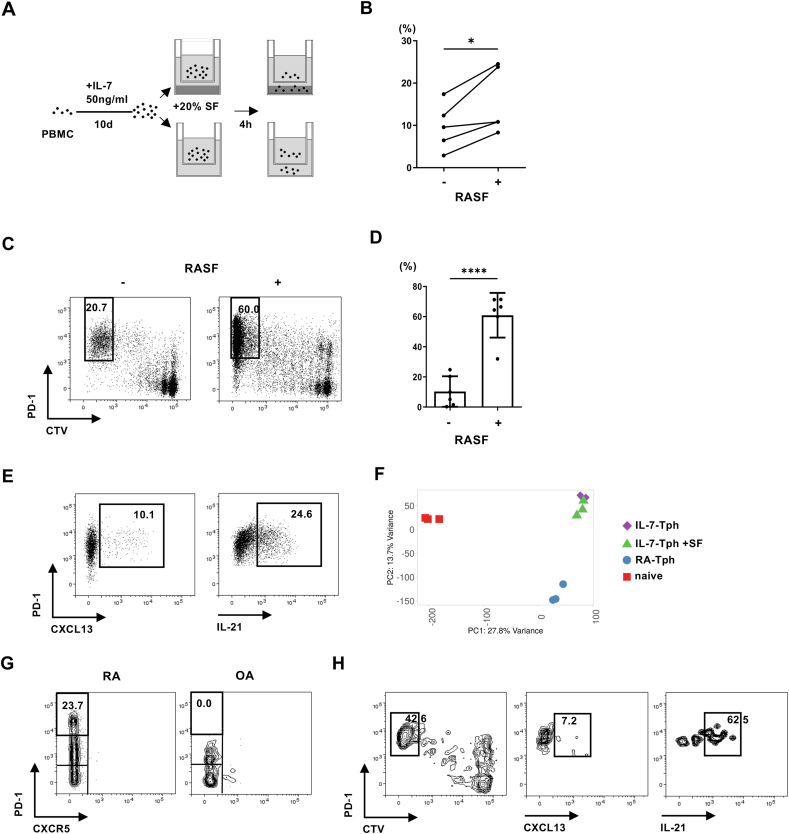
Objective: Dysregulated T cell homeostasis has long been implicated in the pathogenesis of rheumatoid arthritis (RA), in the joint of which peripheral helper T (Tph) cells accumulate and form ectopic lymphoid organs. We examined whether homeostatic signals are involved in the development of Tph cells.

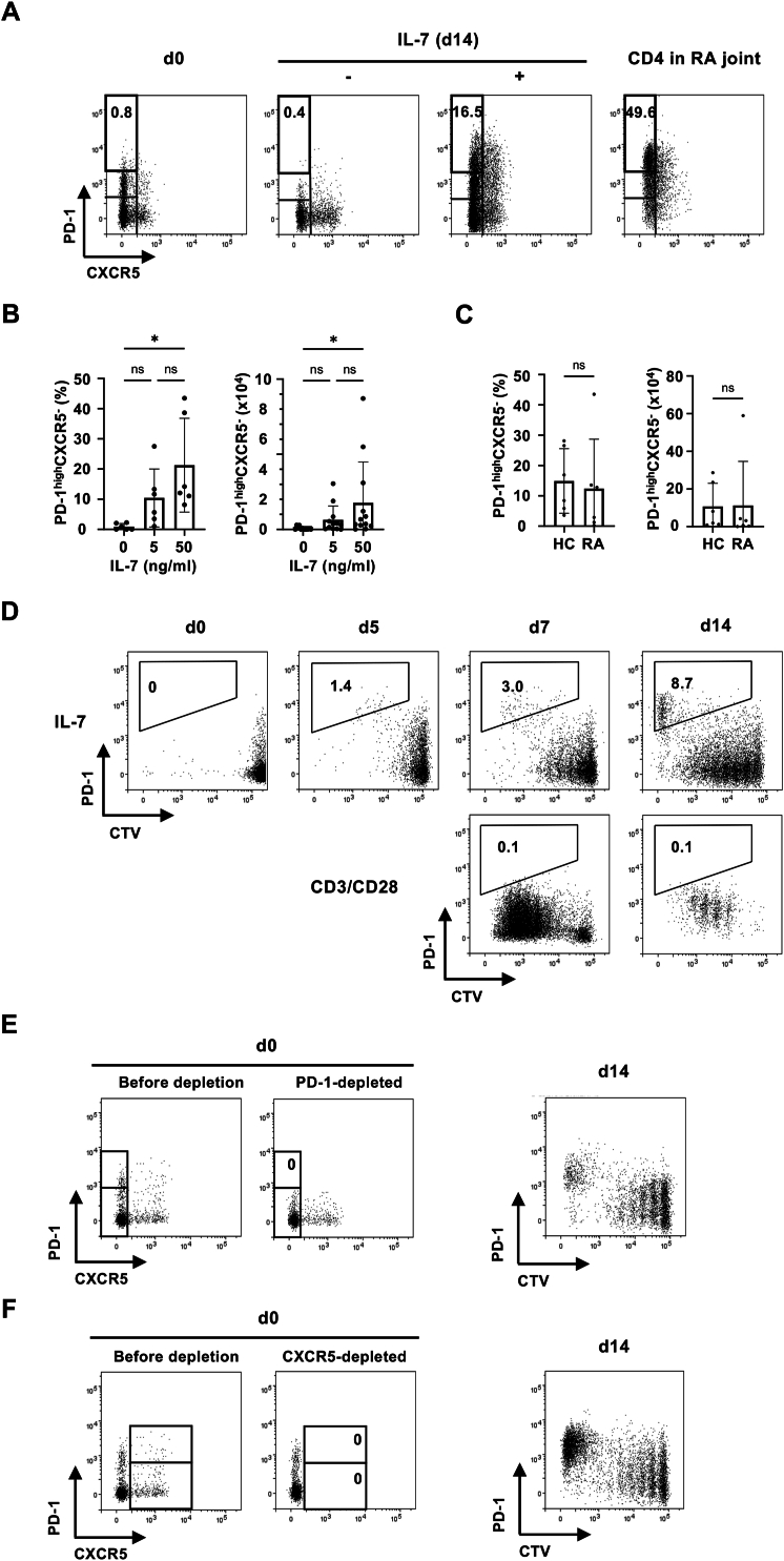
Methods: Human peripheral blood mononuclear cells were cultured with IL-7, the critical cytokine for T cell homeostasis. Development of Tph-like cells was assessed by flow cytometry, gene expression, and functional analysis. Chemotaxis of the Tph-like cells to RA synovial fluid (RASF) and the effect of RASF on the development of Tph-like cells was examined.

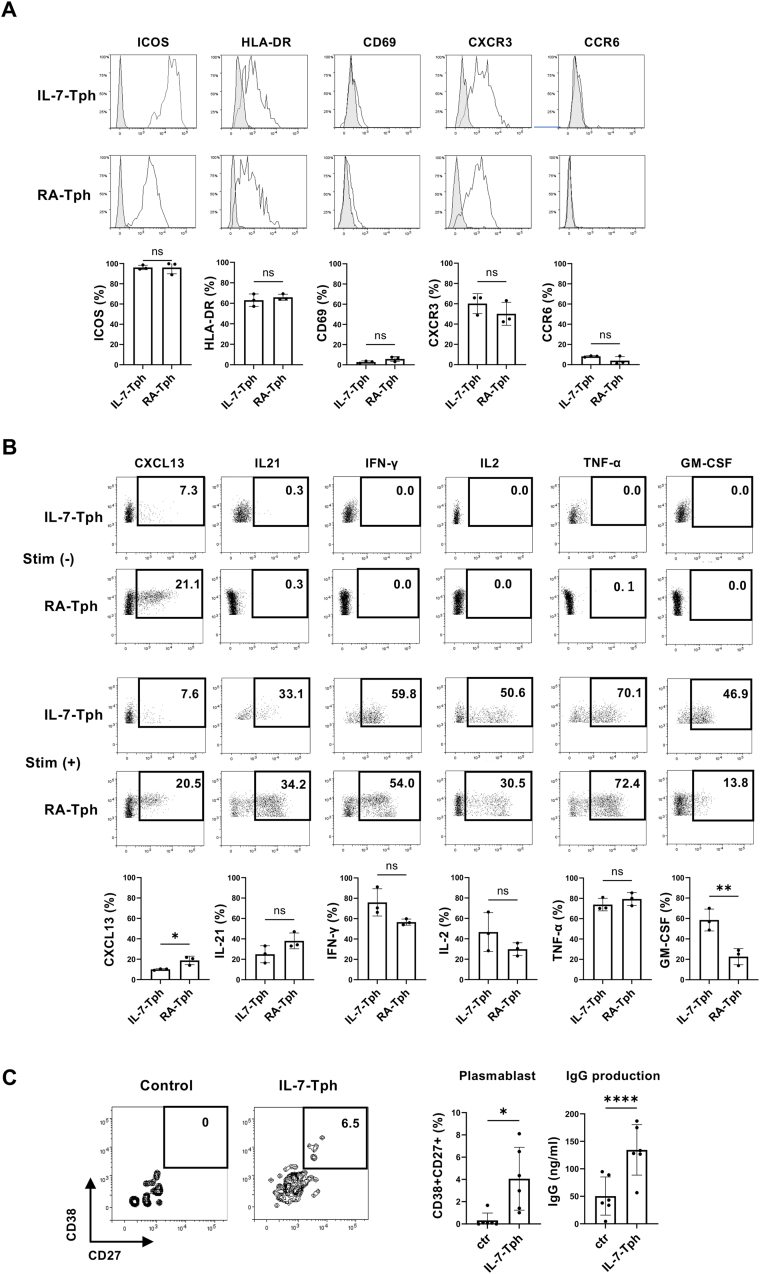
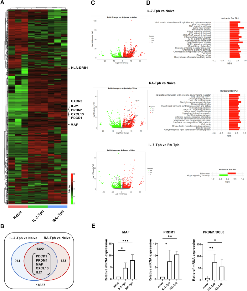
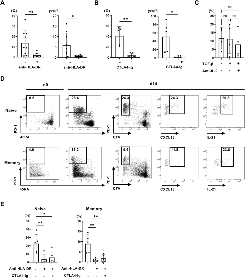
Results: PD-1highCXCR5- Tph-like cells developed from human peripheral blood CD4 T cells after proliferation in response to IL-7. Signals from self-MHC recognition and CD28 co-stimulation were also involved. The IL-7-induced Tph-like (IL-7-Tph) cells produced CXCL13 and IL-21 and helped B cells produce IgG. Comprehensive gene expression analysis further supported the similarity with Tph cells in RA joint. IL-7-Tph cells exhibited chemotaxis toward synovial fluid from RA patients (RASF), and RASF promoted the development of IL-7-Tph cells, which were also induced from CD4 T cells residing in non-inflamed joints.

Conclusions: Our results demonstrate an antigen-nonspecific developmental pathway of Tph cells triggered by homeostatic signals and promoted by the local environment of RA, which accounts for the accumulation of Tph cells in inflamed joints.

Keywords: Interleukin-7; Peripheral helper T cells; Rheumatoid arthritis.


Conflict of interest statement

The authors declare the following financial interests/personal relationships which may be considered as potential competing interests:Hiroaki Niiro reports a relationship with Bristol-Myers Squibb that includes: speaking and lecture fees. If there are other authors, they declare that they have no known competing financial interests or personal relationships that could have appeared to influence the work reported in this paper.

Figures

Fig. 1
Fig. 1
IL-7 induced the development of PD-1highCXCR5- Tph-like cells from human PB CD4 T cells. (A) PBMCs from healthy human subjects were cultured with or without 50 ng/ml of IL-7 for 14 days. Representative dot plots showing the expression of PD-1 and CXCR5 on CD4 T cells are shown. The right panel (CD4 in RA joint) indicates the staining pattern of CD4 T cells isolated from RA joint. (B) The frequency (left) and cell number in a culture well (right) of PD-1highCXCR5- Tph-like cells induced with varying concentration of IL-7. (C) The frequency and number of PD-1highCXCR5- Tph-like cells induced from PBMC of HC and RA are shown (n = 6). (D) CTV-labelled PBMCs were cultured either with IL-7 (upper panels) or in plate wells precoated with anti-CD3 and anti-CD28 mAbs (lower panels). Expression of PD-1 and CTV staining of CD4 T cells cultured for the indicated days is shown. Induction of PD-1highCXCR5- Tph-like cells from PD-1-depleted (E) or CXCR5-depleted (F) PBMCs by IL-7. Left panels (d0) indicate the expression of PD-1 and CXCR5 on CD4 T cells before and after negative selection. ∗p < 0.05.
Fig. 2
Fig. 2
Phenotypical and functional similarities between the IL-7-induced Tph-like (IL-7-Tph) cells and Tph cells in RA joint (RA-Tph cells). (A) Expression of surface molecules (ICOS, CD69, HLA-DR, CXCR3, and CCR6) on IL-7-Tph (n = 3) and RA-Tph cells (n = 3) are compared in histograms. Gray histograms indicate the staining with isotype controls. Graphs indicate the mean percentage of positive cells in PD-1high CD4 T cells (n = 3). (B) Representative dot plots of cytokine production by the IL-7-Tph and RA-Tph cells with (lower panels) or without (upper panels) stimulation with PMA and ionomycin are shown. Graphs indicate the mean percentage of cytokine positive cells in PD-1high CD4 T cells (n = 3). (C) Induction of plasmablasts (CD27+ CD38+) and IgG production from memory B cells cocultured with IL-7-Tph cells(n = 6). Representative dot plots of CD38 and CD27 expression on B cells are shown in left panels. ∗p < 0.05, ∗∗p < 0.01, ∗∗∗∗p < 0.0001.
Fig. 3
Fig. 3
Gene expression analysis of the IL-7-Tph cells. (A) Heatmap showing hierarchical clustering by DEGs among sorted naïve CD4 T (n = 3), IL-7-Tph (n = 3), and RA-Tph cells (n = 3). (B) Venn diagrams showing the number of the upregulated genes overlapped in IL-7-Tph and RA-Tph cells compared to naive CD4 T cells. (C) Volcano plots showing DEGs between IL-7-Tph and naive CD4 (upper), RA-Tph and naive CD4 (middle), and IL-7-Tph and RA-Tph (lower) cells. (D) GSEA analysis showing the pathway of genes upregulated in (C). (E) Expression levels of MAF, PRDM1, and PRDM1/BCL6 ratio in IL-7-Tph and RA-Tph cells by qRT-PCR analysis (n = 5). ∗p < 0.05, ∗∗p < 0.01, ∗∗∗p < 0.001.
Fig. 4
Fig. 4
Signalling requirements for the development of IL-7-Tph cells. The frequency (left) and cell number in a culture well (right) of PD-1high CTVlow cells after culturing PBMC with IL-7 in the presence or absence of anti-DR mAb (A) or CTLA-4 Ig (B) is shown (n = 9). (C) Effect of adding anti-IL-2 mAb and TGF-β was examined (n = 4). (D) Representative dot plots showing the induction of IL-7-Tph cells from either naïve (upper panels) or memory (lower panels) CD4 T cells as depicted by the expression of CD45RA and PD-1 (d0 and d14 left). Expression of CXCL13 and IL-21 in PD-1high CTVlow cells after stimulation was shown in the right panels. (E) The effect of adding anti-DR mAb or CTLA-4 Ig on the induction of IL-7-Tph cells from naive (left) or memory (right) CD4 T cells (n = 5). ∗p < 0.05, ∗∗p < 0.01.
Fig. 5
Fig. 5
Multiple mechanisms account for the accumulation Tph cells in the joint of RA. (A) Experimental system for the chemotaxis assay. (B) The frequency of PD-1high CTVlow IL-7-Tph cells migrated toward wells with or without RA SF (n = 5). (C) Representative dot plots showing the development of IL-7-Tph cells with or without RASF. (D) The frequency of PD-1high CTVlow IL-7-Tph cells induced with or without RA SF (n = 6). (E) Representative dot plots showing the expression of CXCL13 and IL-21 in the IL-7-Tph cells induced with RASF after stimulation. (F) PCA analysis on the gene expression of IL-7-Tph cells with or without RA SF, RA-Tph, and naive CD4 T cells (n = 3). (G) Representative dot plots showing the presence and absence of PD-1highCXCR5- Tph cells in RA and OA joints, respectively. (H) Representative dot plots showing the development of IL-7-Tph cells from CD4 T cells in OA joints. Right panels show the expression of CXCL13 and IL-21 in PD-1high CTVlow cells after stimulation.

References

    1. Catrina A., Krishnamurthy A., Rethi B. Current view on the pathogenic role of anti-citrullinated protein antibodies in rheumatoid arthritis. RMD Open. 2021;7 - PMC - PubMed
    1. Koetz K., Bryl E., Spickschen K., O'Fallon W.M., Goronzy J.J., Weyand C.M. T cell homeostasis in patients with rheumatoid arthritis. Proc Natl Acad Sci U S A. 2000;97:9203–9208. - PMC - PubMed
    1. Liu Q., Zheng Y., Goronzy J.J., Weyand C.M. T cell aging as a risk factor for autoimmunity. J. Autoimmun. 2023;137 - PMC - PubMed
    1. Klareskog L., Gaubitz M., Rodriguez-Valverde V., Malaise M., Dougados M., Wajdula J. A long-term, open-label trial of the safety and efficacy of etanercept (Enbrel) in patients with rheumatoid arthritis not treated with other disease-modifying antirheumatic drugs. Ann. Rheum. Dis. 2006;65:1578–1584. - PMC - PubMed
    1. Kremer J.M., Westhovens R., Leon M., Di Giorgio E., Alten R., Steinfeld S., et al. Treatment of rheumatoid arthritis by selective inhibition of T-cell activation with fusion protein CTLA4Ig. N. Engl. J. Med. 2003;349:1907–1915. - PubMed