Review
. 2024 Nov 7;6(6):fcae396.
doi: 10.1093/braincomms/fcae396. eCollection 2024.

Glial fibrillary acidic protein in Alzheimer's disease: a narrative review

Affiliations
Review

Glial fibrillary acidic protein in Alzheimer's disease: a narrative review

Florine Leipp et al. Brain Commun. .

Abstract

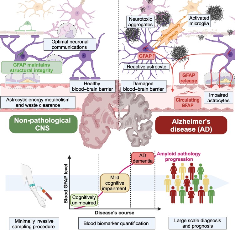
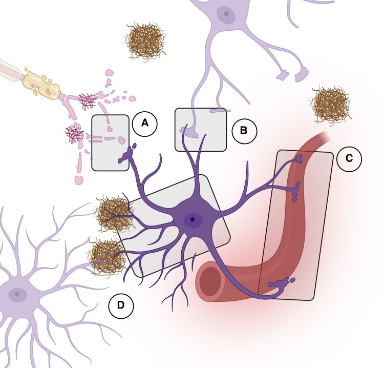
Astrocytes are fundamental in neural functioning and homeostasis in the central nervous system. These cells respond to injuries and pathological conditions through astrogliosis, a reactive process associated with neurodegenerative diseases such as Alzheimer's disease. This process is thought to begin in the early stages of these conditions. Glial fibrillary acidic protein (GFAP), a type III intermediate filament protein predominantly expressed in astrocytes, has emerged as a key biomarker for monitoring this response. During astrogliosis, GFAP is released into biofluids, making it a candidate for non-invasive diagnosis and tracking of neurodegenerative diseases. Growing evidence positions GFAP as a biomarker for Alzheimer's disease with specificity and disease-correlation characteristics comparable to established clinical markers, such as Aβ peptides and phosphorylated tau protein. To improve diagnostic accuracy, particularly in the presence of confounders and comorbidities, incorporating a panel of biomarkers may be advantageous. This review will explore the potential of GFAP within such a panel, examining its role in early diagnosis, disease progression monitoring and its integration into clinical practice for Alzheimer's disease management.

Keywords: Alzheimer’s disease; astrocytes; biomarker; glial fibrillary acidic protein; reactive astrogliosis.


Conflict of interest statement

The authors report no competing interests.

Figures

Graphical Abstract
Graphical Abstract
Figure 1
Figure 1
GFAP alternative splicing. Pre-mRNA and isoform mRNAs of GFAP are represented in the figure. GFAP is the human gene encoding for human glial fibrillary acidic protein (GFAP) and contains nine exons presented in coloured boxes and separated by untranslated introns. Exons 1, 4 and 5 are constitutive for most isoforms (except GFAPµ), while the others undergo alternative splicing leading to 12 human identified isoforms. GFAPα is the canonical transcript and most abundant isoforms of GFAP. Exon 1 contains start codon ATG and encodes N-terminal domain of GFAP, except for GFAPβ where the transcription site is about 169 nucleotides upstream. Several isoforms lack exons. GFAPγ lacks exon 1 in the N-terminal domain, and GFAPµ transcripts contain only exons 1 and 3 lack most of the core coil domains exons. GFAPδ/ε and GFAPκ lack exons 8 and 9 encoding for the last coil and C-terminal domains. Conversely, splicing variant of several isoforms include additional exons like GFAPλ and GFAPδ/ε containing exon 7a after exon 7 and GFAPκ containing exon 7b containing both exon 7, intron 7a and exon 7a. GFAP +1 isoforms category gathers GFAPΔEx6, Δ7, Δ135 andΔ164 isoforms with exhibits several alternatives changes regarding canonical form. Among those, complete or partial deletion of exon 6, exon 7 or exon 3 and exon 9 shortening. Deletions are represented by black-dotted white boxes and intron transcription by black-filled boxes.
Figure 2
Figure 2
Canonical GFAP 3D structure. Canonical GFAP is a 432 amino-acid long protein composed of two core coil domains respectively divided into two subparts (A and B). Together, the four coil subparts structured in alpha helix constitutes the rod-domain situated between head and tail domains. The different coil parts are connected with three linkers (L1, L12 and L2).
Figure 3
Figure 3
From GFAP to intermediate filament. Intermediate filaments are formed through the association of eight GFAP wrapped around tetramers generated from lateral association of coiled-coil structured GFAP dimers. Such dimers successively associate laterally into unit length filaments (ULFs) which are then connected end-to-end to form mature intermediate filaments (IFs).
Figure 4
Figure 4
Astrocytes roles and modifications in Alzheimer’s disease. Due to their multifaced functions and implications in various brain structures, reactive astrocyte modifications deeply impact brain physiology at several levels. (A) Tripartie synapses: Alteration of neurotransmitter and glutamate pathways, modification of water and ion channel transport and release of inflammatory substances leading to synaptic disfunctions, neurotoxicity and neuroinflammation; (B) Astrocytes tight junction: Irregular ion transient (Ca2+, K+) and decreased gap junctions coupling; (C) Astrocytes endfeet surrounding the blood–brain barrier (BBB): Release of overexpressed GFAP due to astrocytes impairment associated with BBB disruption and immune-cell massive infiltration; (D) Astrocytes morphology: Astrocytes structural atrophy and proliferation confining amyloid lesions and inducing Aβ clearance through the glymphatic pathway. Neurofibrillary tangles internalization.
Figure 5
Figure 5
ATNISV classification and current diagnosis tools for Alzheimer’s disease. The ATNIVS Alzheimer’s disease (AD) diagnosis framework draft suggested in 2023 amend former ATN classification described by NIA-AA. It includes four categories to cover a broader spectrum of the pathophysiological complexity of Alzheimer’s disease and relevant biological and imaging related biomarkers. The Core 1 (‘A’) and Core 2 (‘T’) biomarkers categories reflects Alzheimer’s disease main hallmarks: amyloid build-up and neurofibrillary tangles. The measurement in biofluids of Aβ1–42/40 ratio, secreted phosphorylated tau at positions 181, 217, 231 and 205 as well as tau fragments and specific tau regions such as microtubule binding region-243 (MTBR-243), respectively, characterize amyloid and tau pathology. Within this core AD biomarkers, amyloid and tau-PET imaging are used to visualize aggregates location and spreading. Other divisions includes neurodegeneration (‘N’) and neuroinflammation (‘I’) both reflecting non-specific hallmarks related to disease stage. The ‘N’ category stands for neurodegeneration which can be evaluated using CSF or plasma neurofilament light chain (NfL) protein along with anatomic (MRI, CT) or metabolism [18F]-fluorodeoxyglucose (FDG)-PET imaging. The ‘I’ section refers to neuroinflammation stated through glial fibrillary acid protein measurement in biofluids and specifically associated with astrogliosis. The two last additional categories were thought to map potential coexistent co-pathologies adding to the neurodegenerative burden. Hence, ‘S’ category characterize synucleinopathies entwined with aggregated abnormal alpha-synuclein in CSF evidenced with alpha-synuclein seed amplification assay (αsyn-SAA) and ‘V’ refers to vascular damages which can be examined using anatomic imaging.

References

    1. Nichols E, Steinmetz JD, Vollset SE, et al. Estimation of the global prevalence of dementia in 2019 and forecasted prevalence in 2050: An analysis for the Global Burden of Disease Study 2019. Lancet Public Health. 2022;7(2):e105–e125. - PMC - PubMed
    1. Serrano-Pozo A, Frosch MP, Masliah E, Hyman BT. Neuropathological alterations in Alzheimer disease. Cold Spring Harb Perspect Med. 2011;1(1):a006189. - PMC - PubMed
    1. Yiannopoulou KG, Anastasiou AI, Zachariou V, Pelidou SH. Reasons for failed trials of disease-modifying treatments for Alzheimer disease and their contribution in recent research. Biomedicines. 2019;7(4):97. - PMC - PubMed
    1. Kim CK, Lee YR, Ong L, Gold M, Kalali A, Sarkar J. Alzheimer’s disease: Key insights from two decades of clinical trial failures. J Alzheimers Dis. 2022;87(1):83–100. - PMC - PubMed
    1. Hampel H, Hu Y, Cummings J, et al. Blood-based biomarkers for Alzheimer’s disease: Current state and future use in a transformed global healthcare landscape. Neuron. 2023;111(18):2781–2799. - PMC - PubMed

LinkOut - more resources