Review
. 2024 Nov 1;9(45):44876-44890.
doi: 10.1021/acsomega.4c06351. eCollection 2024 Nov 12.

Exploring Hypertension: The Role of AT1 Receptors, Sartans, and Lipid Bilayers

Affiliations
Review

Exploring Hypertension: The Role of AT1 Receptors, Sartans, and Lipid Bilayers

Nikitas Georgiou et al. ACS Omega. .

Abstract

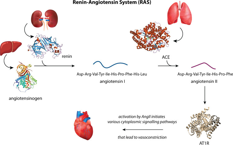
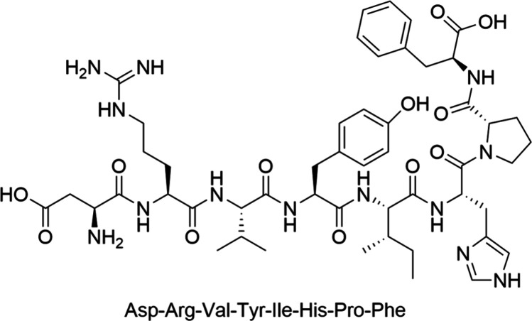
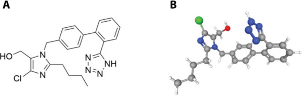
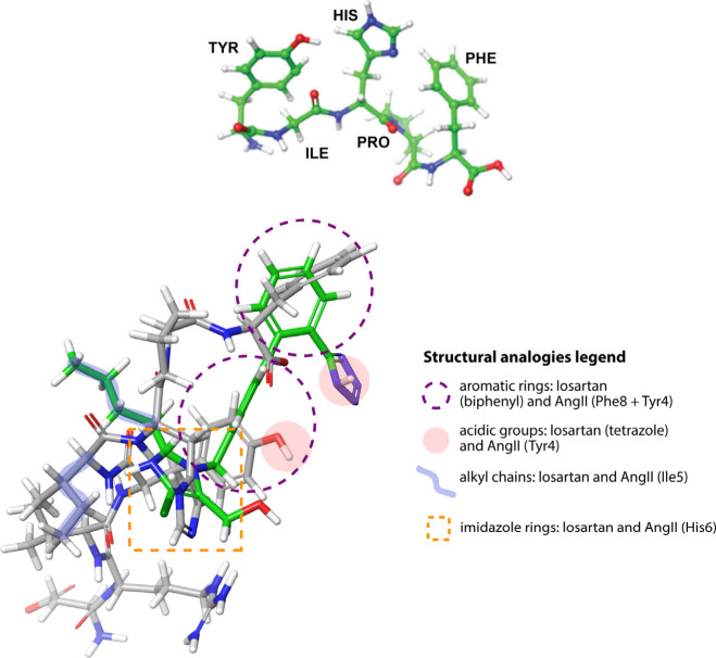
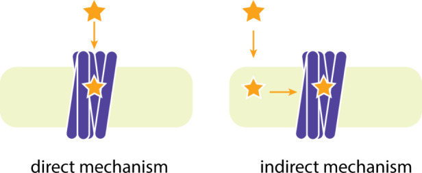
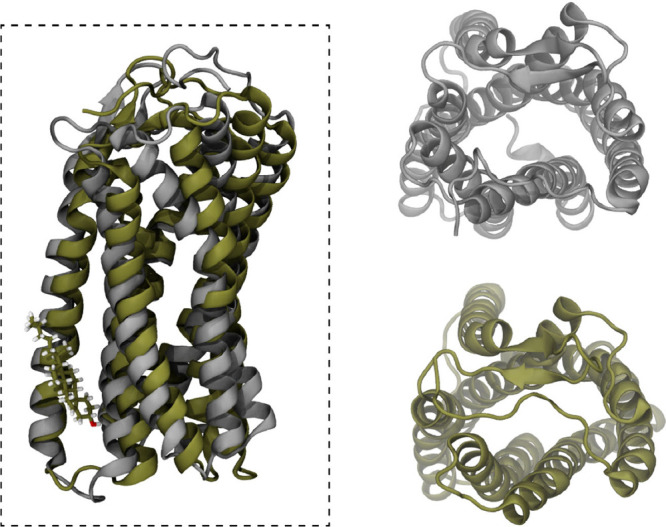
The rational design of AT1 receptor antagonists represents a pivotal approach in the development of therapeutic agents targeting cardiovascular pathophysiology. Sartans, a class of compounds engineered to inhibit the binding and activation of Angiotensin II on the AT1 receptor, have demonstrated significant clinical efficacy. This review explores the multifaceted role of sartans in mitigating hypertension and related complications. We highlight the integration of crystallography, computational simulations, and NMR spectroscopy to elucidate sartan-AT1 receptor interactions, providing a foundation for the next-generation antagonist design. The review also delves into the challenges posed by the high lipophilicity and suboptimal bioavailability of sartans, emphasizing advancements in nanotechnology and novel drug delivery systems. Additionally, we discuss the impact of lipid bilayers on the AT1 receptor conformation and drug binding, underscoring the importance of the lipidic environment in receptor-drug interactions. We suggest that optimizing drug design to account for these factors could enhance the therapeutic potential of AT1 receptor antagonists, paving the way for improved cardiovascular health outcomes.


Conflict of interest statement

The authors declare no competing financial interest.

Figures

Figure 1
Figure 1
A schematic representation of the RAS system consisting of the enzyme renin and its substrate angiotensinogen, the oligopeptide angiotensin I, the ACE which converts angiotensin I to angiotensin II and, finally, the GPCR AT1R to which angiotensin II physiologically binds. The images of liver, lungs, kidneys and heart were downloaded under the Free License of www.vecteezy.com. The images of renin, angiotensinogen, ACE and AT1R are public domain from wikipedia.org.
Scheme 1
Scheme 1. Sequence and 2D Structure of AngII
Scheme 2
Scheme 2. Structure of Losartan in (a) 2D and (b) 3D View
Figure 2
Figure 2
(top) Proposed model of the C-terminus of Angiotensin II. Its major characteristic is the relay system between Tyr4, Hist6, and Phe8 which occurs intramolecularly. (bottom) Superimposition of losartan (green) with AngII (gray). The key analogies between the peptide and the losartan structures are presented in an embedded legend, i.e., the spatial vicinity of the aromatic rings (depicted in purple dashed circles), the acidic groups (in pink circles), the alkyl chains (in blue brush strokes), and the imidazole rings (in orange dashed rectangle).
Figure 3
Figure 3
Relay system derived from DFT calculations at the B3LYP/6-311+g(d,p)/PCM(water) level of theory.
Figure 4
Figure 4
Octapeptide angiotensin II crystallized inside the AT1R. PDB ID: 6OS0. The conformation adopted by Tyr4 and Phe8 is compatible with that of our proposed proton relay system.
Figure 5
Figure 5
(left) EM structure of losartan inside the AT1R/soluble cytochrome b562 complex. The receptor is illustrated in silver, whereas losartan is in green. PDB ID: 8TH4. Interactions of the π-cation nature are taking place between Lys199 (illustrated in silver, licorice representation) and losartan, also suggested by our docking studies to a receptor’s homology model. (right) Detailed ligand interaction diagram of losartan bound to the EM structure of AT1R.
Figure 6
Figure 6
Synthetic AT1R antagonists prepared by our group and collaborators that consist of (A) small, nonpeptide molecules with few synthetic steps, (B) bisartans, (C) losartan-quercetin and losartan-DHA hybrid molecules, and (D) diverse scaffolds resulted from virtual screening of large databases.
Figure 7
Figure 7
The stronger the interactions between 2-hp-β-CD and the drug molecule, the lower is the latter’s activity as the availability of the drug becomes low and cannot exert its beneficial action. The top, highly active drug molecule is candesartan and the lower, less active is candesartan cilexitil, engulfed in 2-hp-β-CD. For more details on our study refer to ref (136).
Figure 8
Figure 8
Schematic representation of the drug:2-hp-β-CD complex, its transport to the lipid bilayer, and the subsequent drug release.
Figure 9
Figure 9
Copolymers and liposomes are used as drug delivery systems.
Figure 10
Figure 10
Candesartan’s relative position with respect to the bilayer center. The drug molecule is found to reside at 1 nm off the bilayer center, at the lipid–water interface. Due to the lower concentration of candesartan with respect to the rest of the system’s components, a smaller graph with its density is embedded in the main plot for scaling.
Figure 11
Figure 11
Schematic representation of the direct and indirect mechanisms of drug-receptor binding.
Figure 12
Figure 12
Most prevalent cluster structure of AT1R embedded in pure DPPC (silver) and DPPC:cholesterol (60:40%mol) bilayers. The top views presented on the right clearly indicate that a conformational change invoked by the allosteric binding of cholesterol on AT1R blocks the entrance of the extracellular site to the binding site through the N-terminus. For more details see refs ( and 12).

Similar articles

Cited by

References

    1. Roth G. A.; Nguyen G.; Forouzanfar M. H.; Mokdad A. H.; Naghavi M.; Murray C. J. L. Estimates of Global and Regional Premature Cardiovascular Mortality in 2025. Circulation 2015, 132 (13), 1270–1282. 10.1161/CIRCULATIONAHA.115.016021. - DOI - PubMed
    1. Staessen J. A.; Wang J.; Bianchi G.; Birkenhäger W. H. Essential Hypertension. Lancet 2003, 361 (9369), 1629–1641. 10.1016/S0140-6736(03)13302-8. - DOI - PubMed
    1. Messerli F. H.; Williams B.; Ritz E. Essential Hypertension. Lancet 2007, 370 (9587), 591–603. 10.1016/S0140-6736(07)61299-9. - DOI - PubMed
    1. Carretero O. A.; Oparil S. Essential Hypertension. Circulation 2000, 101 (4), 446–453. 10.1161/01.CIR.101.4.446. - DOI - PubMed
    1. Weir M. R.; Dzau V. J. The Renin-Angiotensin-Aldosterone System: A Specific Target for Hypertension Management. Am. J. Hypertens. 1999, 12 (4), 205–213. 10.1016/S0895-7061(99)00103-X. - DOI - PubMed

LinkOut - more resources