. 2024 Oct 18;32(4):200894.
doi: 10.1016/j.omton.2024.200894. eCollection 2024 Dec 19.

Combinatorial immunotherapy of anti-MCAM CAR-modified expanded natural killer cells and NKTR-255 against neuroblastoma

Affiliations

Combinatorial immunotherapy of anti-MCAM CAR-modified expanded natural killer cells and NKTR-255 against neuroblastoma

Wen Luo et al. Mol Ther Oncol. .

Abstract

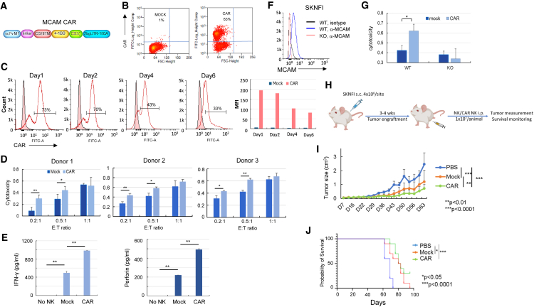
Pediatric patients with recurrent metastatic neuroblastoma (NB) have a dismal 5-year survival. Novel therapeutic approaches are urgently needed. The melanoma cell adhesion molecule (MCAM/CD146/MUC18) is expressed in a variety of pediatric solid tumors, including NB, and constitutes a novel target for immunotherapy. Here, we developed a chimeric antigen receptor (CAR) expressing natural killer (NK) cell-targeting MCAM by non-viral electroporation of CAR mRNA into ex vivo expanded NK cells. Expression of anti-MCAM CAR significantly enhanced NK cell cytotoxic activity compared to mock NK cells against MCAMhigh but not MCAMlow/knockout NB cells in vitro. Anti-MCAM-CAR-NK cell treatment significantly decreased tumor growth and prolonged animal survival in an NB xenograft mouse model. NKTR-255, a polymer-conjugated recombinant human interleukin-15 agonist, significantly stimulated NK cell proliferation and expansion and further enhanced the in vitro cytotoxic activity and in vivo anti-tumor efficacy of anti-MCAM-CAR-NK cells against NB. Our preclinical studies demonstrate that ex vivo expanded and modified anti-MCAM-CAR-NK cells alone and/or in combination with NKTR-255 are promising novel alternative therapeutic approaches to targeting MCAMhigh malignant NB.

Keywords: MCAM; MT: Regular Issue; NKTR-255; chimeric antigen receptor; natural killer cell; neuroblastoma.


Conflict of interest statement

M.S.C. has served as a consultant for Jazz Pharmaceuticals, Omeros Pharmaceuticals, Servier Pharmaceuticals, Abbvie, and Novartis Pharmaceuticals; with the Speakers Bureau for Jazz Pharmaceuticals, Servier Pharmaceuticals, Amgen, Inc., Sanofi, and Sobi; and on the Advisory Board for Astra Zeneca and receives research funding from Celularity, Merck, Miltenyi Biotec, Servier, Omeros, Jazz, and Janssen. D.A.L. reports personal fees and others from Kiadis Pharma, CytoSen Therapeutics, Courier Therapeutics, and Caribou Biosciences outside of the submitted work. In addition, D.A.L. has a patent broadly related to NK cell therapy of cancer with royalties paid to Kiadis Pharma. T.P.C. recently served as a one-time consultant to Blueprint, Incyte, and Oncopeptides and as a DSMB chair for SpringWorks and is a cofounder of Vironexis Biotherapeutics, Inc.

Figures

None
Graphical abstract
Figure 1
Figure 1
Anti-MCAM-CAR-NK cells had superior cytotoxic activity against MCAMhigh NB cells in vitro and enhanced anti-tumor activity in NB xenograft mouse model compared to mock NK cells (A) Schematic of the design of the anti-MCAM CAR construct. (B) CAR expression on ex vivo exNK cells mediated by electroporation of CAR mRNA. Ten micrograms of in vitro-transcribed CAR mRNA was introduced into 5 × 106 exNK cells using electroporation. Forty-eight hours after electroporation, CAR expression was detected by biotinylated MCAM protein, followed by fluorescein isothiocyanate (FITC)-streptavidin staining and flow cytometry. (C) Duration of expression of CAR on ex vivo exNK cells. CAR expression was detected 1, 2, 4, and 6 days post electroporation. Median fluorescence intensity for the CAR+ population was analyzed by FlowJo. (D) Compared to the unmodified exNK cells (mock), expression of anti-MCAM CAR in exNK cells (CAR) significantly enhanced the NK cytotoxic activity against MCAMhigh NB SK-N-FI cells. ∗p < 0.05, ∗∗p < 0.01 (two-tailed Student’s t test). Columns represent the mean values, and error bars indicate the standard deviation (SD) of triplicate samples in a representative experiment. The same trend was seen using three different donor-derived NK cells. (E) Compared to the mock NK cells, the anti-MCAM-CAR-NK cells (CAR) had significantly increased secretion of the cytokines interferon γ (IFN-γ; left) and perforin (right). ∗p < 0.05, ∗∗p < 0.01 (two-tailed Student’s t test). Columns represent the mean values, and error bars indicate the SD of triplicate samples in a representative experiment. The same trend was seen in three independent biological replicates. (F) Flow cytometry analysis showing MCAM KO by the CRISPR-Cas9 approach in SK-N-FI cells. (G) Specific targeting of MCAMhigh SK-N-FI cells by anti-MCAM-CAR-NK cells. Cytotoxic activities of mock and CAR NK cells against MCAM wild-type (WT) and knockout (KO) cells were compared. Columns indicate the mean, and error bars indicate the SD of triplicate samples in a representative experiment. ∗p < 0.05, ∗∗p < 0.01 (two-tailed Student’s t test). The same trend was seen in three independent biological replicates. (H) Schematic showing the in vivo study design and procedure in (I) and (J). NB SK-N-FI cells (4 × 106 cells/site) were implanted subcutaneously into 4- to 6-week-old female NSG mice. After tumor establishment, 1 × 107 of NK or CAR NK cells in PBS were injected intraperitoneally once a week for 5 weeks. Tumor growth was monitored by caliper measurement, and animals were followed until death or sacrificed upon reaching a tumor size of 2 cm in any dimension. (I) Anti-MCAM-CAR-NK treatment significantly decreased tumor growth in an NB xenograft mouse model. Mock NK or anti-MCAM-CAR-NK cells or PBS control were injected into NB xenograft tumor-bearing NSG mice intraperitoneally once a week for 5 weeks. Tumor growth was monitored by caliper measurement twice a week. Tumor size was estimated according to the following formula: tumor size (cm3) = length (cm) × width2 (cm) × 0.5. n = 10 per group. ∗∗p < 0.01, ∗∗∗p < 0.0001 (ANOVA). Growth rates between groups were analyzed using a mixed-effects model. (J) Anti-MCAM-CAR-NK cell treatment significantly prolonged animal survival in the NB xenograft mouse model. Mice were followed until death or sacrificed if any tumor size reached 2 cm in any dimension in (I). Probability of survival was determined by the Kaplan-Meier method using animal death/sacrifice as the terminal event using Prism v.8.0 (GraphPad). ∗p < 0.05, ∗∗∗p < 0.0001 (log rank test).
Figure 2
Figure 2
NKTR-255 treatment increased the expression of NK-activating receptors and stimulated NK cell proliferation and expansion in vitro (A) Expression of NKp30, NKG2D, NKp44, CD94, NKG2A, KIR, NKG2C, and NKp46, detected by flow cytometry on ex vivo exNK cells treated with or without NKTR-255 (40 ng/mL) for 96 h in the absence of IL-2. (B) Ex vivo exNK cells (2.5 × 106 cells per condition) were incubated with increasing concentrations of NKTR-255 (0, 1, 10, and 40 ng/mL) in the absence of IL-2 for various periods of time as indicated (0–144 h). The viable cells were counted every 24 h using the trypan blue staining method. ∗p < 0.05, ∗∗p < 0.01. Columns indicate mean values, and error bars indicate the SD of triplicate samples in a representative experiment. The same trend was seen in three independent biological repeats.
Figure 3
Figure 3
Combination of NKTR-255 significantly increased the cytotoxic activity of anti-MCAM-CAR-NK cells against NB cells in vitro and further enhanced the anti-tumor activity of anti-MCAM-CAR-NK cells in vivo in the NB xenograft mouse model (A–C) Mock NK or MCAM-CAR-NK cells were incubated with or without 40 ng/mL NKTR-255 for 72 h, followed by in vitro cytotoxicity assays against SK-N-FI cells at an E:T of 0.5:1. The number of donors tested is 3. NKTR-255 significantly enhanced the cytotoxic activity of MCAM CAR-engineered NK cells targeting NB SK-N-FI cells (A) and enhanced cytokine IFN-γ (B) and perforin (C) secretion from MCAM-CAR-NK cells. ∗p < 0.05, ∗∗p < 0.01. Columns represent mean values, and error bars indicate the SD of triplicate samples in a representative experiment. The same trend was seen in three independent biological replicates. (D) Schematic showing the in vivo study design and procedure in (E) and (F). NB SK-N-FI cells (4 × 106 cells/site) were implanted subcutaneously into 4- to 6-week-old female NSG mice. After tumor establishment, animals were divided into 4 groups and injected intraperitoneally with PBS or CAR NK cells in PBS (1 × 107/animal once a week for 5 weeks), NKTR-255 (0.3 mg/kg once every 2 weeks 3 times), or CAR NK cells combined with NKTR-255. Tumor growth was monitored by caliper measurement, and animals were followed until death or sacrificed upon reaching a tumor size of 2 cm in any dimension. (E) NKTR-255 further enhanced the efficacy of anti-MCAM-CAR-NK cells in significantly decreasing tumor growth in the NB xenograft mouse model. Tumor growth was monitored by caliper measurement twice a week. Tumor size was estimated according to the following formula: tumor size (cm3) = length (cm) × width2 (cm) × 0.5. n = 7 per group. ∗p < 0.05, ∗∗p < 0.01, ∗∗∗p < 0.001 (ANOVA). Growth rates between groups were analyzed using a mixed-effects model. (F) Anti-MCAM-CAR-NK cell treatment combined with NKTR-255 further significantly prolonged animal survival compared to CAR NK cells alone in the NB xenograft mouse model. Mice were followed until death or sacrificed if any tumor size reached 2 cm in any dimension in (E). Probability of survival was determined by the Kaplan-Meier method using animal death/sacrifice as the terminal event using Prism v.8.0 (GraphPad). ∗p < 0.05, ∗∗p < 0.01, ∗∗∗p < 0.001 (log rank test).

References

    1. Yan P., Qi F., Bian L., Xu Y., Zhou J., Hu J., Ren L., Li M., Tang W. Comparison of Incidence and Outcomes of Neuroblastoma in Children, Adolescents, and Adults in the United States: A Surveillance, Epidemiology, and End Results (SEER) Program Population Study. Med. Sci. Monit. 2020;26 - PMC - PubMed
    1. Whittle S.B., Smith V., Doherty E., Zhao S., McCarty S., Zage P.E. Overview and recent advances in the treatment of neuroblastoma. Expert Rev. Anticancer Ther. 2017;17:369–386. - PubMed
    1. Maris J.M. Recent advances in neuroblastoma. N. Engl. J. Med. 2010;362:2202–2211. - PMC - PubMed
    1. Ozkaynak M.F., Gilman A.L., London W.B., Naranjo A., Diccianni M.B., Tenney S.C., Smith M., Messer K.S., Seeger R., Reynolds C.P., et al. A Comprehensive Safety Trial of Chimeric Antibody 14.18 With GM-CSF, IL-2, and Isotretinoin in High-Risk Neuroblastoma Patients Following Myeloablative Therapy: Children's Oncology Group Study ANBL0931. Front. Immunol. 2018;9:1355. - PMC - PubMed
    1. Park J.R., Kreissman S.G., London W.B., Naranjo A., Cohn S.L., Hogarty M.D., Tenney S.C., Haas-Kogan D., Shaw P.J., Kraveka J.M., et al. Effect of Tandem Autologous Stem Cell Transplant vs Single Transplant on Event-Free Survival in Patients With High-Risk Neuroblastoma: A Randomized Clinical Trial. JAMA. 2019;322:746–755. - PMC - PubMed

LinkOut - more resources