. 2025 Apr;246(4):616-630.
doi: 10.1111/joa.14175. Epub 2024 Nov 18.

Cyclin-dependent kinase 13 is indispensable for normal mouse heart development

Affiliations

Cyclin-dependent kinase 13 is indispensable for normal mouse heart development

Qazi Waheed-Ullah et al. J Anat. 2025 Apr.

Abstract

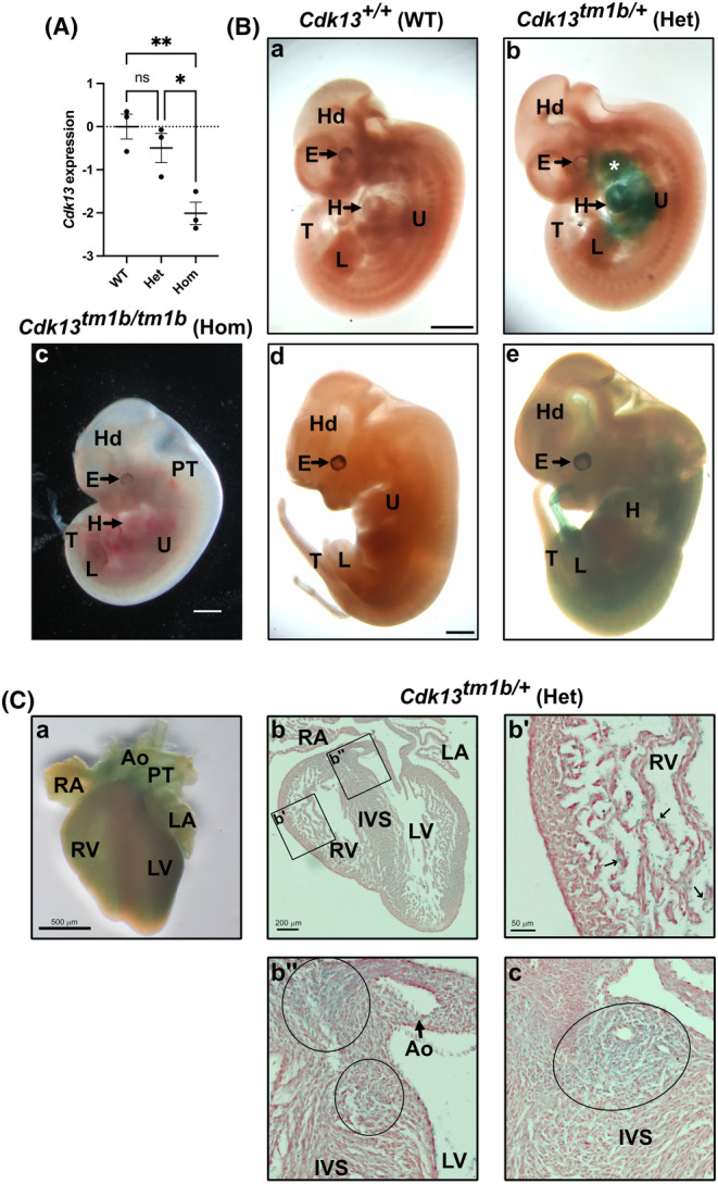
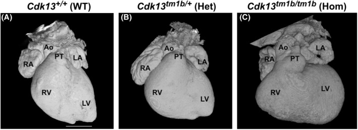
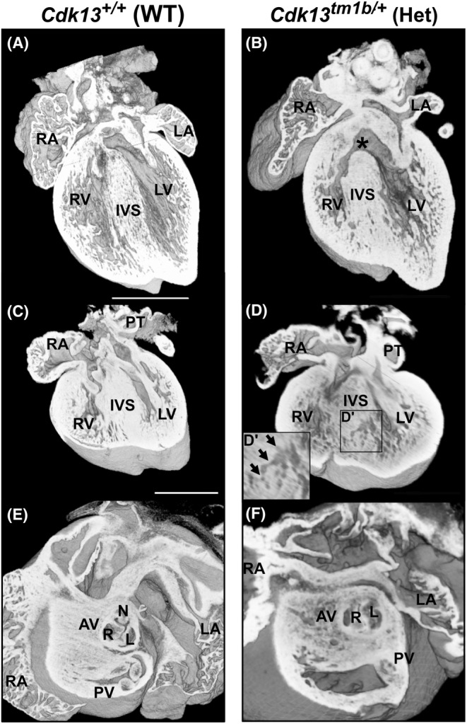
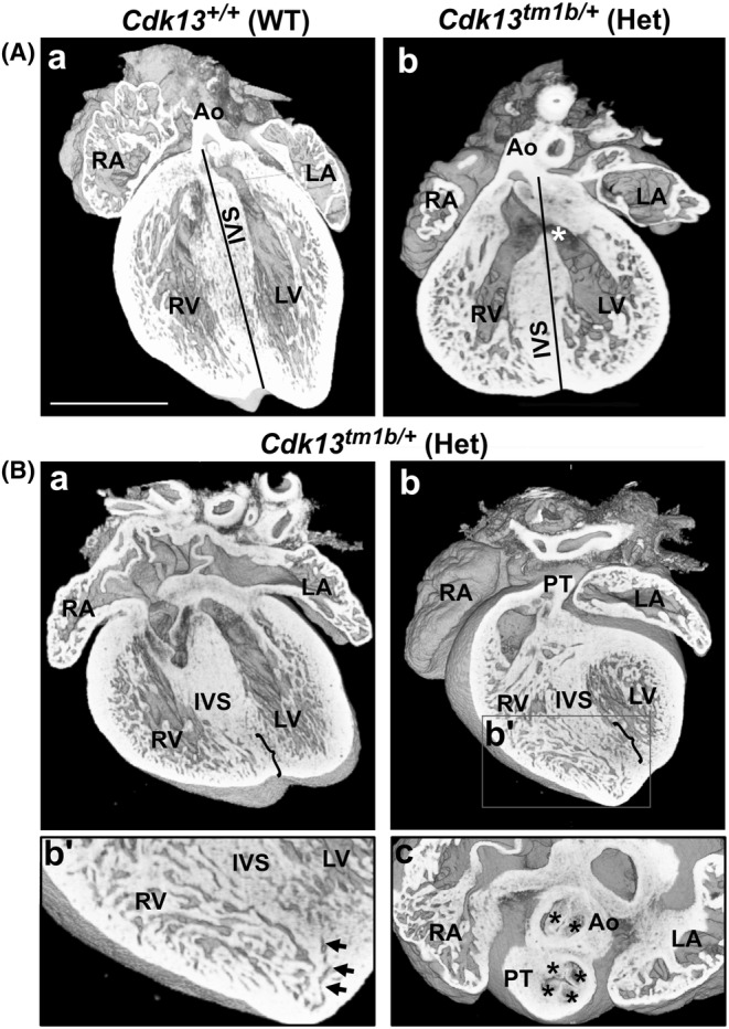
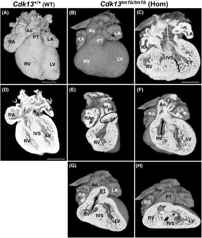
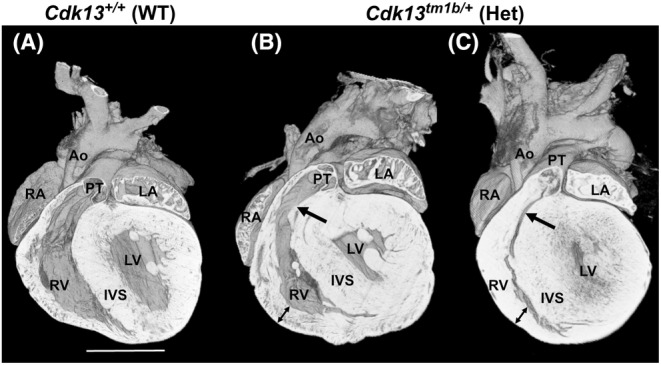
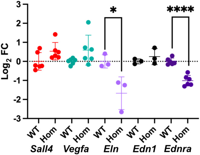
Congenital heart disease (CHD) has an incidence of approximately 1%. Over the last decade, sequencing studies including large cohorts of individuals with CHD have begun to unravel the genetic mechanisms underpinning CHD. This includes the identification of variants in cyclin-dependent kinase 13 (CDK13), in individuals with syndromic CHD. CDK13 encodes a serine/threonine protein kinase. The cyclin partner of CDK13 is cyclin K; this complex is thought to be important in transcription and RNA processing. Pathogenic variants in CDK13 cause CDK13-related disorder in humans, characterised by intellectual disability and developmental delay, recognisable facial features, feeding difficulties and structural brain defects, with 35% of individuals having CHD. To obtain a greater understanding for the role that this essential protein kinase plays in embryonic heart development, we have analysed a presumed loss of function Cdk13 transgenic mouse model (Cdk13tm1b). The homozygous mutants were embryonically lethal in most cases by E15.5. X-gal staining showed Cdk13 expression localised to developing facial regions, heart and surrounding areas at E10.5, whereas at E12.5, it was more widely present. In the E15.5 heart, staining was seen throughout. RT-qPCR showed significant reduction in Cdk13 transcript expression in homozygous compared with WT and heterozygous hearts at E10.5 and E12.5. Detailed morphological 3D analysis of embryonic and postnatal hearts was performed using high-resolution episcopic microscopy, which affords a more detailed analysis of structures such as cardiac valve leaflets and endocardial cushions, compared with more traditional histological techniques. We show that both the homozygous and heterozygous Cdk13tm1b mutants exhibit a range of CHD, including ventricular septal defects, bicuspid aortic valve, double outlet right ventricle and atrioventricular septal defects. 100% (n = 4) of homozygous hearts displayed CHD. Differential expression was seen in Cdk13tm1b homozygous mutants for two genes known to be necessary for normal heart development. The types of defects, and the presence of CHD in heterozygous mice (17.02%, n = 8/47), are consistent with the CDK13-related disorder phenotype in humans. This study provides important insights into the effects of reduced function of CDK13 in the mouse heart and contributes to our understanding of the mechanism behind this disorder as a cause of CHD.

Keywords: Cdk13; CHD; congenital heart disease; congenital heart disorders; cyclin‐dependant kinase 13; high‐resolution episcopic microscopy.


Conflict of interest statement

The authors declare they have no conflicts of interest.

Figures

FIGURE 1
FIGURE 1
Expression of Cdk13 in Cdk13 tm1b mouse line. (A) Expression in E12.5 mouse hearts by RT‐qPCR. The log2FC of Cdk13 (mean with SEM) in heterozygous (Het; Cdk13 tm1b/+ ) and homozygous (Hom; Cdk13 tm1b/tm1b ) hearts collected at E12.5 compared to WT (Cdk13 +/+ ). Using one way‐ANOVA and post hoc Tukey test, no statistically significant difference is present in the level of Cdk13 transcript in heterozygous compared to WT (p = 0.5049) hearts. In contrast, the difference is significant in homozygous compared to WT (p = 0.0073) and heterozygous (p = 0.0263) hearts. Het, heterozygous; Hom, homozygous; ns, not significant; WT, wild type. (B) X‐gal staining to show Cdk13 expression in Cdk13 tm1b/+ (Het) embryos. At E10.5, in comparison to WT control (a), X‐gal staining is localised to the developing facial regions (asterisk denotes maxillary and mandibular processes of 1st pharyngeal arch), and the heart (H) and surrounding area (b). A whole E12.5 embryo is shown (c) to allow comparison to WT (d) and heterozygous (e); note that the WT and heterozygous are littermates, but the homozygous is not. At E12.5, X‐gal staining is more widely present with staining seen to the heart (H) and abdominal regions, and faint staining to the head (d). X‐gal staining was not seen in WT control (c). To visualise the heart, the upper limb in (d) has been removed. E, eye; H, heart; Hd, head; Het, heterozygous; U, upper limb bud; L, lower limb bud; T, tail; WT, wild type. Scale in a, c and d 1 mm, same magnifications in b and d respectively. (C) Anterior view of a E15.5 X‐gal stained Cdk13 tm1b/+ (Het) heart shows widespread staining (a). Upon sectioning, expression could be seen to all regions (b), with noticeably punctate staining in the trabeculae of the ventricular chambers (b'). Slightly more intense areas of staining were seen at the superior aspect of the interventricular septum (within small and large circles in b″). Another section also shows this more intense staining at the superior aspect of the interventricular septum (denoted in oval in c); two lumens of the left coronary artery branches can be seen within. Ao, aorta; IVS, interventricular septum; LA, left atrium; LV, left ventricle; PT, pulmonary trunk; RA, right atrium; RV, right ventricle. Scale bar: A, 500 μm; b, 200 μm; b', 50 μm (same magnification for b″ and c).
FIGURE 2
FIGURE 2
External analysis of E15.5 Cdk13 tm1b homozygous, heterozygous and wild type hearts. (A–C): 3D reconstruction of wild type (a), heterozygous (b) and homozygous (c) whole hearts. Scale bar in (A) = 500 μm; same magnification for (B, C). Ao, aorta; Het; heterozygous; Hom, homozygous; LA, left atrium; LV, left ventricle; PT, pulmonary trunk; RA, right atrium; RV, right ventricle; WT, wild type.
FIGURE 3
FIGURE 3
Isolated defects in heterozygous (Cdk13 tm1b/+ ) E15.5 mouse hearts. (A,B) Four chamber view of WT (Cdk13 +/+ ) heart (a) showing the left and right ventricular chambers separated by a complete interventricular septum (IVS). In comparison, a heterozygous (Cdk13 tm1b/+ ) heart (b) has a perimembranous outlet ventricular septal defect (denoted by asterisk). The trabeculae also appear hypertrophic in comparison to the control heart in A. (C, D) Coronal section of WT (Cdk13 +/+ ) mouse heart (c) showing the left and right ventricular chambers separated from each other by an IVS in comparison to a heterozygous (Cdk13 tm1b /+ ) heart (D and inset D′) which had a muscular ventricular septal defect (denoted by black arrows). The myocardium also appears spongy in D compared to C. (E, F): Superior view of the WT heart (E) showing aortic valve having three leaflets and corresponding three sinuses—right (R), left (L) and non‐coronary (NC). In comparison, a heterozygous (Cdk13 tm1b/+ ) heart (f) has bicuspid aortic valve, with just two leaflets and two sinuses. Scale bar in (A) = 500 μm; same magnification for (B). Scale bar in (C) = 500 μm; same magnification for (D). AV, aortic valve; Het; heterozygous; IVS, interventricular septum; L, left leaflet and sinus; LA, left atrium; LV, left ventricle; NC, non‐coronary leaflet and sinus; PT, pulmonary trunk; PV, pulmonary valve; R, right leaflet and sinus; RA, right atrium; RV, right ventricle; WT, wild type.
FIGURE 4
FIGURE 4
Complex defects are seen in heterozygous (Cdk13 tm1b /+ ) E15.5 mouse hearts. (A) A coronal section of E15.5 WT (Cdk13 +/+ ) mouse heart (a) showing the left and right ventricular chambers separated from each other by the interventricular septum (IVS), and the aorta (Ao) originating from left ventricle (LV), as denoted by the black line. In contrast, a coronal section of a heterozygous (Cdk13 tm1b/+ ) heart (b) shows double outlet right ventricle and a single interventricular communication (perimembranous ventricular septal defect, denoted by asterisk) with interconnected inlet and outlet components. The black line denotes that more than 50% of the diameter of the aorta connects to the RV instead of the LV. The trabeculae are also hypertrophic. Scale bar in a = 500 μm; same magnification for b. (B) A four‐chamber view of a heterozygous (Cdk13 tm1b/+ ) heart (a) appears to have recesses in the lower region of the ventricular septum (denoted by bracket). This can also be seen in a more ventral view of the same heart (b; see bracket). The trabeculae had a sponge‐like appearance. In addition, the boxed area (b') denotes a small muscular ventricular septal defect (shown by black arrows). This heart also had a two‐sinus bicuspid aortic valve (c; denoted by two black asterisks in aorta) and a quadricuspid pulmonary valve (c; denoted by 4 black asterisks in pulmonary trunk). Ao, aorta; LA, left atrium, LV, left ventricle; PT, pulmonary trunk; RA, right atrium; RV, right ventricle; IVS, interventricular septum. Scale bar in a = 500 μm; same magnification for b.
FIGURE 5
FIGURE 5
Homozygous (Cdk13 tm1b /tm1b ) E15.5 heart with AVSD and DORV. Externally, this homozygous heart (B) has an abnormal shape with a rounded appearance compared with WT (A). Coronal view shows that the Cdk13 tm1b homozygous heart (C) has AVSD with apparent absence of atrial septation (white line) in comparison to WT heart (D) where an atrial septum (AS) is present. The trabeculae are hypertrophic, and recesses are present in the interventricular septum (IVS) (C; denoted by bracket). A sagittal view from the right of the heart shows that there are aortic and pulmonary valve leaflets (encircled by black oval) present, but there is no underlying outlet septum (E); the expected number of leaflets were seen. In the homozygous heart, a ventral view shows both the aorta and pulmonary trunk arising from the right ventricle (F and G, respectively; denoted by black arrows). An outlet ventricular communication can be seen (G; denoted by small white arrow). Viewed in anteroinferior plane (H), the outlet (small white arrow) and inlet VSD (small black arrow) can both be seen and were found to be interconnected. The inlet ventricular communication (black arrow) is there as the ventral (*) endocardial cushion has failed to fuse with the dorsal endocardial cushion and muscular interventricular septum (IVS). Scale bar in (A) = 500 μm; same magnification for (B). Scale bar in (C, D) = 500 μm. Ao, aorta; AS, atrial septum; AVSD, atrioventricular septal defect; DORV, double outlet right ventricle; LA, left atrium; LV, left ventricle; PT, pulmonary trunk; RA, right atrium; RV, right ventricle; IVS, interventricular septum; VSD, ventricular septal defect.
FIGURE 6
FIGURE 6
Increased ventricular wall thickness and narrowed right ventricle outflow region in P6 heterozygous (Cdk13 tm1b /+ ) hearts. Coronal sections of two P6 neonatal heterozygous (Cdk13 tm1b/+ ) hearts (B, C) with thickened ventricular walls (denoted by double headed arrows) compared to wild‐type control (A). Narrowing of the right ventricular outflow region (pulmonary trunk; PT) is denoted by a black arrow. Scale bar in (A) = 1 mm; same magnification for (B and C). Ao, aorta; LA, left atrium; LV, left ventricle; PT, pulmonary trunk; RA, right atrium; RV, right ventricle; IVS, interventricular ventricular septum; WT, wild type.
FIGURE 7
FIGURE 7
Modulated expression of genes related to cardiogenesis in E12.5 mouse homozygous (Cdk13 tm1b /tm1b ) heart. Modulated expression of genes related to cardiogenesis in homozygous Cdk13 tm1b mouse hearts compared to WT was seen, shown as mean log2FC ± SD. The difference was statistically significant only in case of Eln and Ednra (p < 0.05, unpaired t‐test). Sall4, Sal‐like 4; Vegfa, Vascular endothelial growth factor A; Edn1, Endothelin 1; Ednra, Endothelin A receptor; Eln, Elastin; SD, standard deviation; FC, fold change. Single asterisk (*) denotes p = 0.0352 whereas four asterisks (****) denote p < 0.0001.

Similar articles

  • The Black Book of Psychotropic Dosing and Monitoring.
    DeBattista C, Schatzberg AF. DeBattista C, et al. Psychopharmacol Bull. 2024 Jul 8;54(3):8-59. Psychopharmacol Bull. 2024. PMID: 38993656 Free PMC article. Review.
  • Signs and symptoms to determine if a patient presenting in primary care or hospital outpatient settings has COVID-19.
    Struyf T, Deeks JJ, Dinnes J, Takwoingi Y, Davenport C, Leeflang MM, Spijker R, Hooft L, Emperador D, Domen J, Tans A, Janssens S, Wickramasinghe D, Lannoy V, Horn SRA, Van den Bruel A; Cochrane COVID-19 Diagnostic Test Accuracy Group. Struyf T, et al. Cochrane Database Syst Rev. 2022 May 20;5(5):CD013665. doi: 10.1002/14651858.CD013665.pub3. Cochrane Database Syst Rev. 2022. PMID: 35593186 Free PMC article.
  • Systemic pharmacological treatments for chronic plaque psoriasis: a network meta-analysis.
    Sbidian E, Chaimani A, Garcia-Doval I, Doney L, Dressler C, Hua C, Hughes C, Naldi L, Afach S, Le Cleach L. Sbidian E, et al. Cochrane Database Syst Rev. 2021 Apr 19;4(4):CD011535. doi: 10.1002/14651858.CD011535.pub4. Cochrane Database Syst Rev. 2021. Update in: Cochrane Database Syst Rev. 2022 May 23;5:CD011535. doi: 10.1002/14651858.CD011535.pub5. PMID: 33871055 Free PMC article. Updated.
  • Cardiac Surgery.
    Senst B, Kumar A, Diaz RR. Senst B, et al. 2024 Sep 3. In: StatPearls [Internet]. Treasure Island (FL): StatPearls Publishing; 2025 Jan–. 2024 Sep 3. In: StatPearls [Internet]. Treasure Island (FL): StatPearls Publishing; 2025 Jan–. PMID: 30422530 Free Books & Documents.
  • CDK13-Related Disorder: Novel Insights From A Series of 27 Cases and Recommendations for Clinical Management.
    Contrò G, Baroni MC, Caraffi SG, Napoli M, Artuso R, Giliberti A, Bargiacchi S, Mancano G, Traficante G, Mucciolo M, Radio FC, Cordeddu V, Mancini C, Bottillo I, Pirro FA, Bonati MT, Becker CC, Carli D, Mussa A, Gonzalez MIA, Ruiz-Arana IL, Kumps C, Maystadt I, Moortgat S, Peker A, Piccione M, Grammatico P, Rostomashvili N, Lévy J, Scala M, Capra V, Torella A, van Eyk C, Isidor B, Cogne B, Srivastava S, Quinlan A, Vaisfeld A, Licchetta L, Frattini D, Graziano C, Severi G, Bacchi I, Soliani L, Sherr EH, Argilli E, Goel H, De Luca C, Leonardi S, Brancati F, Faletra F, Mio C, Braibanti S, Gargano G, Fusco C, Novelli A, Tartaglia M, Garavelli L. Contrò G, et al. Clin Genet. 2025 Aug;108(2):146-155. doi: 10.1111/cge.14726. Epub 2025 Feb 19. Clin Genet. 2025. PMID: 39971730 Free PMC article.

References

    1. Acharya, A. , Raza, S.I. , Anwar, M.Z. , Bharadwaj, T. , Liaqat, K. , Khokhar, M.A.S. et al. (2021) Wolfram‐like syndrome with bicuspid aortic valve due to a homozygous missense variant in CDK13. Journal of Human Genetics, 66, 1009–1018. - PMC - PubMed
    1. Ackerman, C. , Locke, A.E. , Feingold, E. , Reshey, B. , Espana, K. , Thusberg, J. et al. (2012) An excess of deleterious variants in VEGF‐A pathway genes in Down‐syndrome‐associated atrioventricular septal defects. American Journal of Human Genetics, 91, 646–659. - PMC - PubMed
    1. Anderson, R.H. , Mohun, T.J. , Spicer, D.E. , Bamforth, S.D. , Brown, N.A. , Chaudhry, B. et al. (2014) Myths and realities relating to development of the arterial valves. Journal of Cardiovascular Development and Disease, 1, 177–200.
    1. Anderson, R.H. , Wessels, A. & Vettukattil, J.J. (2010) Morphology and morphogenesis of atrioventricular septal defect with common atrioventricular junction. World J Pediatr Congenit Heart Surg, 1, 59–67. - PubMed
    1. Angelini, P. (2002) Coronary artery anomalies—current clinical issues: definitions, classification, incidence, clinical relevance, and treatment guidelines. Texas Heart Institute Journal, 29, 271–278. - PMC - PubMed

Publication types

Substances

LinkOut - more resources