. 2025 Jul 8;121(7):1076-1090.
doi: 10.1093/cvr/cvae244.

C-C Motif chemokine receptor-2 blockade ameliorates pulmonary hypertension in rats and synergizes with a pulmonary vasodilator

Affiliations

C-C Motif chemokine receptor-2 blockade ameliorates pulmonary hypertension in rats and synergizes with a pulmonary vasodilator

Naoki Tsuboya et al. Cardiovasc Res. .

Abstract

Aims: We investigated whether the disruption of C-C motif chemokine receptor (CCR) 2 may attenuate the development of pulmonary arterial hypertension (PAH) in any rat models with the reversal of the associated pro-inflammatory state and vascular dysfunction, and synergize with a conventional pulmonary vasodilator.

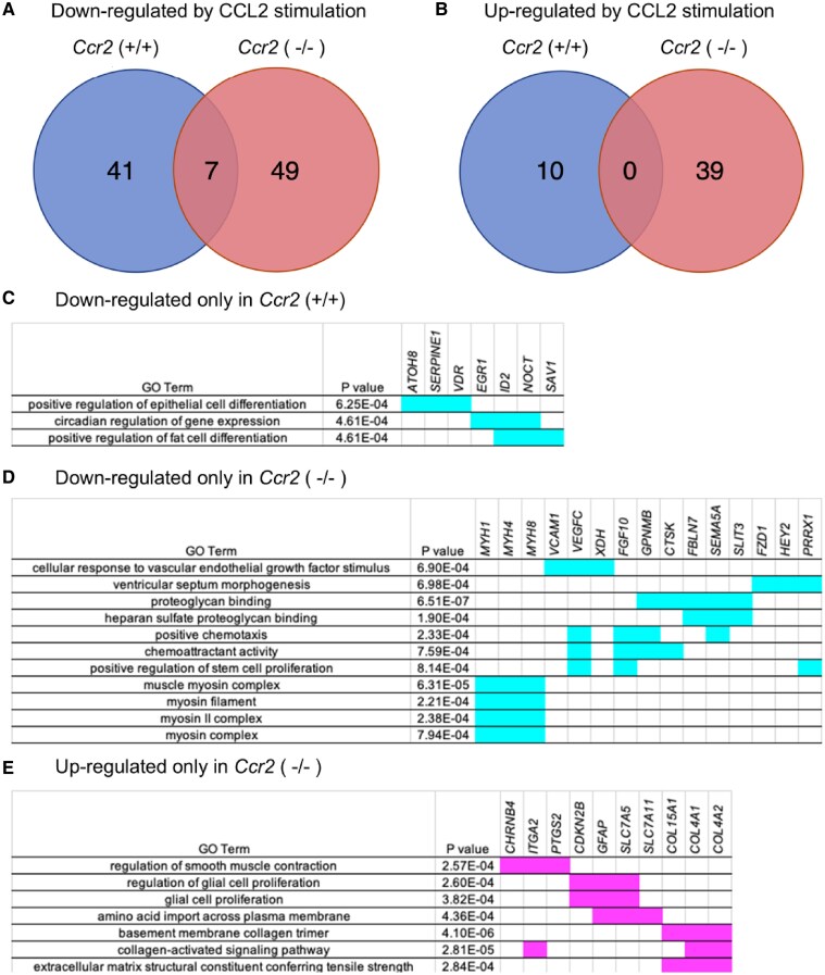
Methods and results: Using Ccr2(-/-) rats generated by CRISPR/Cas9, we investigated pulmonary hypertension (PH) in Ccr2(+/+) or Ccr2(-/-) rats treated with monocrotaline (MCT), SU5416/hypoxia (SuHx) and chronic hypoxia (CH). Ccr2(-/-) decreased the right ventricular systolic pressure, an index of right ventricular hypertrophy and mortality rate, and reversed increased expression of inflammatory cytokines/chemokines [interleukin-6, tumour necrosis factor-α, C-C motif chemokine receptor (CCL)-2, interleukin-1β, transforming growth factor-β] in rats 3weeks after MCT injection, but not in SuHx or CH models. Consistently, Ccr2(-/-) decreased indices of pulmonary vascular diseases (PVDs) and perivascular macrophage infiltration, as well as reversed impaired bone morphogenetic protein receptor type 2 signalling, increased endothelial apoptosis and impaired nitric oxide signalling and decreased phosphodiesterase-5 (PDE5) expression in lungs in MCT-treated rats. Gene expression of receptors for prostaglandin I2 and endothelin was not changed by Ccr2(-/-) in MCT-treated rats. In cultured pulmonary arterial smooth muscle cells (PASMCs), Ccr2(-/-) suppressed CCL2-induced hyperproliferation and dedifferentiation as well as reversed CCL2-induced decrease in PDE5 expression. The whole-genome RNA sequencing analysis identified differentially expressed genes in CCL2-stimulated Ccr2(-/-) PASMCs, which are related to the regulation of cellular differentiation and contraction. Based on studies in rats and cultured PASMCs, we investigated whether a PDE5 inhibitor, tadalafil, synergizes with Ccr2(-/-). Tadalafil administration ameliorated PH and PVDs in MCT-treated Ccr2(-/-) rats but not in Ccr2(+/+) rats. Tadalafil further improved survival in MCT-treated Ccr2(-/-) rats.

Conclusion: The present findings demonstrated that CCR2 disruption ameliorated PAH in MCT-treated rats, which was associated with the reversal of dysregulated inflammatory pathways and vascular dysfunction and synergized with tadalafil. These findings suggest that CCR2 may be a therapeutic target in intractable PAH patients with a certain CCR2-related inflammatory phenotype and refractory to conventional pulmonary vasodilators.

Keywords: Combination therapy; C–C Chemokine receptor type 2; Inflammation; Pulmonary hypertension.


Conflict of interest statement

Conflict of interest: none declared.

Figures

Graphical Abstract
Graphical Abstract
Figure 1
Figure 1
Ccr2 disruption ameliorated pulmonary hypertension in monocrotaline-treated rats. (A) Schematic illustration of the study protocols. Seven-week-old male rats with Ccr2(+/+), Ccr2(+/−), or Ccr2(−/−) background (n = 10/group) were injected with monocrotaline (MCT: 60 mg/kg); then, pulmonary hemodynamics and lung histology were assessed 3 weeks after MCT injection. (B) Mean pulmonary arterial pressure, (C) right ventricular systolic pressure measured by catheter, (D) the ratio of acceleration time/ejection time in the main pulmonary artery by echocardiography, (E) the weight ratio of the right ventricle to the left ventricle plus septum, (F) the cardiac index measured by echocardiography, (G) body weight gain after injection of MCT in rats of Ccr2(+/+), Ccr2(+/−) and Ccr2(−/−), respectively with injection of MCT or vehicle, n = 10/group. One-way ANOVA followed by Bonferroni’s multiple comparisons test was used for the analysis in the panel A-F. *P < 0.05, vs. Ccr2(+/+)/vehicle. †P < 0.05, vs. Ccr2(+/−)/vehicle. ‡P < 0.05, vs. Ccr2(−/−)/vehicle. §P < 0.05, vs. Ccr2(+/+)/MCT. Values are presented as the mean ± standard error. n = number of rats. MCT, monocrotaline; mPAP, mean pulmonary arterial pressure; RVSP, right ventricular systolic pressure; Act/ET, acceleration time/ejection time; RV/(LV + S), the weight ratio of the right ventricle to the left ventricle plus septum; CI, Cardiac index.
Figure 2
Figure 2
Ccr2 disruption ameliorated pulmonary vasculopathy and reduced perivascular infiltration of macrophages in monocrotaline-treated rats. (A) Representative morphological images of barium filled distal pulmonary arteries (PAs) (ϕ15–50 µm) and (C) proximal PAs (ϕ100–200 µm) assessed on week 3 after monocrotaline (MCT) injection for Ccr2(+/+) and Ccr2(−/−) rats (scale bar: 50 µm), and the quantification of (B) muscularized distal PAs and (D) the medial wall thickness, n = 5/group. (E) Representative images for CD68 or (G) CD163 immunohistochemistry in distal PAs (scale bar: 25 µm), and the quantification of the number of (F) CD68 or (H) CD163-positive cells infiltrating around the distal PAs (ϕ15–50 µm), n = 5/group. The negative controls were shown in Supplementary material online, Figure S3. One-way ANOVA followed by Bonferroni's multiple comparisons test was used for the analysis in the panel E-H. *P < 0.05, vs. Ccr2(+/+)/vehicle. †P < 0.05, vs. Ccr2(+/+)/MCT. Values are presented as the mean ± standard error. n = number of rats. MCT, monocrotaline.
Figure 3
Figure 3
Ccr2 disruption reversed pro-inflammatory cytokine responses and an imbalance of the expression of BMPR2 and TGF-ß in lungs from monocrotaline-treated rats. (A) The mRNA expression associated with inflammatory cytokines, IL-6, TNF-ɑ, CCL2, IL-1ß and TGF-ß, and BMPR2 and its main ligands, BMP-4, in lung extracts at 3 weeks after monocrotaline (MCT) or vehicle administration, n = 6/group. (B) Representative immunoblots and the quantification of relative expression of BMPR2 protein in lung extracts at week 3 of MCT or vehicle treatment, (C, D) and its downstream substrate proteins (SMAD1/5/9, phosphorylated SMAD1/5/9, ID1) and (E) SMAD2/3 and phosphorylated SMAD2/3, the downstream substrates of TGF-ß, n = 3/group. One-way ANOVA followed by Bonferroni's multiple comparisons test was used for the analysis in the panel A-E. *P < 0.05, vs. Ccr2(+/+)/vehicle. †P < 0.05, vs. Ccr2(+/+)/MCT. Values are presented as the mean ± standard error. n = number of rats. MCT, monocrotaline; BMPR2, bone morphogenetic protein receptor type 2. ID1, inhibitor of DNA binding 1.
Figure 4
Figure 4
Ccr2 disruption decreased apoptotic endothelial cells and reversed eNOS dephosphorylation and reduced PDE5 expression in lungs from monocrotaline-treated rats. (A) Representative images for Cleaved Caspase3 immunohistochemistry in distal pulmonary arteries (ϕ15–50 µm) on week 3 after monocrotaline (MCT) or vehicle injection (scale bar: 25 µm), and (B) quantitative analysis of vessels with Cleaved Caspase3-positive cells in the endothelial layer, n = 5/group. The negative control was shown in supplemental Figure 3. (C–G) Representative immunoblots of proteins and quantitative analysis of Cleaved Caspase3, Bcl-2, PAI-1, eNOS, peNOS and phosphodiesterase-5A (n = 3/group), and (H) the mRNA expression of Pde5a (n = 6/group) in whole lung extracts at week 3 of MCT or vehicle administration. One-way ANOVA followed by Bonferroni's multiple comparisons test was used for the analysis in the panel B-H. *P < 0.05, vs. Ccr2(+/+)/vehicle. †P < 0.05, vs. Ccr2(+/+)/MCT. Values are presented as the mean ± standard error. n = number of rats. MCT, monocrotaline; Bcl2, B-cell lymphoma-2; PAI1, plasminogen activator inhibitor-1; eNOS, endothelial nitric oxide synthase; peNOS, phosphorylated endothelial nitric oxide synthase; PDE5A, phosphodiesterase-5A.
Figure 5
Figure 5
Ccr2 disruption suppressed smooth muscle cell proliferation and dedifferentiation in lungs from MCT-treated rats and in cultured cells. (A) Representative images for Ki67 immunohistochemistry (scale bar: 50 µm) and (B) immunofluorescent images (scale bar: 25 µm) of distal pulmonary arteries (PAs) stained for Ki67 (green) and ɑ-smooth muscle actin (red) on week 3 after monocrotaline or vehicle injection. The negative controls were shown in Supplementary material online, Figure S3. (C) The quantification of vessels with Ki67-positive cells in the distal PAs on 3 weeks after MCT or vehicle injection, n = 4/group. (D) The mRNA expression of differentiation markers (Myh11, Cnn1, Acta2, Tagln, Myocd) in lung extracts at 3 weeks after MCT or vehicle administration, n = 6/group. (E) The ratio of absorbance by BrdU assay of pulmonary artery smooth muscle cells isolated from Ccr2(+/+) or Ccr2(−/−) rats stimulated with CCL2, platelet-derived growth factor (PDGF)-BB, or CCL2 + PDGF-BB to control, n = 4/group. (F) The ratio of mRNA expression of Myh11, Cnn1, Acta2, Tagln, Myocd, Pde5a, Ccnd1 and Pcna in isolated PASMCs upon stimulation with CCL2 relative to unstimulated control, n = 4/group. One-way ANOVA followed by Bonferroni's multiple comparisons test was used for the analysis in the panels C–E. The unpaired two-tailed Student’s t-test was used for the analysis in the panel F. *P < 0.05, vs. Ccr2(+/+)/vehicle. †P < 0.05, vs. Ccr2(+/+)/MCT. ‡P < 0.05, vs. Ccr2(+/+)/CCL2(−), §P < 0.05, vs. Ccr2(+/+)/CCL2(+), Values are presented as the mean ± standard error. n = number of rats. MCT, monocrotaline; ɑ-SMA, ɑ-smooth muscle actin; CCL2, C–C motif chemokine ligand 2, PDGF, platelet-derived growth factor.
Figure 6
Figure 6
Tadalafil treatment synergized with Ccr2 disruption in ameliorating pulmonary hypertension and vasculopathy in monocrotaline-treated rats. (A) Schematic illustration of the study protocols. Survival was assessed 6 weeks after MCT injection in the 7-week-old Ccr2(+/+) and Ccr2(−/−) male rats. (B) Tadalafil (10 mg/kg/day) was started 3 weeks after MCT injection in the Ccr2(+/+) and Ccr2(−/−) rats. Survival was assessed 6 weeks, and haemodynamics were assessed 4 weeks after MCT injection. (C) Survival outcomes of Ccr2(+/+) and Ccr2(−/−) rats after monocrotaline (MCT) injection (n = 40), and of (D) Ccr2(+/+) rats (n = 36), and (E) Ccr2(−/−) rats (n = 37) after MCT injection treated with tadalafil or sham from week 3 to 6. (F) The right ventricular systolic pressure, (G) the weight ratio of the right ventricle to the left ventricle plus septum, (H) the cardiac index, and (I) the quantification of the number of muscularized distal pulmonary arteries or (J) the medial wall thickness, n = 5/group, for MCT-injected Ccr2(+/+) or Ccr2(−/−) rats treated with 1 week of tadalafil or sham. Kaplan–Meier curves for survival were compared among treatment groups using the log-rank test for the panel A-C. One-way ANOVA followed by Bonferroni's multiple comparisons test was used for the panel D-H. *P < 0.05, vs. Ccr2(+/+)/MCT. †P < 0.05, vs. Ccr2(+/+)/MCT/sham. ‡P < 0.05, vs. Ccr2(−/−)/MCT/sham. §P < 0.05, vs. Ccr2(+/+)/MCT/tadalafil. Values are presented as the mean ± standard error. n = number of rats. Filled circles denote Ccr2(+/+)/MCT/sham; filled squares, Ccr2(−/−)/MCT/sham; empty circles, Ccr2(+/+)/MCT/tadalafil; and empty squares, Ccr2(−/−)/MCT/tadalafil. MCT, monocrotaline; RVSP, right ventricular systolic pressure; RV/(LV + S), the weight ratio of the right ventricle to the left ventricle plus septum; CI, Cardiac index.
Figure 7
Figure 7
Differentially expressed gene profiles in CCL2-stimulated pulmonary artery smooth muscle cells in the Ccr2(+/+) and Ccr2(−/−) rats. The transcriptome profile was assessed in the pulmonary artery smooth muscle cells (PASMCs) from three Ccr2(+/+) and three Ccr2(−/−) rats after treatment with rat recombinant C–C motif chemokine ligand 2 (CCL2: 100 ng/mL) for 24 h. Venn diagram for the number of genes (A) down-regulated or (B) up-regulated by CCL2 in Ccr2(+/+) and Ccr2(−/−) PASMCs. Gene ontology analysis for (C) the genes down-regulated only in Ccr2(+/+), (D) the genes down-regulated only in Ccr2(−/−) and (E) the genes up-regulated only in Ccr2(+/+), indicating down-regulated genes (cyan) and up-regulated genes (magenta) corresponding to GO terms. CCL2, C–C motif chemokine ligand 2.

Comment in

References

    1. Humbert M, Guignabert C, Bonnet S, Dorfmüller D, Klinger JR, Nicolls MR, Olschewski AJ, Pullamsetti SS, Schermuly RT, Stenmark KR, Rabinovitch M. Pathology and pathobiology of pulmonary hypertension: state of the art and research perspectives. Eur Respir J 2019;53:1801887. - PMC - PubMed
    1. Noordegraaf AV, Chin KM, Haddad F, Hassoun PM, Hemnes AR, Hopkins SR, Kawut SM, Langleben D, Lumens J, Naeije R. Pathophysiology of the right ventricle and of the pulmonary circulation in pulmonary hypertension: an update. Eur Respir J 2019;53:1801900. - PMC - PubMed
    1. Galiè N, Channick RN, Frantz RP, Grünig E, Jing ZC, Moiseeva O, Preston IR, Pulido T, Safdar Z, Tamura Y, McLaughlin VV. Risk stratification and medical therapy of pulmonary arterial hypertension. Eur Respir J 2019;53:1801889. - PMC - PubMed
    1. Zelt JGE, Sugarman J, Weatherald J, Partridge ACR, Liang JC, Swiston J, Brunner N, Chandy G, Stewart DJ, Contreras-Dominguez V, Thakrar M, Helmersen D, Varughese R, Hirani N, Umar F, Dunne R, Doyle-Cox C, Foxall J, Mielniczuk L. Mortality trends in pulmonary arterial hypertension in Canada: a temporal analysis of survival per ESC/ERS guideline era. Eur Respir J 2021;59:2101552. - PMC - PubMed
    1. Pogoriler JE, Rich S, Archer SL, Husain AN. Persistence of complex vascular lesions despite prolonged prostacyclin therapy of pulmonary arterial hypertension. Histopathology 2012;61:597–609. - PMC - PubMed

MeSH terms