. 2024 Nov 18;20(11):e1012711.
doi: 10.1371/journal.ppat.1012711. eCollection 2024 Nov.

Polyamines mediate cellular energetics and lipid metabolism through mitochondrial respiration to facilitate virus replication

Affiliations

Polyamines mediate cellular energetics and lipid metabolism through mitochondrial respiration to facilitate virus replication

Yazmin E Cruz-Pulido et al. PLoS Pathog. .

Abstract

Polyamines are critical cellular components that regulate a variety of processes, including translation, cell cycling, and nucleic acid metabolism. The polyamines, putrescine, spermidine, and spermine, are found abundantly within cells and are positively-charged at physiological pH. Polyamine metabolism is connected to distinct other metabolic pathways, including nucleotide and amino acid metabolism. However, the breadth of the effect of polyamines on cellular metabolism remains to be fully understood. We recently demonstrated a role for polyamines in cholesterol metabolism, and following these studies, we measured the impact of polyamines on global lipid metabolism. We find that lipid droplets increase in number and size with polyamine depletion. We further demonstrate that lipid anabolism is markedly decreased, and lipid accumulation is due to reduced mitochondrial fatty acid oxidation. In fact, mitochondrial structure and function are largely ablated with polyamine depletion. To compensate, cells depleted of polyamines switch from aerobic respiration to glycolysis in a polyamine depletion-mediated Warburg-like effect. Finally, we show that inhibitors of lipid metabolism are broadly antiviral, suggesting that polyamines and lipids are promising antiviral targets. Together, these data demonstrate a novel role for polyamines in mitochondrial function, lipid metabolism, and cellular energetics.


Conflict of interest statement

The authors have declared that no competing interests exist.

Figures

Fig 1
Fig 1. Lipid droplets accumulate in polyamine-depleted cells.
Huh7 cells were treated with 1 mM DFMO for four days and (A) visualized for lipid droplets by staining for BODIPY (green) and nuclei by DAPI (blue). (B) Polyamine depletion was confirmed in cells treated with increasing doses of DFMO for four days by thin layer chromatography. (C) Huh7 cells were treated as in (A) but stained with Nile Red to differentiate neutral/nonpolar lipids (green) from polar lipids (red). (D) Lipid droplets from (A) were quantified for droplet number per cells and droplet volume. (E) siRNA targeting ODC1 (“siODC1”) or a scrambled siRNA (“scrambled”)were transfected into Huh7 cells and lipid droplets visualized four days later as in (A). Knockdown of ODC1 was verified by (F) qPCR for ODC1 mRNA expression, comparing control transfected cells to scrambled and ODC1-specific siRNA, and (G) TLC of polyamines in transfected cells. Values listed below indicate the relative quantity of total polyamines in the sample normalized to the control cells, as measured in ImageJ. (H) Quantitation of the number of lipid droplets within scramble siRNA and siODC1 samples from (E) as measured in ImageJ. (I) Murine embryonic fibroblasts (MEFs) were treated with 1 mM DFMO for four days prior to visualizing lipid droplets as in (A). (J) Quantitation of corrected total cell fluorescence in MEFs from (I) as measured in ImageJ. Bars indicate 5 μm in immunofluorescent images. **p<0.01; ***p<0.001; ****p<0.0001 by Student’s T-test, n≥3.
Fig 2
Fig 2. Expression of lipid biosynthetic genes is decreased with polyamine depletion.
(A) Diagram of the initial steps in lipid synthesis. Lipids and their products are centered, and the enzymes participating in lipid metabolism are highlighted in gray. (B-F) Huh7 cells were treated with DFMO at 0.5 or 1 mM for four days prior to collection for qPCR (B-E) and cells were treated with 0.1, 0.5, and 1 mM DFMO prior to collection for western blot (F) for the genes and proteins indicated, as highlighted in (A). qPCR data were normalized to β-actin and untreated controls to calculate relative gene expression. ****p<0.0001 by Student’s T-test, n = 3.
Fig 3
Fig 3. SREBF1 requires polyamines for translation.
(A) Diagram of polyamine synthesis, hypusination, and inhibitors of the pathway. Pertinent enzymes are highlighted in gray. GC7 and DFMO inhibit hypusination and polyamine synthesis by inhibiting ODC and DHPS, respectively. (B) Huh7 cells were treated with 0.1, 0.5, and 1 mM DFMO for four days or 500 μM GC7 for 24h prior to collection of samples for western blot, probing for SREBP1. (C) Cells were treated with increasing doses of GC7 and transcription of srebf1 measured by qPCR using specific primers and normalizing to β-actin and untreated controls to calculate relative gene expression (n = 2). Western blot is representative of n≥3.
Fig 4
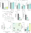
Fig 4. Polyamine depletion reroutes energy production to glycolysis.
(A) Huh7 cells were treated with 1 mM DFMO for four days, 500 μM GC7 for 24h or 40 μM etomoxir (ETM) for 24h, and fatty acid oxidation (FAO) activity was measured by fluorescent assay. Standard curves were used to quantify activity. (B) Huh7 cells were treated with increasing doses of DFMO (0.5, 1, and 5 mM) as indicated for four days prior to being assayed for cellular lipase activity by commercial kit. Raw relative luciferase values were measured. (C) Huh7 cells were treated with increasing doses of DFMO (0.5, 1, and 5 mM) as indicated for four days prior to measuring lipid uptake by cells by measuring incorporation of BODIPY from the supernatant into cells. Briefly, cells were incubated with BODIPY for 15m to allow for lipid uptake. Cells were then washed and media replenished. Fluorescent BODIPY levels in the cell were measured 24h later via fluorescent plate reader. (D) Supernatant from cells described in (C) was assayed for BODIPY levels to measure secretion of BODIPY after 24h incubation. (E) The ratio of cell-associated (C) to extracellular (D) BODIPY signal was measured by dividing the cell-associated fluorescence from BODIPY by the extracellular BODIPY fluorescence. (F) Huh7 cells were treated with 1 mM DFMO for four days or 500 μM GC7 for 16h and oxygen consumption rate (OCR) was measured by Seahorse XF Cell Mito Stress Test. Red arrows indicate the time of addition of inhibitors of the mitochondrial electron transport chain to measure basal, maximal, and minimum oxygen consumption through the electron transport chain. (G) Basal respiration and (H) ATP production-coupled respiration were derived from data in (B). (I) Huh7 cells were treated with increasing doses of GC7 for 16h and cell viability measured by MTT assay. Values were normalized to untreated controls. (J) Metabolic state was derived from data in (F) to differentiate between the impact of DFMO and GC7 on Huh7 energetics. (K) Glycolytic activity was measured by Lactate-GLO assay after treatment with increasing doses of DFMO (0.5, 1, and 5 mM) for four days prior to assay. “-”indicates blank control; “Lactate” indicates sample spiked with lactate. Values represent raw luciferase values measured. *p<0.05; ***p<0.001 by Student’s T-test, n = 2 (A-D; F, G-K) or n = 3 (E).
Fig 5
Fig 5. Mitochondrial structure is compromised by polyamine depletion.
Huh7 cells were treated with 1 mM DFMO, 2 μg/mL TOFA, or 500 μM GC7 and cellular structures were analyzed by electron microscopy. Bar indicates 2 μm. Black outlines indicate zoomed areas to highlight detail in mitochondrial structure.
Fig 6
Fig 6. Virus infection relies on lipid production.
Huh7 cells were treated with increasing doses of (A) C75 (5, 10, and 30 μM), a FASN inhibitor, and (B) TOFA (1, 1.5, and 2 μg/mL), an ACC1 inhibitor, and subsequently infected with chikungunya virus (CHIKV) at multiplicity of infection (MOI) of 0.1 PFU/cell for 24h. Viral titers were determined by plaque assay on Vero cells. (C) Murine embryonic fibroblasts (MEFs) were treated with 500 μM DFMO for four days, 500 μM GC7 for 16h, 100 μM C75, or 2 μg/mL TOFA, as indicated, infected with CHIKV at MOI 0.1, and viral titers determined at 24 hpi by plaque assay on Vero cells. Cells were treated with (D) 100 μM C75 or (E) 2 μg/mL TOFA and infected with human rhinovirus 1A (HRV1A), human rhinovirus 2 (HRV2), La Crosse virus (LACV), Sindbis virus (SINV), or Mayaro virus (MAYV). Titers were determined at 24h by plaque assay. (F) Huh7 cells were treated with 1 mM DFMO for 4 days and subsequently infected with CHIKV at MOI 1 for 24h. Cells were fixed and stained for dsRNA (red; viral replication compartments) and lipid droplets (green, BODIPY). Overlap of dsRNA and lipid droplets is shown through orange/yellow staining. Boxed image is magnified to the right of the images. *p<0.05; **p<0.01; ***p<0.001 by Student’s T test, n≥3.

References

    1. Celano P, Baylin SB, Casero RA. Polyamines Differentially Modulate the Transcription of Growth-associated Genes in Human Colon Carcinoma Cells. J Biol Chem. 1989;264: 8922–8927. doi: 10.1016/S0021-9258(18)81881-6 - DOI - PubMed
    1. Frugier M, Florentz C, Hosseini MW, Lehn JM, Giegé R. Synthetic polyamines stimulate in vitro transcription by T7 RNA polymerase. Nucleic Acids Res. 1994;22: 2784–2790. doi: 10.1093/nar/22.14.2784 - DOI - PMC - PubMed
    1. Yoshida M, Kashiwagi K, Shigemasa A, Taniguchi S, Yamamoto K, Makinoshima H, et al.. A Unifying Model for the Role of Polyamines in Bacterial Cell Growth, the Polyamine Modulon *. J Biol Chem. 2004;279: 46008–46013. doi: 10.1074/jbc.M404393200 - DOI - PubMed
    1. Sakamoto A, Terui Y, Uemura T, Igarashi K, Kashiwagi K. Polyamines regulate gene expression by stimulating translation of histone acetyltransferase mRNAs. J Biol Chem. 2020;295: 8736–8745. doi: 10.1074/jbc.RA120.013833 - DOI - PMC - PubMed
    1. Dever TE, Ivanov IP. Roles of polyamines in translation. J Biol Chem. 2018;293: 18719–18729. doi: 10.1074/jbc.TM118.003338 - DOI - PMC - PubMed

LinkOut - more resources