. 2024 Nov 26;121(48):e2416827121.
doi: 10.1073/pnas.2416827121. Epub 2024 Nov 18.

In vivo photoreceptor base editing ameliorates rhodopsin-E150K autosomal-recessive retinitis pigmentosa in mice

Affiliations

In vivo photoreceptor base editing ameliorates rhodopsin-E150K autosomal-recessive retinitis pigmentosa in mice

Samuel W Du et al. Proc Natl Acad Sci U S A. .

Abstract

Rhodopsin, the prototypical class-A G-protein coupled receptor, is a highly sensitive receptor for light that enables phototransduction in rod photoreceptors. Rhodopsin plays not only a sensory role but also a structural role as a major component of the rod outer segment disc, comprising over 90% of the protein content of the disc membrane. Mutations in RHO which lead to structural or functional abnormalities, including the autosomal recessive E150K mutation, result in rod dysfunction and death. Therefore, correction of deleterious rhodopsin mutations could rescue inherited retinal degeneration, as demonstrated for other visual genes such as RPE65 and PDE6B. In this study, we describe a CRISPR/Cas9 adenine base editing strategy to correct the E150K mutation and demonstrate precise in vivo editing in a Rho-E150K mouse model of autosomal recessive retinitis pigmentosa (RP). Using ultraviolet-visible spectroscopy, mass spectrometry, and the G-protein activation assay, we characterized wild-type rhodopsin and rhodopsin variants containing bystander base edits. Subretinal injection of dual-adeno-associated viruses delivering our base editing strategy yielded up to 44% Rho correction in homozygous Rho-E150K mice. Injection at postnatal day 15, but not later time points, restored rhodopsin expression, partially rescued retinal function, and partially preserved retinal structure. These findings demonstrate that in vivo base editing can restore the function of mutated structural and functional proteins in animal models of disease, including rhodopsin-associated RP and suggest that the timing of gene-editing is a crucial determinant of successful treatment outcomes for degenerative genetic diseases.

Keywords: base editing; prime editing; retinitis pigmentosa; rhodopsin.


Conflict of interest statement

Competing interests statement:K.P. is a consultant for Polgenix Inc. and serves on the Scientific Advisory Board at Hyperion Eye Ltd. D.R.L. is a consultant and/or equity owner for Prime Medicine, Beam Therapeutics, Pairwise Plants, Chroma Medicine, and Nvelop Therapeutics, companies that use or deliver genome-editing or epigenome-engineering agents. One reviewer, A.V.C. is listed on a patent that is potentially competing to the work described in this paper. All other authors have declared that no conflict of interest exists.

Figures

Fig. 1.
Fig. 1.
Establishment of rhodopsin-E150K cell line and base editing screening in vitro. (A, Left) The ROS is composed of stacked disc membranes with a high concentration of rhodopsin (red) (adapted from Gulati and Palczewski) (1). When rhodopsin encounters photons of light, it is photoactivated (yellow) and initiates phototransduction. (A, Right) 2-D protein structure of mouse rhodopsin. Locations of Schiff-base Lys296 and E150K mutation are indicated in red. (B) Coding sequence of the E150K-rhodopsin mutation. (C) Design of base editing SpCas9 sgRNAs, each named for the location where the adenine of interest is positioned within the protospacer. (D) Schematic diagram of the HEK293T-RhoE150K (HEK-E150K) cell line generated by retroviral transduction, used for in vitro screening. IRES-GFP downstream of the Rho fragment enables FACS purification of transduced cells. (E) Base editing outcomes via next-generation sequencing (NGS) after cotransfection of HEK-E150K cells with plasmids expressing ABEmax and sgRNAs. (F) Base editing outcomes via NGS after cotransfection of HEK-E150K cells with various ABEs and sgRNAs with the target adenine placed at positions 5, 7, 9, 10, or 11 within the protospacer.
Fig. 2.
Fig. 2.
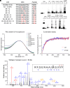
Characterization of bystander-edited variants of E150K-rhodopsin. (A) List of all potential coding variants of rhodopsin resulting from on-target and bystander editing observed by next-generation sequencing. (B) Anti-1D4 Western blot of HEK293T cells transfected with expression plasmids for all rhodopsin variants, loaded either undiluted (1:1) or diluted (1:10). Purified bovine rhodopsin is used as a positive control (concentration indicated in µg/mL). (C) UV-vis absorbance spectra of purified rhodopsin variants in LMNG after reconstitution with 11-cis-retinal. Absorbances are normalized to the absorbance of rhodopsin at its λmax, 500 nm. (D) Gt activation assay of WT rhodopsin and the E150K mutant. The results are plotted as normalized increase of fluorescence intensity at 345 nm of Gt upon addition of GTPγS. The curves represent the fitting of a pseudo-first-order association model. In this experiment, the rate constants (turnover numbers) were determined to be 16.2 ± 0.4 × 10⁻³ s−1 for WT and 17.8 ± 0.4 × 10⁻³ s−1 for the E150K mutant. (E) Tandem MS/MS spectrum of a unique peptide from purified WT-rhodopsin and its fragmentation pattern.
Fig. 3.
Fig. 3.
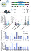
In vivo ABE of Rho-E150K mice treated at P21. (A) Schematic diagram (Upper) depicting the dual-AAV strategy for in vivo base editing. The gene coding for ABEmax and the sgRNA are split into a N-terminal and a C-terminal AAV. When a cell is transduced by both AAVs, the full-length ABEmax is reconstituted via Npu intein splicing. Schematic diagram (Lower) illustrating the P21 animal-treatment protocol: E150K mice were treated at postnatal day 21 (P21) by subretinal injection of dual-AAV8 and analyzed by ERG 10 wk later (P91). Retinas were then collected and analyzed by NGS (51). (B) NGS analysis of bulk retinal genomic DNA for base editing outcomes after dual-AAV8 treatment. (C) NGS analysis of bulk retinal cDNA (synthesized from retinal RNA) for base editing outcomes after dual-AAV8 treatment. (D) ERG a-wave amplitudes from E150K mice after dual-AAV8 treatment, compared with E150K mice treated with PBS (controls). P = 0.1411 by Student’s t test. (E) CIRCLE-seq analysis of A>G editing of on- and off-target genomic DNA within the ABE activity window (protospacer positions 4 to 8) for retinas from E150K dual-AAV8-treated mice versus untreated mice. (F) CIRCLE-seq analysis of all indels within the entire NGS amplicon for retinas from E150K dual-AAV8-treated mice versus untreated mice. All results are represented as mean ± SD.
Fig. 4.
Fig. 4.
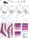
In vivo ABE of Rho-E150K mice treated at P15. (A) Schematic diagram of P15 treatment protocol: E150K mice were treated at postnatal day 15 (P15) by subretinal injection of ABE-expressing dual-AAV8, and analyzed by ERG after 10 wk (P86) or 14 wk (P116). Retinas were then collected and analyzed by NGS. (B) ERG a-wave (Left) and b-wave (Right) amplitudes from E150K mice 10 wk after dual-AAV8 treatment. P = 0.2268 and 0.2084 by Student’s t test. (C) ERG a-wave (Left) and b-wave (Right) amplitudes from E150K mice 14 wk after dual-AAV8 treatment. P = 0.1131 and 0.0122 by Student’s t test. (D) Representative ERG waveforms at −0.3 log cd s m−2 from three dual-AAV8-treated mice (Left) and three untreated mice (Right). The ERG a- and b-wave markers are indicated on trace 1 (treated mice). (E) Representative hematoxylin and eosin sections from dual-AAV8-treated mice (Left) and untreated mice (Right). ONH, optic nerve head. Scale bar represents 100 µm. (F) Representative hematoxylin and eosin sections from the thickest region of the retina from dual-AAV8-treated and untreated mice. Quantification of photoreceptor nuclei per ONL, from thickest and thinnest regions of retinas. Scale bar represents 50 µm. ONL, outer nuclear layer; INL, inner nuclear layer; GCL, ganglion cell layer. All results are represented as mean ± SD.
Fig. 5.
Fig. 5.
Rhodopsin expression in E150K mice after base editing at P15. Representative immunohistochemistry of retinal sections stained with DAPI and 1D4 (anti-Rho) of (A) WT, (B) untreated Rho-E150K mice, and (C) Rho-E150K mice treated with ABE-expressing dual-AAV8 at P15. Each row represents an individual and independent eye taken from mice 15 wk posttreatment or the equivalent age for WT and untreated mice. ONL, outer nuclear layer; OS, outer segment. Scale bar represents 20 µm.

References

    1. Gulati S., Palczewski K., Structural view of G protein-coupled receptor signaling in the retinal rod outer segment. Trends Biochem. Sci. 48, 172–186 (2023). - PMC - PubMed
    1. Palczewski K., et al. , Crystal structure of rhodopsin: A G protein-coupled receptor. Science 289, 739–745 (2000). - PubMed
    1. Ripps H., The color purple: Milestones in photochemistry. FASEB J. 22, 4038–4043 (2008). - PubMed
    1. Böll F., Zur Anatomie und Physiologie der Retina. Monatsber Akad Wiss Berlin 783–788 (1876).
    1. Hong J. D., Palczewski K., A short story on how chromophore is hydrolyzed from rhodopsin for recycling. Bioessays 45, e2300068 (2023). - PMC - PubMed