. 2024 Nov 18;45(3):e2027232024.
doi: 10.1523/JNEUROSCI.2027-23.2024. Online ahead of print.

Human iPSC-derived microglia sense and dampen hyperexcitability of cortical neurons carrying the epilepsy-associated SCN2A-L1342P mutation

Affiliations

Human iPSC-derived microglia sense and dampen hyperexcitability of cortical neurons carrying the epilepsy-associated SCN2A-L1342P mutation

Zhefu Que et al. J Neurosci. .

Abstract

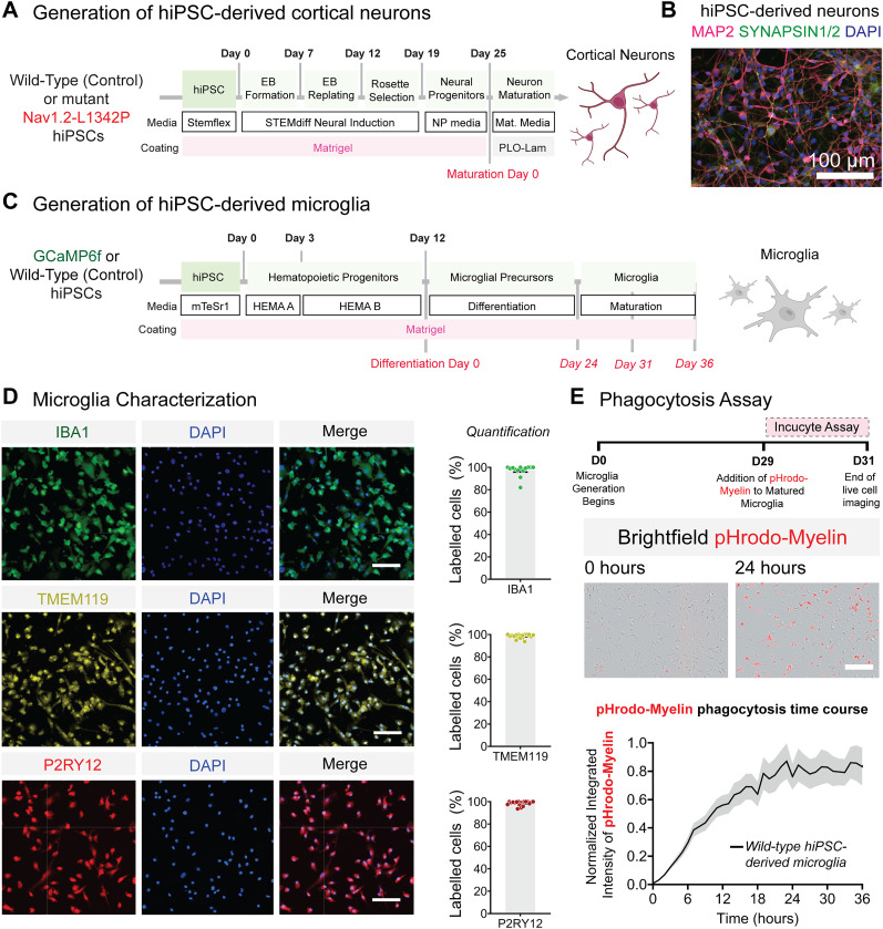
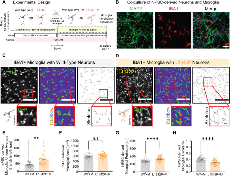
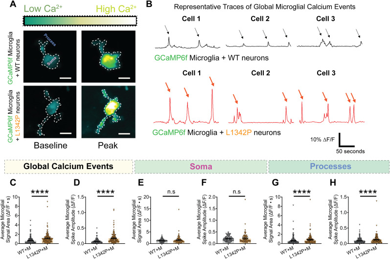
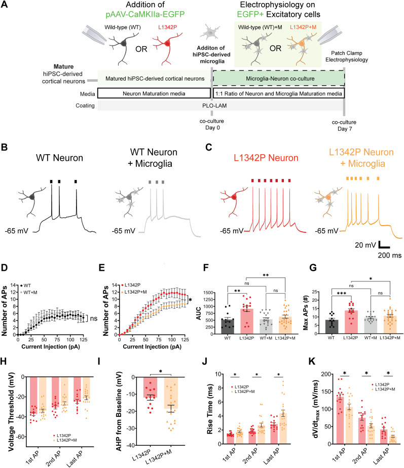
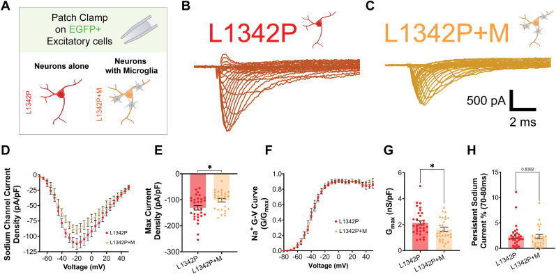
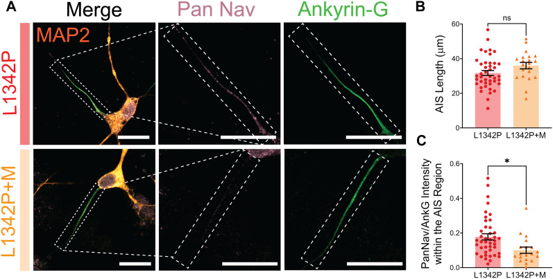
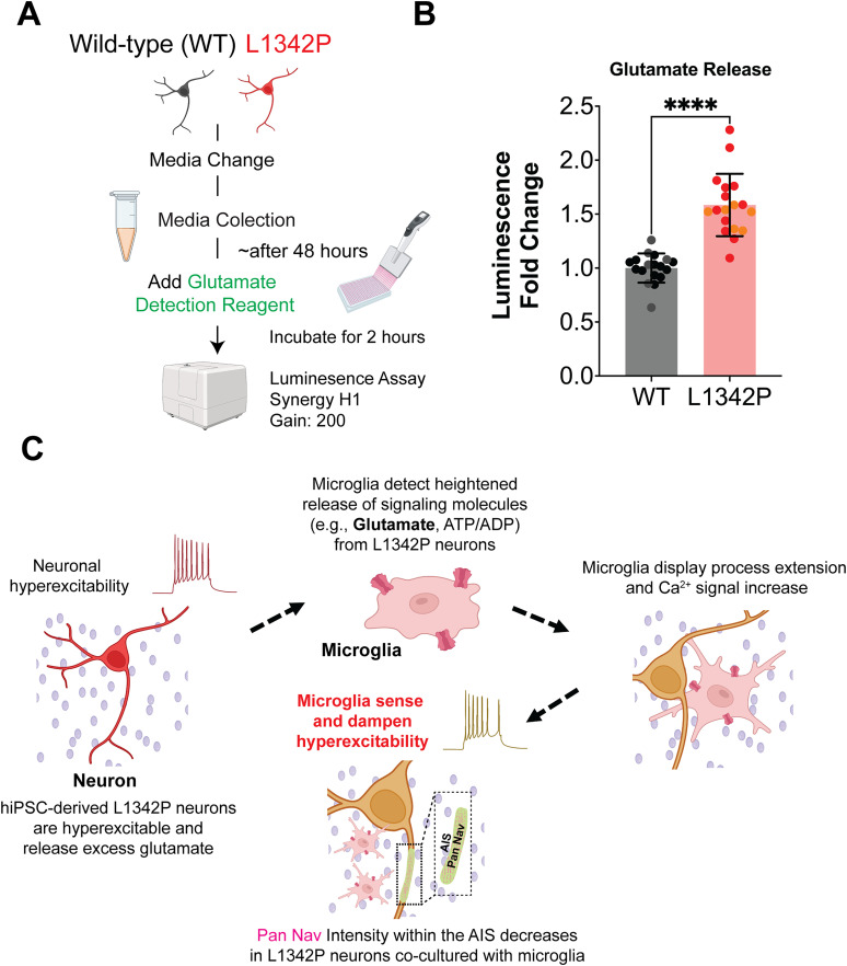
Neuronal hyperexcitability is a hallmark of epilepsy. It has been recently shown in rodent models of seizures that microglia, the brain's resident immune cells, can respond to and modulate neuronal excitability. However, how human microglia interact with human neurons to regulate hyperexcitability mediated by an epilepsy-causing genetic mutation found in patients is unknown. The SCN2A gene is responsible for encoding the voltage-gated sodium channel Nav1.2, one of the leading contributors to monogenic epilepsies. Previously, we demonstrated that the recurring Nav1.2-L1342P mutation leads to hyperexcitability in a male donor (KOLF2.1) hiPSC-derived cortical neuron model. Microglia originate from a different lineage (yolk sac) and are not naturally present in hiPSCs-derived neuronal cultures. To study how microglia respond to neurons carrying a disease-causing mutation and influence neuronal excitability, we established a co-culture model comprising hiPSC-derived neurons and microglia. We found that microglia display increased branch length and enhanced process-specific calcium signal when co-cultured with Nav1.2-L1342P neurons. Moreover, the presence of microglia significantly lowered the repetitive action potential firing and current density of sodium channels in neurons carrying the mutation. Additionally, we showed that co-culturing with microglia led to a reduction in sodium channel expression within the axon initial segment of Nav1.2-L1342P neurons. Furthermore, we demonstrated that Nav1.2-L1342P neurons release a higher amount of glutamate compared to control neurons. Our work thus reveals a critical role of human iPSCs-derived microglia in sensing and dampening hyperexcitability mediated by an epilepsy-causing mutation.Significance Statement Seizure studies in mouse models have highlighted the role of microglia in modulating neuronal activity, particularly in the promotion or suppression of seizures. However, a gap persists in comprehending the influence of human microglia on intrinsically hyperexcitable neurons carrying epilepsy-associated pathogenic mutations. This research addresses this gap by investigating human microglia and their impact on neuronal functions. Our findings demonstrate that microglia exhibit dynamic morphological alterations and calcium fluctuations in the presence of neurons carrying an epilepsy-associated SCN2A mutation. Furthermore, microglia suppressed the excitability of hyperexcitable neurons, suggesting a potential beneficial role. This study underscores the role of microglia in the regulation of abnormal neuronal activity, providing insights into therapeutic strategies for neurological conditions associated with hyperexcitability.


Conflict of interest statement

The authors declare no competing financial interests. ChatGPT was used to improve the readability and language of this work.

Figures

Figure 1.
Figure 1.
Characterization of hiPSC-derived cortical neurons and microglia. A, Schematic illustrating the protocol for generating hiPSC-derived cortical neurons. B, Representative fluorescent image of hiPSC-derived neurons stained for somatodendritic marker MAP2 (magenta), synaptic vesicle proteins SYN1/2 (green), and DAPI (blue). C, Schematic illustrating the protocol for generating hiPSC-derived microglia. hiPSCs are differentiated into HPCs for 12 d and cultured in microglia differentiation media for 24 d. The microglia maturation process is then carried out for up to 12 d. D, Representative images of hiPSC-differentiated microglia expressing microglial-specific markers: IBA1 (D, top panel, green, n = 13 fields of view, two differentiations), TMEM119 (D, middle panel, yellow, n = 18 fields of view, two differentiations), P2RY12 (D, lower panel red, n = 19 fields of view, two differentiations). DAPI was used to stain nuclei. Data are presented as mean ± SEM. Scale bar, 100 μm. E, Phagocytosis of pHrodo-myelin by WT (control) hiPSC-derived microglia. Data were obtained from one differentiation of three wells (48 images per well). Representative images at 0 and 24 h after the addition of pHrodo-myelin. hiPSC-derived microglia phagocytosed the pHrodo-labeled bioparticles, showing a gradually increasing red fluorescent signal over time. Scale bar, 25 μm. hiPSC, human-induced pluripotent stem cells; EB, embryoid body; NP, neural progenitors; MAP2, microtubule-associated protein 2; SYN1/2, synapsin1/2; IBA1, ionized calcium-binding adaptor molecule 1; TMEM119, transmembrane protein 119; P2RY12, purinergic receptor P2Y12.
Figure 2.
Figure 2.
Human microglia in coculture with Nav1.2-L1342P neurons display morphological changes. A, The hiPSC-derived neurons and microglia were matured separately, and then microglia were seeded on top of neurons for 7 d before imaging. B, Representative images of cocultured neurons stained for neuron-specific marker MAP2 (green), microglia stained for IBA1 (red), and DAPI (blue) as a nuclear stain. C, D, hiPSC-derived microglia in coculture with control (WT) neurons (WT + M, C) and Nav1.2-L1342P neurons (L1342P + M, D). IBA1 + microglia cocultured with the hyperexcitable Nav1.2-L1342P neurons displayed an extended ramified process compared with coculture with control (WT) neurons. Images are pseudocolored in a rainbow gradient to facilitate identification, and the skeletonized view was included to detail branches. E, The microglial average branch length increases in coculture with Nav1.2-L1342P cortical neurons (WT + M, n = 43 fields of view, and L1342 + M, n = 49 fields of view; three differentiations; two clones per condition). F, The total microglial area shows a trend toward being increased in coculture with Nav1.2-L1342P neurons (WT + M, n = 43 fields of view, and L1342 + M, n = 49 fields of view; three differentiations; two clones per condition). G, Microglial perimeter is enhanced in coculture with Nav1.2-L1234P neurons, indicating extended processes (WT + M, n = 43 fields of view, and L1342 + M, n = 49 fields of view; three differentiations; two clones per condition). H, Microglial circularity is decreased in coculture with Nav1.2-L1342P neurons, indicating that they are less ameboid-like (WT + M, n = 43 fields of view, and L1342 + M, n = 49 fields of view; three differentiations; two clones per condition). Each dot represents the mean value of a parameter per field of view. Data are presented as mean ± SEM. Scale bar, 50 μm. Data was pooled from three differentiations. Data were analyzed by nested t test; **p < 0.01 and ****p < 0.0001.
Figure 3.
Figure 3.
Calcium signal is enhanced in human microglia processes when cocultured with hyperexcitable hiPSC-derived Nav1.2-L1342P neurons. A, Fluorescent images of hiPSC-derived microglia expressing GCaMP6 cocultured with WT (top) and mutant Nav1.2-L1342P (bottom) hiPSC-derived neurons. The calcium signal is pseudocolored, with dark green indicating low signal and yellow hues depicting high signal. B, Representative ΔF/F traces of global calcium activity of microglia in coculture with control (WT) neurons (top, black) or Nav1.2-L1342P neurons (bottom, red). Three representative cells per condition are shown. C, The global average microglia calcium signal area increases in coculture with Nav1.2-L1342P neurons (WT + M, n = 209 cells, and L1342P + M, n = 168 cells). D, The global average microglial calcium spike amplitude increases in coculture with Nav1.2-L1342P neurons (WT + M, n = 204 cells, and L1342P + M, n = 168 cells). E, The average microglial signal area in the soma microdomain is not statistically different in the two coculture conditions (WT + M, n = 100 cells; L1342P + M, n = 92 cells). F, The average microglial spike amplitude in the soma microdomain is not statistically different in the two coculture conditions (WT + M, n = 100 cells; L1342P + M, n = 92 cells). G, The average microglial calcium signal area of the processes microdomain is increased in coculture with Nav1.2-L1342P neurons (WT + M, n = 250 processes; L1342P + M, n = 183 processes). H, The processes' average microglial calcium spike amplitude increases in coculture with L1342P neurons (WT + M, n = 250 processes; L1342P + M, n = 183 processes). Data were collected from three coverslips from two independent differentiations. Data in C–H were analyzed with Mann–Whitney's U test. ****p < 0.0001 and n.s. (not significant).
Figure 4.
Figure 4.
The repetitive firing of hiPSC-derived Nav1.2-L1342P neurons is reduced when cocultured with microglia. A, hiPSC-derived cortical neurons were transduced with an AAV-CaMKIIa-EGFP to allow the detection of excitatory neuronal populations for patch-clamp electrophysiology. Neurons and microglia were cocultured for 7 d before patch-clamp measurements. B, Representative AP firings from hiPSC-derived control (WT) cortical neurons alone (left) and with microglia (right). C, Representative AP firings from hiPSC-derived Nav1.2-L1342P cortical neurons alone (left) and with microglia (right). D, In WT neurons, current injection-triggered AP firing remains relatively unchanged regardless of microglia coculture (WT, n = 12 neurons; two differentiations; WT + M, n = 18 neurons; two differentiations). E, In Nav1.2-L1342P neurons, current injection-triggered AP firing is reduced when cocultured with microglia (L1342P, n = 14 neurons; two differentiations; L1342P + M, n = 25 neurons; two differentiations). F, AUC summary of the number of APs versus current injection for all conditions. Each dot corresponds to one neuron. G, Maximum AP number per cell across increasing current injections until 125 pA. H, Voltage thresholds for first, second, and last APs exhibited a consistent trend toward depolarization in the L1342P + M group. I, AHP from the baseline was decreased in the L1342P + M conditions. J, The rise time was increased in each successive AP in the L1342P + M conditions, indicating a longer time for the AP to take place. K, The dV/dtmax (mV/ms) as a function of AP# measured at 85 pA current injection decreased with each AP firing in the L1342P + M conditions. Each dot represents an individual neuron. Data are presented as mean ± SEM. Data in panels D and E were analyzed by repeated-measures of two-way ANOVA, with data pooled from at least two differentiations per condition. Data in F and G were analyzed using two-way ANOVA with Tukey's multiple comparisons. Data in panels H, J, and K were analyzed using multiple t tests with Holm–Šídák correction for multiple comparisons, while data in I was analyzed with an unpaired Student's t test. *p < 0.05; **p < 0.01; ***p < 0.001; and n.s. (not significant).
Figure 5.
Figure 5.
hiPSC-derived Nav1.2-L1342P neurons display reduced sodium current density with microglia coculture. A, The patch clamp was performed on hiPSC-derived cortical neurons transduced with CaMKII virus to selectively label excitatory neuron populations. B, Representative sodium current trace from Nav1.2-L1342P cortical neurons. C, Representative sodium current trace from Nav1.2-L1342P cortical neurons cocultured with hiPSC-derived microglia. In both panels B and C, the outward current was blocked using tetraethylammonium chloride in the bath solution. D, Average peak sodium current density versus voltage (IV) curves for Nav1.2-L1342P neurons with or without microglia coculture. E, The maximum current density was significantly reduced in Nav1.2-L1342P neurons in coculture with microglia (right) (L1342P: n = 31 neurons, three differentiations: L1342P + M, n = 28 neurons from two differentiations). F, Normalized G/Gmax versus voltage curves, showing no notable difference between L1342P and L1342P + M (with microglia) conditions. G, The maximum conductance (Gmax), normalized to cell capacitance, was significantly decreased in L1342P neurons cocultured with microglia. H, The persistent current, measured as a percentage of the peak amplitude at 70–80 ms after the peak sodium transient current, showed no change between the L1342P and L1342P + M conditions. Data are presented as mean ± SEM. Data in E, G, and H was analyzed by unpaired Student's t test, with each dot representing an individual neuron. Data were collected from at least two independent differentiations per genotype. *p < 0.05.
Figure 6.
Figure 6.
The intensity of the PanNav/AnkG in the AIS is reduced in the hiPSC-derived Nav1.2-L1342P neurons cocultured with microglia. A, Representative images of L1342P alone (top) and L1342P neurons cocultured with microglia (bottom) were shown. MAP2 (orange), Pan Nav (magenta), and ANK-G (green). Scale bar, 20 μm. B, No notable change was observed for AIS length in L1342P neurons with or without microglia coculture. C, The normalized PanNav/AnkG intensity in the AIS region was reduced in the hiPSC-derived L1342P neurons in microglia coculture conditions. Each dot corresponds to one neuron. Data are presented as mean ± SEM. Data in B–C were analyzed by unpaired Student's t test. *p < 0.05 and n.s. (not significant)
Figure 7.
Figure 7.
hiPSC-derived Nav1.2-L1342P neuron cultures produce excessive extracellular glutamate. A, Experimental design schematic. B, Glutamate release from hiPSC-derived WT or L1342P neurons over 48 h, presented as fold change in luminescence (WT, n = 18 wells, from two differentiations; L1342P, n = 18 wells, from two differentiations). Data are presented as mean ± SEM. Statistical analysis conducted using unpaired Student's t test. C, Proposed possible mechanism illustrating microglial sensing and dampening hyperexcitability of neurons carrying epilepsy-associated SCN2A mutation.

Update of

Similar articles

Cited by

References

    1. Abud EM, et al. (2017) iPSC-derived human microglia-like cells to study neurological diseases. Neuron 94:278–293.e9. 10.1016/j.neuron.2017.03.042 - DOI - PMC - PubMed
    1. Asadollahi R, et al. (2023) Pathogenic SCN2A variants cause early-stage dysfunction in patient-derived neurons. Hum Mol Genet 32:2192–2204. 10.1093/hmg/ddad048 - DOI - PMC - PubMed
    1. Badimon A, et al. (2020) Negative feedback control of neuronal activity by microglia. Nature 586:417–423. 10.1038/s41586-020-2777-8 - DOI - PMC - PubMed
    1. Barker-Haliski M, White HS (2015) Glutamatergic mechanisms associated with seizures and epilepsy. Cold Spring Harb Perspect Med 5:a022863. 10.1101/cshperspect.a022863 - DOI - PMC - PubMed
    1. Baxter PS, Dando O, Emelianova K, He X, McKay S, Hardingham GE, Qiu J (2021) Microglial identity and inflammatory responses are controlled by the combined effects of neurons and astrocytes. Cell Rep 34:108882. 10.1016/j.celrep.2021.108882 - DOI - PMC - PubMed

LinkOut - more resources