Elotuzumab-mediated ADCC with Th1-like Vγ9Vδ2 T cells to disrupt myeloma-osteoclast interaction
- PMID: 39557586
- PMCID: PMC11786308
- DOI: 10.1111/cas.16401
Elotuzumab-mediated ADCC with Th1-like Vγ9Vδ2 T cells to disrupt myeloma-osteoclast interaction
Abstract
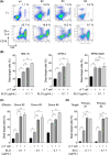
Multiple myeloma (MM) cells and osteoclasts (OCs) activate with each other to cause drug resistance. Human Th1-like Vγ9Vδ2 (γδ) T cells, important effectors against tumors, can be expanded and activated ex vivo by the aminobisphosphonate zoledronic acid in combination with IL-2. We previously reported that the expanded γδ T cells effectively targeted and killed OCs as well as MM cells. Because the expanded γδ T cells expressed CD16 on their surface, we investigated the utilization of the expanded γδ T cells for antibody-dependent cellular cytotoxicity (ADCC). Although the expanded γδ T cells alone induced cell death in MM cell lines, the addition of the anti-SLAMF7 monoclonal antibody elotuzumab (ELO) further enhanced their cytotoxic activity only against SLAMF7-expressing MM cell lines and primary MM cells. Intriguingly, ELO was also able to enhance γδ T cell-induced cell death against OCs cultured alone, and against both MM cells and OCs in their coculture settings. SLAMF7 was found to be highly expressed in OCs differentiated in vitro from monocytes by receptor activator of nuclear factor-κ B ligand and M-CSF, although monocytes only marginally expressed SLAMF7. These results demonstrate that SLAMF7 is highly expressed in both MM cells and OCs, and that the ex vivo-expanded γδ T cells can exert ELO-mediated ADCC against SLAMF7-expressing MM cells and OCs besides their direct cytotoxic activity. Further study is warranted for the innovative utilization of γδ T cells.
Keywords: Elotuzumab; SLAMF7; Vγ9Vδ2 T cell; multiple myeloma; osteoclast.
© 2024 The Author(s). Cancer Science published by John Wiley & Sons Australia, Ltd on behalf of Japanese Cancer Association.
Conflict of interest statement
T.H. received research funding and honoraria from Bristol‐Myers. M.A. received research funding from Bristol‐Myers Squibb and honoraria from Bristol‐Myers Squibb, Janssen Pharma K.K., and Sanofi K.K. The other authors declare no competing financial interests.
Figures


References
-
- Nakamura K, Smyth MJ, Martinet L. Cancer immunoediting and immune dysregulation in multiple myeloma. Blood. 2020;136:2731‐2740. - PubMed
-
- Thompson K, Rojas‐Navea J, Rogers MJ. Alkylamines cause Vgamma9Vdelta2 T‐cell activation and proliferation by inhibiting the mevalonate pathway. Blood. 2006;107:651‐654. - PubMed
-
- Cui Q, Shibata H, Oda A, et al. Targeting myeloma‐osteoclast interaction with Vgamma9Vdelta2 T cells. Int J Hematol. 2011;94:63‐70. - PubMed
-
- Mensurado S, Blanco‐Domínguez R, Silva‐Santos B. The emerging roles of γδ T cells in cancer immunotherapy. Nat Rev Clin Oncol. 2023;20:178‐191. - PubMed
MeSH terms
Substances
Grants and funding
LinkOut - more resources
Full Text Sources
Medical
Research Materials
Miscellaneous

