

Quantitative distribution of essential elements and non-essential metals in breast cancer tissues by LA-ICP-TOF-MS
- PMID: 39557687
- PMCID: PMC11698889
- DOI: 10.1007/s00216-024-05652-8
Quantitative distribution of essential elements and non-essential metals in breast cancer tissues by LA-ICP-TOF-MS
Abstract
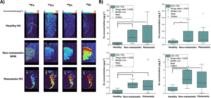
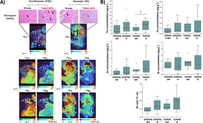
Breast cancer (BC) is the leading cause of cancer death among women worldwide, making the discovery and quantification of new biomarkers essential for improving diagnostic and preventive strategies to limit dissemination and improve prognosis. Essential trace metals such as Fe, Cu, and Zn may play critical roles in the pathophysiology of both benign and malignant breast tumors. However, due to the high metabolic activity and reduced element selectivity of cancer cells, also non-essential elements may be taken up and may even be implicated with disease progression. This study investigates the spatial distribution and concentrations of both essential and non-essential elements in breast tissues, assessing their potential for diagnostic applications. Laser ablation (LA)-inductively coupled plasma-mass spectrometry (ICP-MS) with a time-of-flight (ToF) mass analyzer (LA-ICP-ToF-MS) was used to inquire the distribution of almost all elements across the periodic table and their abundance in metastatic (n = 11), non-metastatic (n = 7), and healthy (n = 4) breast tissues. Quantification was achieved using gelatine-based standards for external calibration to quantitatively map various elements. Overall, the Fe, Cu, Zn, Sr, and Ba levels were significantly increased in tumor samples with Sr and Ba showing strong correlation, likely due to their similar chemistry. Comparison of calibrated LA-ICP-ToF-MS data with a histologic staining demonstrated the possibility to clearly differentiate between various tissue types and structures in breast tissues such as tumor niche and stroma. The levels of the studied elements were significantly higher in the tumor niche areas compared to the stroma, and for Fe, a significant accumulation was observed in the tumor niche areas from the metastatic patient group relative to the levels found in the same areas of the non-metastatic group.
Keywords: Elemental bioimaging; Elemental quantification; Hyphenated techniques; LA-ICP-MS; Time of flight.
© 2024. The Author(s).
Conflict of interest statement
Declarations. Ethical approval: This study was conducted in accordance with national regulations and received approval from the Ethics and Investigation Committee of the Hospital de Jove Foundation (PI02/2018). Competing interests: The authors declare no competing interests.
Figures


References
-
- Geneva, Lyon (2024) Global cancer burden growing, amidst mounting need for services. https://www.who.int/news/item/01-02-2024-global-cancer-burden-growing--a.... Accessed 2 Oct 2024 - PMC - PubMed
-
- Cabré N, Luciano-Mateo F, Arenas M, Nadal M, Baiges-Gaya G, Hernández-Aguilera A, Fort-Gallifa I, Rodríguez E, Riu F, Camps J, Joven J, Domingo JL. Trace element concentrations in breast cancer patients. Breast. 2018;42:142–9. 10.1016/j.breast.2018.09.005. - PubMed
-
- Singer P, Manzanares W, Berger MM. What’s new in trace elements? Intensive Care Med. 2018;44:643–5. 10.1007/s00134-017-4955-1. - PubMed
MeSH terms
Substances
Grants and funding
LinkOut - more resources
Full Text Sources
Medical
Research Materials
