. 2024 Nov 18;15(11):843.
doi: 10.1038/s41419-024-07128-0.

YTHDF1 boosts the lactate accumulation to potentiate cervical cancer cells immune escape

Affiliations

YTHDF1 boosts the lactate accumulation to potentiate cervical cancer cells immune escape

Jing Xiong et al. Cell Death Dis. .

Abstract

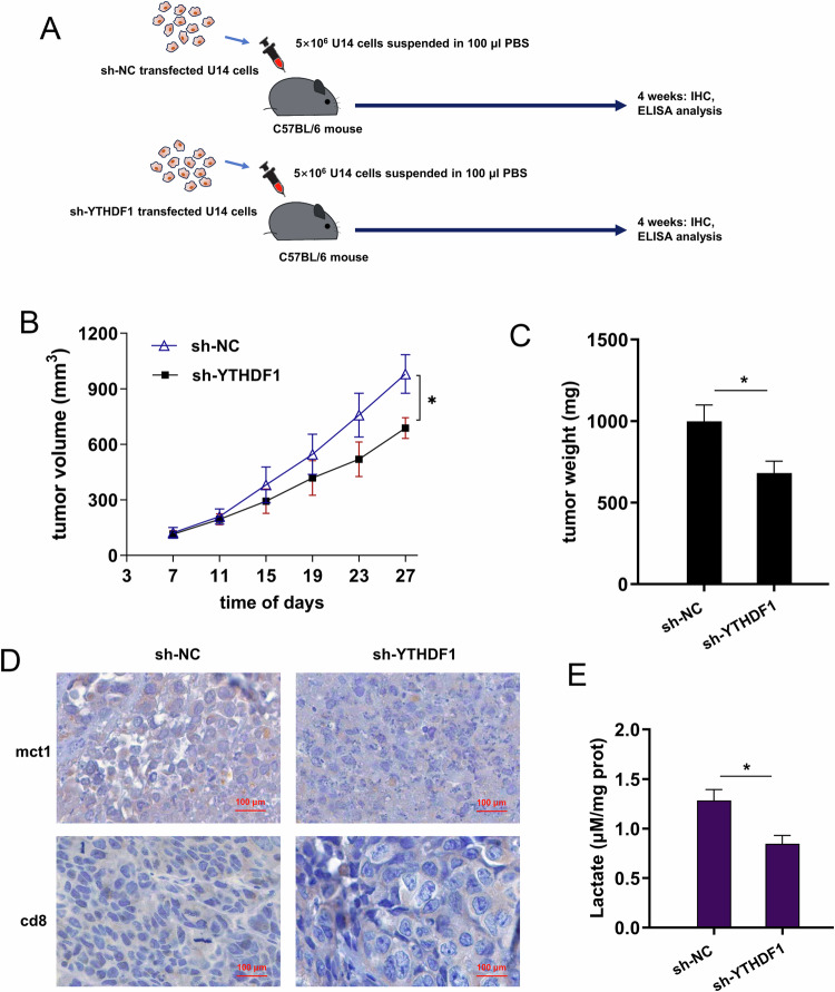
Lactate is a major metabolic product of tumor cells in microenvironment. Increasing evidence has indicated that lactate accumulation could alter the immune response in human cancers, including cervical cancer. However, the function and significance of N6-methyladenosine (m6A) reader YTHDF1 in cervical cancer cells' lactate metabolism and immunotherapy remain obscure. Results illustrated that YTHDF1 predicted unfavorable clinical outcomes of cervical cancer, which was negatively correlated with CD8+ T cell infiltration. In the co-culture of tumor cells with CD8+ T cells, YTHDF1 overexpression promoted the lactate accumulation and attenuated the cytotoxic CD8+ T cell's killing effect. Correspondingly, YTHDF1 knockdown exerted the opposite effects. Mechanistically, YTHDF1 targeted the m6A site on SLC16A1 gene (MCT1) to determine its fate. YTHDF1 upregulated MCT1 expression by enhancing MCT1 stability mediated by m6A-modified manner. Collectively, our results revealed an oncogenic role played by YTHDF1 in cervical cancer through m6A/MCT1-dependent manner. In conclusion, these findings unveil the immune escape-promoting effect of YTHDF1 in cervical cancer by boosting the lactate accumulation, which might illuminate a novel target for more precise immunotherapy.


Conflict of interest statement

Competing interests The authors declare no competing interests. Ethics approval and consent to participate Written informed consent was obtained from each patient, and the study had been approved by The Second Xiangya Hospital of Central South University.

Figures

Fig. 1
Fig. 1. Elevated YTHDF1 indicated the poor prognosis and tumor infiltrating lymphocyte.
A The level of YTHDF1 was detected in the cervical cancer samples. B In the public dataset (http://gepia.cancer-pku.cn/index.html), the YTHDF1 level increased the cervical cancer individuals. C In the TIMER database (http://timer.cistrome.org/), the YTHDF1 copy number altered the infiltration levels of CD8+ T cells. D TIMER were used to predict the correlation of YTHDF1 with CD8+ T cells in cervical cancer. E RT-PCR was performed to illustrate the YTHDF1 mRNA in the cervical cancer cells. *p < 0.05; p < 0.01.
Fig. 2
Fig. 2. YTHDF1 promoted the glycolysis, lactate accumulation and surface PD-L1 expression.
AD Extracellular acidification rate (ECAR) was performed to detected the extracellular acidification rate of glycolysis. E The supernatant lactate expression was detected by l-lactatic acid colorimetric assay kit in cervical cancer cells with YTHDF1 silencing and control group. F The cellular surface PD-L1 expression on cervical cancer cells were detected using flow cytometry. *p < 0.05; **p < 0.01.
Fig. 3
Fig. 3. YTHDF1 hampered the cytotoxic CD8+ T cell’s killing effect to cervical cancer cells.
A The coculture within activated CD8+ T cells and cervical cancer cells was constructed. B Survival analysis was performed by CCK-8 assay illustrated the survival rate of cervical cancer cells (HeLa, SiHa) in the coculture system in different E:T ratio (effector:target ratio) ranging from 1:1 to 10:1. CF In the coculture within activated CD8+ T cells and cervical cancer cells (HeLa, SiHa), the quantitative analysis of immunosuppressive cytokines (IL-10, TGF-β) and immune effector cytokines (IFN-γ, IL-2) from activated CD8+ T cells was identified by ELISA kit. G The production of cytolytic granzyme B from activated CD8+ T cells was tested by ELISA kit. H The CD8+ T cells’ cytotoxicity assay based on LDH release was performed to determine the release of LDH into the coculture supernatants. *p < 0.05; p < 0.01.
Fig. 4
Fig. 4. MCT1 was targeted by YTHDF1 in cervical cancer microenvironment.
A The m6A modification on MCT1 (SLC16A1 gene) was shown by IGV (Integrative Genomics Viewer). B The m6A methylation motifs of cervical cancer and normal samples were shown. C Distribution of new m6A peaks in mRNA detected derived from the NCBI GEO dataset (Series GSE242071). D The m6A methylation distribution was showed, including 5′-UTR, CDS, 3′-UTR and start/stop codon. E The precise location of m6A methylation site on MCT1 (SLC16A1 gene). F RNA fluorescence in situ hybridization (FISH) reflected the co-location of YTHDF1 and MCT1 in the cervical cancer cells.
Fig. 5
Fig. 5. YTHDF1 potentiated MCT1 expression by enhancing MCT1 mRNA stability.
A RIP-PCR assay was performed to test the binding within anti-YTHDF1 anti-body and MCT1 mRNA in cervical cancer. B The precipitated MCT1 mRNA enrichment was quantificationally analyzed by PCR. C RIP-PCR assay was performed in cervical cells (SiHa, HeLa) transfected with YTHDF1 overexpression (YTHDF1 or vector) or YTHDF1 knockdown (sh-YTHDF1 or sh-NC). D RNA pull-down assay was performed to confirm the binding of YTHDF1 and MCT1. E, F RNA stability analysis was performed upon Act D treatment. *p < 0.05; p < 0.01.
Fig. 6
Fig. 6. YTHDF1/MCT1 boosted the lactate accumulation and potentiated cervical cancer cells immune escape.
A Lactate was detected by l-lactatic acid colorimetric assay kit in cervical cancer (SiHa) cells transfected with YTHDF1 overexpression plasmids (YTHDF1), MCT1 silencing (si-MCT1), MCT1 specific inhibitor (AZD3965) and exogenous lactate (Lactate, 10 mM l-lactate). B The ECAR was detected in the cervical cancer (SiHa) cells. C Survival analysis by CCK-8 exhibited the survival rate of cervical cancer cells (SiHa) in the coculture system in different E:T ratio (effector:target ratio) ranging from 1:1 to 10:1. D The CD8+ T cells’ cytotoxicity assay based on LDH release was performed to determine the release of LDH into the coculture supernatants. E, F The cellular surface PD-L1 expression on cervical cancer cells were detected using flow cytometry. *p < 0.05; p < 0.01.
Fig. 7
Fig. 7. YTHDF1 silencing repressed the tumor growth and lactate in vivo.
A The in vivo mice assay was performed using mouse cervical cancer cells (U14 cells with sh-NC, sh-YTHDF1). B Tumor volume and C tumor weight of cervical cancer were detected and calculated. D Immumohistochemical (IHC) staining revealed the positive mct1 and cd8 protein in the YTHDF1 silencing group and control group. E The in vivo lactate expression was detected by l-lactatic acid colorimetric assay kit in YTHDF1 silencing and control group. *p < 0.05.
Fig. 8
Fig. 8
YTHDF1 deteriorates cervical cancer immune escape by inhibiting cytotoxic CD8+ T cells antitumor effect.

References

    1. Aranda-Gutierrez A, Ferrigno Guajardo AS, Vaca-Cartagena BF, Gonzalez-Sanchez DG, Ramirez-Cisneros A, Becerril-Gaitan A, et al. Obstetric and neonatal outcomes following taxane use during pregnancy: a systematic review. BMC Cancer. 2024;24:9. - PMC - PubMed
    1. Corbeau A, Heemsbergen WD, Kuipers SC, Godart J, Creutzberg CL, Nout RA. et al. Predictive factors for toxicity after primary chemoradiation for locally advanced cervical cancer: a systematic review. Int J Radiat Oncol Biol Phys. 2024;119:127–42. - PubMed
    1. D'Oria O, Bogani G, Cuccu I, D'Auge TG, Di Donato V, Caserta D. et al. Pharmacotherapy for the treatment of recurrent cervical cancer: an update of the literature. Expert Opin Pharmacother. 2024;25:55–65. - PubMed
    1. Jia Y, Zou K, Zou L. Research progress of metabolomics in cervical cancer. Eur J Med Res. 2023;28:586. - PMC - PubMed
    1. Ahmadi M, Abbasi R, Rezaie J. Tumor immune escape: extracellular vesicles roles and therapeutics application. Cell Commun Signal : CCS. 2024;22:9. - PMC - PubMed

MeSH terms

LinkOut - more resources