. 2024 Nov 18;9(1):325.
doi: 10.1038/s41392-024-02018-5.

Renal remodeling by CXCL10-CXCR3 axis-recruited mesenchymal stem cells and subsequent IL4I1 secretion in lupus nephritis

Affiliations

Renal remodeling by CXCL10-CXCR3 axis-recruited mesenchymal stem cells and subsequent IL4I1 secretion in lupus nephritis

Qixiang Zhang et al. Signal Transduct Target Ther. .

Abstract

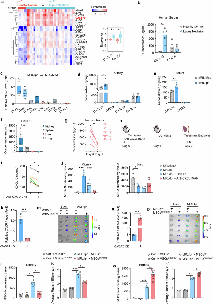
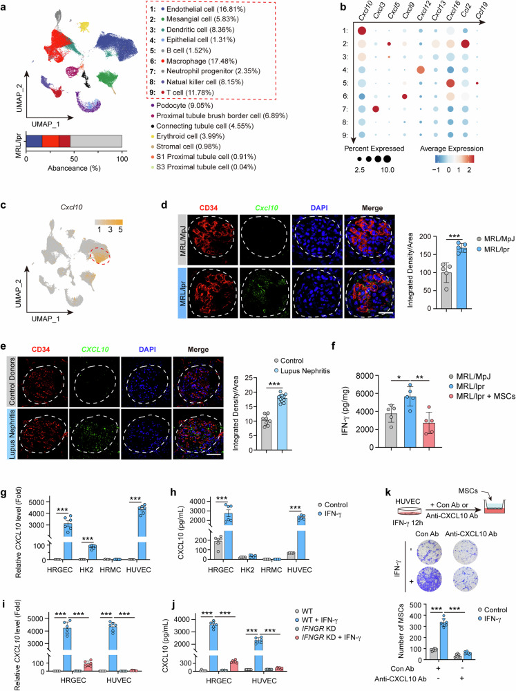
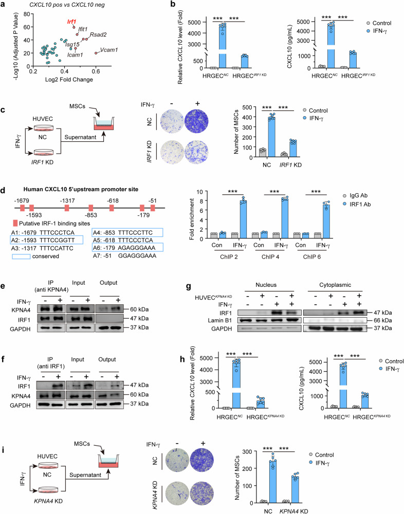
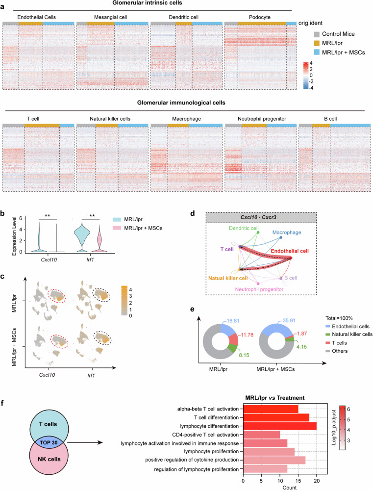
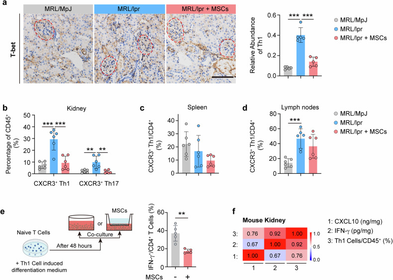
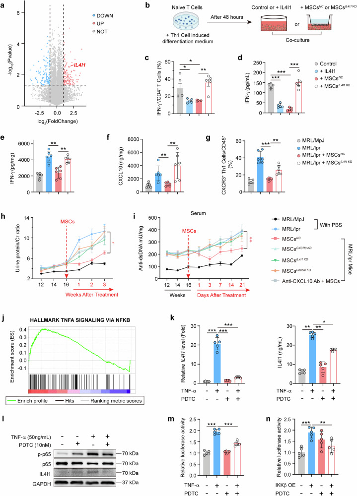
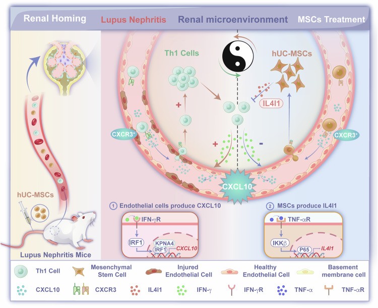
Human umbilical cord mesenchymal stem cells (hUC-MSCs) have shown potential as a therapeutic option for lupus nephritis (LN), particularly in patients refractory to conventional treatments. Despite extensive translational research on MSCs, the precise mechanisms by which MSCs migrate to the kidney and restore renal function remain incompletely understood. Here, we aim to clarify the spatiotemporal characteristics of hUC-MSC migration into LN kidneys and their interactions with host cells in microenvironment. This study elucidates that the migration of hUC-MSCs to the LN kidney is driven by elevated levels of CXCL10, predominantly produced by glomerular vascular endothelial cells through the IFN-γ/IRF1-KPNA4 pathway. Interestingly, the blockade of CXCL10-CXCR3 axis impedes the migration of hUC-MSCs to LN kidney and negatively impacts therapeutic outcomes. Single cell-RNA sequencing analysis underscores the importance of this axis in mediating the regulatory effects of hUC-MSCs on the renal immune environment. Furthermore, hUC-MSCs have been observed to induce and secrete interleukin 4 inducible gene 1 (IL4I1) in response to the microenvironment of LN kidney, thereby suppressing Th1 cells. Genetically ablating IL4I1 in hUC-MSCs abolishes their therapeutic effects and prevents the inhibition of CXCR3+ Th1 cell infiltration into LN kidneys. This study provides valuable insights into the significant involvement of CXCL10-CXCR3 axis in hUC-MSC migration to the LN kidneys and the subsequent remodeling of renal immune microenvironment. Regulating the CXCL10-CXCR3 axis and IL4I1 secretion may be developed as a novel therapeutic strategy to improve treatment outcomes of LN.


Conflict of interest statement

Competing interests The authors declare no competing interests.

Figures

Fig. 1
Fig. 1
Increased recruitment of hUC-MSCs to kidney in MRL/lpr mice alleviates renal of lupus nephritis. a The quantities of hUC-MSCs were assessed by Q-PCR in the lungs and kidneys of MRL/lpr and MRL/MpJ mice at 1 day, 3 days, 7 days after intravenous transplantation of hUC-MSCs (n = 4–5). b Proportional distribution of hUC-MSCs in the lungs, kidneys, and other tissues at 1 day after intravenous transplantation. c The line plots illustrating the quantity of MRL/lpr mice at 1 day, 3 days, 7 days, 14 days and 21 days after intravenous injection (n = 4–5). BLD means below the low detection. d Fluorescence intensity of RFP-hUC-MSCs (the hUC-MSCs expressing RFP fluorescent protein) in kidneys is determined by IVIS Imaging System (n = 3). e Representative images of RFP-hUC-MSCs distribution in kidney sections from MRL/lpr mice. FITC-LTL is administered intravenously. Scale bar: full scan of kidney section (1000 µm), Glomerulus (50 µm), Cortex (10 µm). f Dynamic curves of urine protein/creatinine levels in MRL/MpJ and MRL/lpr mice treated with or without hUC-MSCs (n = 7). g Dynamic curves of serum anti-dsDNA antibody levels in MRL/MpJ and MRL/lpr mice treated with or without hUC-MSCs (n = 7). h Glomerular pathological sections of MRL/lpr and MRL/MpJ mice treated with or without hUC-MSCs (n = 5). Scale bar: 50 µm. i Deposition of immune complexes IgG in glomerulus of MRL/lpr mice treated with or without hUC-MSCs (n = 5). Scale bar: 30 µm. jl SLEDAI-2K Score (Systemic Lupus Erythematosus Disease Activity Score) (j), urine protein/creatinine levels (k), anti-dsDNA antibodies levels (l) in 5 lupus nephritis patients before and 30 days after treatment with hUC-MSCs. *P < 0.05, ***P < 0.001
Fig. 2
Fig. 2
Disruption of CXCL10-CXCR3 axis suppresses hUC-MSCs recruitment to LN kidney. a Differential analysis of mRNA expression in the kidneys of LN patients compared to that of healthy donors based on ERCB Lupus TubInt Dataset Summary database. Each square represents one patient. b Concentrations of CXCL10 and CXCL9 in serum from LN patients (n = 5) and healthy donors (n = 6). c Relative mRNA expression levels of chemokines are determined by Q-PCR in the kidney of MRL/lpr and MRL/MpJ mice (n = 5). d The concentrations of chemokines in kidneys from MRL/MpJ and MRL/lpr mice (n = 5). e The concentrations of CXCL9 and CXCL10 in serum from MRL/MPJ and MRL/lpr mice (n = 5). f The concentration of CXCL10 in the kidney, spleen, liver, and lung from MRL/lpr mice (n = 5). g CXCL10 concentrations in the serum of five LN patients in whom treatment was effective before and 1 day after treatment with hUC-MSCs. h Scheme illustrating MSC tissue distribution after pretreatment with anti-CXCL10 antibodies in MRL/lpr and MRL/MpJ mice. Mice were pretreated with 20 µg isotype control antibodies or anti-CXCL10 antibodies after 1 day they were injected intravenously with hUC-MSCs. i Concentration of CXCL10 in serum from MRL/lpr mice before and after pretreated with anti-CXCL10 antibodies (n = 5). j Number of hUC-MSCs was determined by Q-PCR in the lungs and kidneys of MRL/lpr and MRL/MpJ mice at 1 day after intravenous injection of hUC-MSCs (n = 5). k CXCR3 mRNA expression are measured in hUC-MSCs with or without CXCR3 knockdown (n = 3). hUC-MSCs are transfected with CXCR3 siRNA or negative control (NC) for 24 h, respectively. Each data point represents an independent experiment. l Number of hUC-MSCs was determined by Q-PCR in the kidneys of control and MRL/lpr mice at 1 day after intravenous injection of hUC-MSCsNC and hUC-MSCsCXCR3 KD (n = 6). m Fluorescence intensity of RFP-hUC-MSCs (the hUC-MSCs expressing RFP fluorescent protein) with or without CXCR3 knockdown in kidneys is determined by IVIS Imaging System (n = 6). n CXCR3 mRNA expression is measured in hUC-MSCs with or without CXCR3 overexpression (n = 3). hUC-MSCs are transfected with CXCR3 overexpression lentivirus or negative control (NC) for 48 h, respectively. Each data point represents an independent experiment. o Number of hUC-MSCs were determined by Q-PCR in the kidneys of control and MRL/lpr mice at 1 day after intravenous injection of hUC-MSCsNC and hUC-MSCsCXCR3 OE (n = 6). p Fluorescence intensity of RFP-hUC-MSCs with or without CXCR3 overexpression in kidneys is determined by IVIS Imaging System (n = 6). *P < 0.05, **P < 0.01, ***P < 0.001
Fig. 3
Fig. 3
Renal CXCL10 is primarily derived from glomerular vascular endothelial cells via IFN-γ/IFN-γ R pathway in LN. a UMAP showing cell clusters from glomerulus of MRL/lpr mice. b Air bubble diagram showing mRNA expression of chemokines in cells as indicated. c UMAP plot showing expression of Cxcl10 from glomerulus of MRL/lpr mice. d In situ mRNA expression of Cxcl10 in the glomeruli of MRL/lpr and MRL/MpJ mice. Cxcl10 in green, CD34 in red (n = 5). Scale bar: 20 µm. e In situ mRNA expression of CXCL10 mRNA in the glomeruli of patients with lupus nephritis and controls, CXCL10 in green, CD34 in red (n = 5). Scale bar: 50 µm. f Concentration of IFN-γ in kidneys from MRL/MpJ and MRL/lpr mice and MRL/lpr mice at 7 days treated with hUC-MSCs (n = 5). g, h HRGECs, HK-2 cells, HRMCs, and HUVECs were stimulated with or without IFN-γ (50 ng/mL) for 12 h. The mRNA level (g) and secretory protein level (h) of CXCL10 were detected (n = 6). i, j HRGECsNC and HRGECsIFNGR KD, HUVECsNC, and HUVECsIFNGR KD were stimulated with or without IFN-γ (50 ng/mL) for 12 h. The mRNA level (i) and secretory protein level (j) of CXCL10 were detected (n = 6). k Representative images and numbers of hUC-MSCs recruited by culture medium supernatants which were collected after 12 h of IFN-γ (50 ng/mL) stimulation of HUVECs and with or without anti-CXCL10 antibody (5 µg/mL) (n = 3). Cell migration was assessed by transwell assays. Data represent mean ± SD. *P < 0.05, **P < 0.01, ***P < 0.001
Fig. 4
Fig. 4
Nuclear transport of IRF1-KPNA4 mediates IFN-γ-induced secretion of CXCL10 from endothelial cells. a Fold change in gene expression levels of Cxcl10 positive expressing endothelial cells compared to Cxcl10 negative expressing endothelial cells from single-cell sequencing of MRL/lpr mouse glomeruli. b HRGECsNC and HRGECsIRF1 KD were stimulated with or without IFN-γ (50 ng/mL) for 12 h. The mRNA level and secretory protein level of CXCL10 were detected (n = 6). c Representative images and numbers of hUC-MSCs recruited by culture medium supernatants collected after 12 h of IFN-γ (50 ng/mL) stimulation in HUVECsNC or HUVECsIRF1 KD (n = 3), cell migration was assessed by transwell assays. d Schematic representation of IRF1 binding sites in the human CXCL10 promoter region as predicted by PROMO bioinformatics software. ChIP assay is performed with IgG or anti-IRF1 antibody in cell lysis from HUVEC cells which are induced by human IFN-γ (50 ng/mL) for 8 h. The Q-PCR analysis of immunoprecipitated DNA is conducted using the primers which are designed to amplify the indicated region of the CXCL10 promoter. Each data point represents an independent experiment (n = 3). e, f The lysates of HUVEC cells with or without human IFN-γ (50 ng/mL) for 8 h were subjected to immunoprecipitation (IP) using anti-KPNA4 (e) antibody or anti-IRF1 antibody (f) and the enriched proteins were identified by western blotting to determine the protein level of IRF1 (e) or KPNA4 (f). g Total protein, cytoplasmic protein, and nucleoprotein expression of IRF1 are respectively analyzed by western blotting in HUVECsNC and HUVECsKPNA4 KD cells with or without human IFN-γ (50 ng/mL) for 8 h. h HRGECsNC and HRGECsKPNA4 KD were stimulated with or without IFN-γ (50 ng/mL) for 12 h. The mRNA level and secretory protein level of CXCL10 were detected (n = 6). i Representative images and number of hUC-MSCs recruited by culture medium supernatants which were collected after 12 h of IFN-γ (50 ng/mL) stimulation of HUVECsNC or HUVECsKPNA4 KD (n = 3). Cell migration was assessed by transwell assays. HRGECs or HUVECs are transfected with IRF1 or KPNA4 siRNA or negative control (NC) for 24 h, respectively. Data represent mean ± SD. ***P < 0.001
Fig. 5
Fig. 5
Amelioration of endothelial inflammation in glomeruli of lupus nephritis by hUC-MSCs. a Heatmaps showing the top variably expressed genes in glomerular intrinsic cells and glomerular immunological cells of control mice, MRL/lpr mice and MRL/lpr mice treated with hUC-MSCs. Each column represents a cell, each row represents a gene. b, c Violin diagram (b) and UMAP plot (c) of Cxcl10 and Irf1 gene levels in glomerular endothelial cells of MRL/lpr mice at 21 days treated with or without hUC-MSCs. d Cell chat analysis between endothelial cells and immune cells of MRL/lpr mouse glomeruli through the Cxcl10-Cxcr3 axis. e Distribution of the percentage of cells, as indicated in glomeruli of MRL/lpr mice at 21 days, treated with or without hUC-MSCs. f Inflammatory pathway activation levels of T cells and NK cells in glomeruli of MRL/lpr mice at 21 days treated with or without hUC-MSCs. Data represent mean ± SD. **P < 0.01, ***P < 0.001
Fig. 6
Fig. 6
hUC-MSCs reduce the infiltration of CXCR3+ Th1 cells into lupus nephritis kidney. a Number of Th1 cells in glomeruli of MRL/MpJ and MRL/lpr mice at 21 days treated with or without hUC-MSCs (n = 6). Th1 cells were detected using T-bet immunohistochemical labeling. Scale bar: 50 µm. b Frequencies of CXCR3+ Th1/CD45+ and CXCR3+ Th17/CD45+ cells in kidneys from MRL/MpJ mice and MRL/lpr mice at 7 days treated with or without hUC-MSCs were detected by flow cytometry (n = 6). c, d Frequencies of CXCR3+ Th1/CD4+ cells in the spleen (c), lymph nodes (d) from MRL/MpJ mice and MRL/lpr mice at 7 days treated with or without hUC-MSCs were detected by flow cytometry (n = 6). e Representative flow cytometry plots and frequencies of CD4+ IFN-γ+/CD4+ cells in Th1 cells with or without Co-culture with hUC-MSCs by transwell (n = 5). f Heatmap of the correlation between protein levels of CXCL10, IFN-γ and the frequencies of Th1 cells in the kidney of MRL/lpr mice, respectively. Data represent mean ± SD. **P < 0.01, ***P < 0.001
Fig. 7
Fig. 7
IL4I1 derived from hUC-MSCs inhibits renal CXCL10 level and the infiltration of CXCR3+ Th1 cell in LN. a Genome sequencing of hUC-MSCs with renal homogenate stimulation for 8 h in MRL/lpr and MRL/MpJ mice and analysis of differential genes in the MRL/lpr group relative to the MRL/MpJ group. b Schematic diagram of the method for co-culturing mouse Th1 cells with hUC-MSCs, and flow assay strategy for IFN-γ+ immune cells. c IL4I1 functional protein was added to the culture medium or co-culturing hUC-MSCsNC, and hUC-MSCsIL4I1 KD with Th1 cells, respectively. And frequencies of IFN-γ+/CD4+ cells were detected by flow cytometry after 72 h of culture (n = 5). d Concentration of IFN-γ in Th1 cell culture medium supernatant in (c) (n = 5). e, f IFN-γ (e) and CXCL10 (f) concentrations in the kidneys of MRL/MpJ or MRL/lpr mice at 7 days treated with or without hUC-MSCsNC (Negative Control) or hUC-MSCsIL4I1 KD (IL4I1 knockdown). g The frequencies of CXCR3+ Th1/CD45+ cells in kidneys from mice of (e) were detected by flow cytometry (n = 6). h Dynamic curves of urine protein/creatinine levels in MRL/MpJ and MRL/lpr mice before and after treatment of hUC-MSCs with different gene-knockdown phenotypes (n = 5). i Dynamic curves of serum anti-dsDNA antibody levels in MRL/MpJ and MRL/lpr mice before and after treatment of hUC-MSCs with different gene-knockdown phenotypes (n = 5). j Enrichment analysis of IL4I1-related pathways in (a) genome sequencing result. k hUC-MSCs were pretreated with DMSO or PDTC (10 µM) for 2 h and then stimulated with or without TNF-α (50 ng/mL) for a further 4 h. The mRNA level and secretory protein level of IL4I1 were detected (n = 5). l hUC-MSCs were treated as in (k), cell lysates were collected and protein expression levels of p-p65, p65, and IL4I1 were respectively measured by western blotting. p65 and GAPDH as internal reference for p-p65 and IL4I1, respectively. m HEK293T cells were pretreated with DMSO or PDTC (10 µM) for 2 h and then treated with or without TNF-α (50 ng/mL) for 4 h, followed by a luciferase reporter gene assay to examine the luciferase activity of the transcription factor RelA and IL4I1 promoter binding in HEK293T cells (n = 5). n Luciferase reporter gene assay to examine the luciferase activity of transcription factor RelA and IL4I1 promoter binding in HEK293T cells after overexpression of IκKβ and pretreatment with DMSO or PDTC (10 µM) for 2 h (n = 5). Data represent mean ± SD. *P < 0.05, **P < 0.01, ***P < 0.001
Fig. 8
Fig. 8
hUC-MSCs are recruited to LN kidneys via the CXCL10-CXCR3 axis and subsequently the production of IL4I1 to inhibit the infiltration of CXCR3+ Th1 cells. The significantly increased levels of IFN-γ stimulate glomerular vascular endothelial cells to produce CXCL10, which facilitates the infiltration of CXCR3+ Th1 cells into kidneys, thereby aggravating the progression of disease. Interestingly, when hUC-MSCs are administered intravenously, they can be activated through the CXCL10-CXCR3 axis, leading to their migration to LN kidneys. Subsequently, hUC-MSCs exert an inhibitory effect on the infiltration of CXCR3+ Th1 cells by TNF-α-induced secretion of IL4I1 and contribute to the restructure of the renal immune microenvironment

References

    1. Anders, H. J. et al. Lupus nephritis. Nat. Rev. Dis. Prim.6, 7 (2020). - DOI - PubMed
    1. Gasparotto, M. et al. Lupus nephritis: clinical presentations and outcomes in the 21st century. Rheumatology59, v39–v51 (2020). - DOI - PMC - PubMed
    1. Parikh, S. V., Almaani, S., Brodsky, S. & Rovin, B. H. Update on lupus nephritis: core curriculum 2020. Am. J. Kidney Dis.76, 265–281 (2020). - DOI - PubMed
    1. Yo, J. H., Barbour, T. D. & Nicholls, K. Management of refractory lupus nephritis: challenges and solutions. Open Access Rheumatol.11, 179–188 (2019). - DOI - PMC - PubMed
    1. Li, W., Chen, W. & Sun, L. An update for mesenchymal stem cell therapy in lupus nephritis. Kidney Dis.7, 79–89 (2021). - DOI - PMC - PubMed

Publication types

MeSH terms