. 2024 Dec 11;52(22):13544-13560.
doi: 10.1093/nar/gkae1034.

Selective recognition of RNA G-quadruplex in vitro and in cells by L-aptamer-D-oligonucleotide conjugate

Affiliations

Selective recognition of RNA G-quadruplex in vitro and in cells by L-aptamer-D-oligonucleotide conjugate

Haizhou Zhao et al. Nucleic Acids Res. .

Abstract

RNA Guanine-quadruplexes (rG4s) are important nucleic acid structures that govern vital biological processes. Although numerous tools have been developed to target rG4s, few specific tools are capable of discerning individual rG4 of interest. Herein, we design and synthesize the first L-aptamer-antisense oligonucleotide (ASO) conjugate, L-Apt.4-1c-ASO15nt(APP), with a focus on recognizing the amyloid precursor protein (APP) rG4 region as an example. The L-aptamer module binds with the rG4 structure, whereas ASO hybridizes with flanking sequences. Together, these two modules enhance the precise recognition of APP rG4. We demonstrate that the L-Apt.4-1c-ASO15nt(APP) conjugate can interact with the APP rG4 region with sub-nanomolar binding affinity, and distinguish APP rG4 from other G4s and non-G4s in vitro and in cells. We also show that L-Apt.4-1c-ASO15nt(APP) can inhibit APP protein expression. Notably, we investigate the inhibitory mechanism of this newly developed tool, and reveal that it controls gene expression by hindering DHX36 protein from unraveling the rG4, as well as by promoting translational inhibition and RNase H-mediated mRNA knockdown activity. Our novel L-aptamer-ASO conjugate tool not only enables the specific recognition of rG4 region of interest, but also allows efficient gene control via targeting rG4-containing transcripts in cells.


Figures

Graphical Abstract
Graphical Abstract
Figure 1.
Figure 1.
Rational design of L-aptamer–antisense oligonucleotide (ASO) conjugate for recognizing specific rG4 region of interest. (A) Schematic illustration of an rG4 structure. The G-quartets are further stabilized by K+. (B) The components of the rG4 region of interest and L-aptamer–ASO conjugate, and the principle for molecular recognition. The L-aptamer module is used to recognize the rG4 motif via structural recognition, and the ASO module is employed to recognize the flanking sequence near the rG4 motif via sequence recognition. By combining the L-aptamer module with the ASO module, an L-aptamer–ASO conjugate that utilizes both the structural recognition and sequence recognition mechanisms can be generated for better target binding affinity and selectivity. (C) The conjugation of the L-aptamer module and ASO module via chemical ligation. L-aptamer and ASO are modified with 5′ hexynyl and 3′ azide functional groups respectively, followed by Cu (I)-catalyzed azide–alkyne click reaction to produce the L-aptamer–ASO conjugate.
Figure 2.
Figure 2.
L-Apt.4–1c-ASO conjugates show enhanced binding affinity to APP D-rG4 region. (A) The synthesis of L-Apt.4–1c-ASO(APP) conjugates. Different lengths of ASO (10, 15 and 20 nt) that are reverse complementary to the APP transcript were designed. They are referred to as L-Apt.4–1c-ASO10nt(APP), L-Apt.4–1c-ASO15nt(APP) and L-Apt.4–1c-ASO20nt(APP), respectively. From the denaturing polyacrylamide gel, a new band with decreased electrophoretic mobility was observed after reaction, suggesting the successful chemical ligation of L-aptamer and ASO. (B) Binding of L-Apt.4–1c-ASO10nt(APP) against APP rG4 wt region analyzed by EMSA. (C) Binding of L-Apt.4–1c-ASO15nt(APP) against APP rG4 wt region analyzed by EMSA. (D) Binding of L-Apt.4–1c-ASO20nt(APP) against APP rG4 wt region analyzed by EMSA. (E) Binding curve of L-Apt.4–1c-ASO10nt(APP), L-Apt.4–1c-ASO15nt(APP) and L-Apt.4–1c-ASO20nt(APP) against APP rG4 wt region from the EMSA results in panels (B–D). The Kds for L-Apt.4–1c-ASO10nt(APP), L-Apt.4–1c-ASO15nt(APP) and L-Apt.4–1c-ASO20nt(APP) were determined to be 11.0 ± 1.2 nM, 0.4 ± 0.1 nM and 0.6 ± 0.1 nM, respectively. Compared with L-Apt.4–1c (Kd = 60.4 ± 2.6 nM), L-Apt.-4–1c-ASO(APP) conjugates bound to the APP rG4 wt region more strongly with lower Kd values. Values were obtained from three replicates and error bars display standard errors of the mean. P values were calculated using two-tailed t-tests with P < 0.05 considered to indicate significant differences. The HEX_APP rG4 wt region used had a concentration of 10 nM.
Figure 3.
Figure 3.
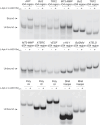
L-Apt.4–1c-ASO15nt(APP) conjugate displays strong binding affinity and specificity toward the APP rG4 wt region over other non-targets. Strong binding is observed between L-Apt.4–1c-ASO15nt(APP) and APP rG4 wt region. Weak binding is observed between L-Apt.4–1c-ASO15nt(APP) and Bcl2 rG4 region, TRF2 rG4 region and MT3-MMP rG4 region. No binding is observed between L-Apt.4–1c-ASO15nt(APP) and dG4 regions, namely Bcl2 dG4 region, TRF2 dG4 region, MT3-MMP dG4 region, hTERC dG4 region, VEGF dG4 region, c-Kit 1 dG4 region, Bcl2Mid dG4 region or hTELO dG4 region. No binding is observed between L-Apt.4–1c-ASO15nt(APP) and non-G4 motifs, including DNA and RNA hairpins and single-stranded poly rA/rC/rU RNAs. L-Apt.4–1c-ASO15nt(APP) preferentially interacts with APP rG4 wt region over other constructs tested. All the FAM_rG4 regions, FAM_dG4 regions and FAM_non-G4 motifs used had a concentration of 10 nM, and the L-Apt.4–1c-ASO15nt(APP) conjugate had a concentration of 5 nM.
Figure 4.
Figure 4.
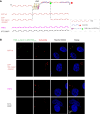
L-Apt.4–1c-ASO15nt(APP) conjugate preferentially colocalizes with the APP rG4-containing transcript over other rG4-containing transcripts in cells. (A) Schematic illustration of the cell-imaging assay. The RNA constructs (APP rG4 wt region, APP rG4 and flanking region deleted, TRF2 rG4 region, MT3-MMP rG4 region) were transfected into HeLa cells independently. After cell membrane permeabilization induced by Triton X-100, Cy3-probe and FAM_L-Apt.4–1c-ASO15nt(APP) were used to stain the RNA and rG4 region separately. The Cy3-containing DNA probe was used to identify the location of the transfected RNA construct. The L-Apt.4–1c-ASO15nt(APP) was labeled with FAM to identify its colocalization with the corresponding RNA construct in cells. (B) Confocal microscopy of cells that were transfected with different RNA constructs, followed by staining with FAM-labeled L-Apt.4–1c-ASO15nt(APP) conjugate and Cy3-containing DNA probe. Nuclei were stained with Hoechst 33342. Only APP rG4 wt region showed colocalization with L-Apt.4–1c-ASO15nt(APP).
Figure 5.
Figure 5.
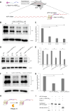
L-Apt.4–1c-ASO15nt(APP) conjugate inhibits endogenous APP protein in cells. (A) Schematic illustration of cell treatment with L-Apt.4–1c-ASO15nt(APP). (B) Effect of different concentrations (0, 30, 60, and 90 nM) of L-Apt.4–1c-ASO15nt(APP) addition on APP expression in HeLa cells. The APP expression decreased with increasing L-Apt.4–1c-ASO15nt(APP) concentration. GAPDH was used as loading control. Cells were treated for 22 h. (C) Normalized APP expression results from western blotting obtained from (B). (D) Kinetic experiments (4, 10 and 22 h) of L-Apt.4–1c-ASO15nt(APP) addition on APP expression in HeLa cells. The APP expression decreased with increasing time of treatment. The concentration of L-Apt.4–1c-ASO15nt(APP) was 60 nM. (E) Normalized APP expression results from western blotting obtained from (D). (F) Comparison between L-Apt.4–1c-ASO15nt(APP) and L-Apt.4–1c_5′hexynyl. The APP expression of both was decrease, with L-Apt.4–1c-ASO15nt(APP) having a stronger inhibitory effect. (G) Normalized APP expression results from western blotting obtained from (F). (H) Schematic illustration of RNase H treatment assay in vitro. (I) RNase H treatment assay detected by denaturing polyacrylamide gel. RNase H cleaved the conjugate–APP rG4 region complex. Results were obtained from three replicates. Error bars represent the standard error of the mean. P values were calculated using two-tailed t-tests, with P < 0.05 considered to indicate significant differences.
Figure 6.
Figure 6.
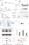
L-Apt.4–1c-ASO15nt(APP) can dissociate APP rG4–DHX36 interactions, and control the DHX36-dependent APP rG4-mediated gene expression. (A) Schematic illustration of APP rG4 unwinding by DHX36 protein in an ATP-dependent manner. The ssRNA trap was designed to complementarily pair with the APP rG4 sequence. In the folded rG4 conformation, the ssRNA trap was unable to bind to APP rG4. Only when the rG4 was unwound by DHX36 did base paring occur between APP rG4 sequence and ssRNA trap. (B) DHX36 unwound APP rG4 in the presence of ATP, while not in the ATP non-hydrolysable analog, AMP-PNP. The ssRNA trap hybridized with the unwound FAM_APP rG4 region, showing unwound APP rG4–trap duplex shift bands. M: ssRNA trap was boiled together with FAM_APP rG4 region to form the duplex as a shift marker in the native polyacrylamide gel. (C) Schematic illustration of L-Apt.4–1c-ASO15nt(APP) conjugate dissociating APP rG4–DHX36 protein interactions. (D) Inhibition assay showing that L-Apt.4–1c-ASO15nt(APP) conjugate can effectively disrupt the interaction between HEX_APP rG4 wt region and DHX36 protein. (E) Inhibition curve obtained from (D). The IC50 value was determined to be 13.1 ± 1.2 nM. (F) Schematic illustration of RNA immunoprecipitation (RIP) assay. (G) L-Apt.4–1c-ASO15nt(APP) conjugate can disrupt APP rG4–DHX36 interactions according to RIP assay (100 nM L-Apt.4–1c-ASO15nt(APP)). (H) DHX36 overexpression up-regulated APP expression, while L-Apt.4–1c-ASO15nt(APP) rescued the effect of DHX36 according to western blotting. GAPDH was used as loading control. (I) Normalized APP expression results obtained from (H). (J) Schematic illustration of the mechanism by which L-Apt.4–1c-ASO15nt(APP) regulates APP expression. Results were obtained from three replicates. Error bars represent the standard error of the mean. P values were calculated using two-tailed t-tests, with P < 0.05 considered to indicate significant differences.

References

    1. Varshney D., Spiegel J., Zyner K., Tannahill D., Balasubramanian S.. The regulation and functions of DNA and RNA G-quadruplexes. Nat. Rev. Mol. Cell Biol. 2020; 21:459–474. - PMC - PubMed
    1. Dong J., O’Hagan M.P., Willner I.. Switchable and dynamic G-quadruplexes and their applications. Chem. Soc. Rev. 2022; 51:7631–7661. - PubMed
    1. Kwok C.K., Merrick C.J.. G-quadruplexes: prediction, characterization, and biological application. Trends Biotechnol. 2017; 35:997–1013. - PubMed
    1. Banco M.T., Ferre-D’Amare A.R.. The emerging structural complexity of G-quadruplex RNAs. RNA. 2021; 27:390–402. - PMC - PubMed
    1. Farag M., Mouawad L.. Comprehensive analysis of intramolecular G-quadruplex structures: furthering the understanding of their formalism. Nucleic Acids Res. 2024; 52:3522–3546. - PMC - PubMed

MeSH terms

LinkOut - more resources