. 2024 Dec 11;52(22):14205-14228.
doi: 10.1093/nar/gkae1107.

Intrinsically disordered RNA-binding motifs cooperate to catalyze RNA folding and drive phase separation

Affiliations

Intrinsically disordered RNA-binding motifs cooperate to catalyze RNA folding and drive phase separation

Annika Niedner-Boblenz et al. Nucleic Acids Res. .

Abstract

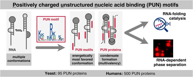
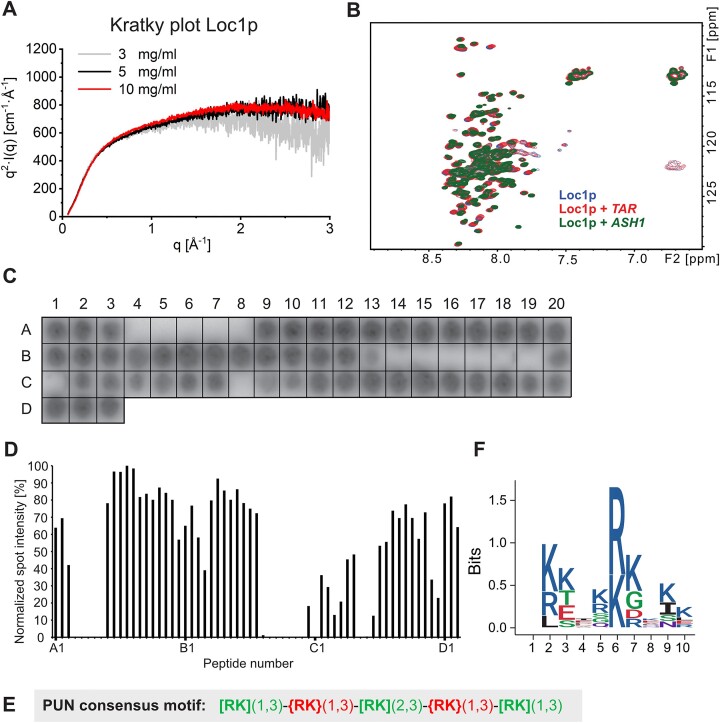
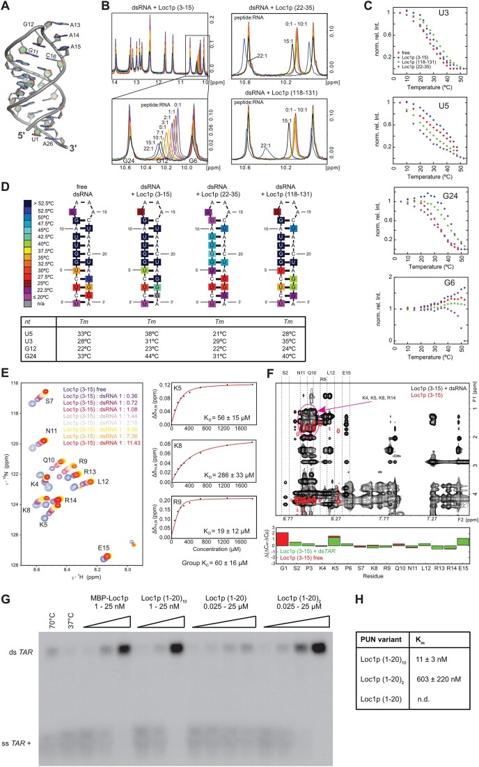
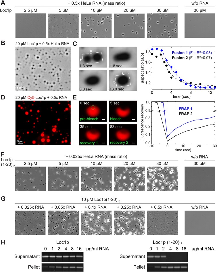
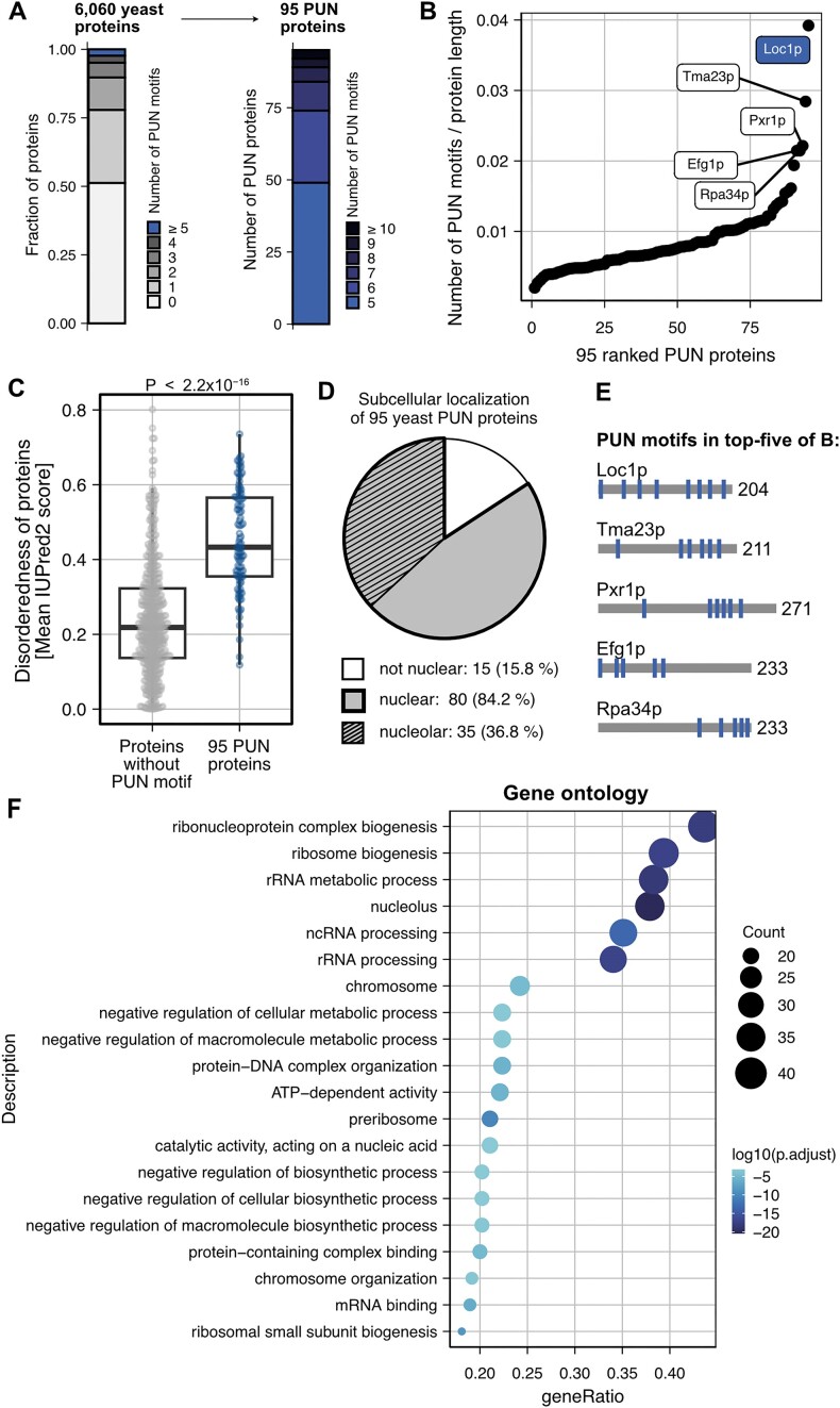
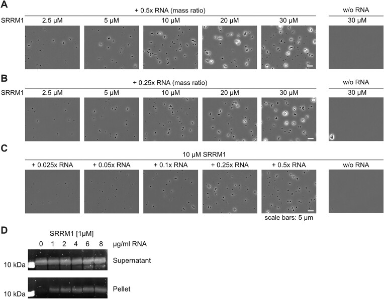
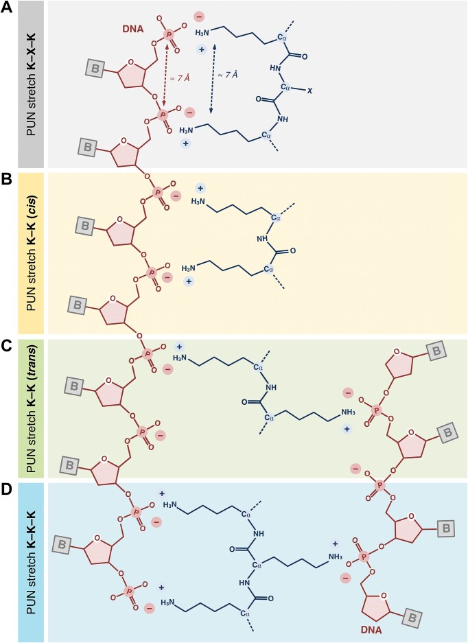
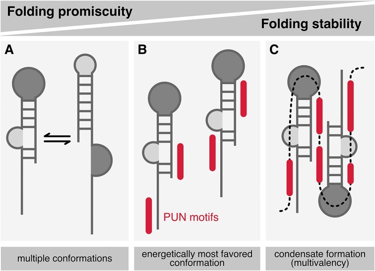
RNA-binding proteins are essential for gene regulation and the spatial organization of cells. Here, we report that the yeast ribosome biogenesis factor Loc1p is an intrinsically disordered RNA-binding protein with eight repeating positively charged, unstructured nucleic acid binding (PUN) motifs. While a single of these previously undefined motifs stabilizes folded RNAs, multiple copies strongly cooperate to catalyze RNA folding. In the presence of RNA, these multivalent PUN motifs drive phase separation. Proteome-wide searches in pro- and eukaryotes for proteins with similar arrays of PUN motifs reveal a strong enrichment in RNA-mediated processes and DNA remodeling. Thus, PUN motifs are potentially involved in a large variety of RNA- and DNA-related processes by concentrating them in membraneless organelles. The general function and wide distribution of PUN motifs across species suggest that in an ancient 'RNA world' PUN-like motifs may have supported the correct folding of early ribozymes.


Figures

Graphical Abstract
Graphical Abstract
Figure 1.
Figure 1.
Loc1p binds directly to ribosomes and unspecifically to RNA. (A) Comparison of doubling time of wild-type (S288C) and loc1Δ strains. loc1Δ cells show a severe growth delay (n = 3). Analytical ultracentrifugation of S. cerevisiae (B) and M. smegmatis (C) ribosomes and His6-tagged Loc1p reveals a co-enrichment of Loc1p with ribosomes in the pellet. In the absence of ribosomes, Loc1p is mainly found in the supernatant fraction of the sucrose cushion. (D) EMSAs with Loc1p and radioactively labeled ASH1 E3 localization element (LE), EAR1 LE, HIV-TAR stem–loop and poly-A60 RNA reveal unspecific binding in the nanomolar range. (EF) Competition EMSAs with 100 nM MBP-Loc1p, 5 nM radioactively labeled RNA, and 10-, 100- and 500-fold excess of cold competitor RNA. Experiments show that ASH1 E3, TAR-57 and poly-A60 can compete for Loc1p interaction with ASH1 E3 (E) or TAR-57 (F).
Figure 2.
Figure 2.
Loc1p induces conformational changes in nucleic acids. (A) Fluorescence intensity of a stem–loop molecular beacon (MB-D16) is not changed upon titration with MBP-Loc1p, indicating that Loc1p is unable to change the secondary structure of this RNA. (B) Fluorescence intensity of a single-stranded molecular beacon (MB-A5) is decreased upon titration of Loc1p, indicating the induction of conformational changes. (C) In the presence of antisense RNA (anti-MB-D16), titration of Loc1p changes the fluorescence intensity of the stem–loop shown in panel (A). This suggests that Loc1p promotes the formation of the energetically favored structure. Left: For steady-state experiments, the molecular beacon was pre-incubated with antisense RNA before Loc1p was added. Right: In time-course experiments, Loc1p, MBP or buffer alone was pre-incubated with the molecular beacon prior to addition of antisense RNA. Addition of 1 μM Loc1p increases the reaction rate ∼7-fold, suggesting that Loc1p promotes the formation of the energetically most favored state. (D) Comparative TAR annealing assay showing that MBP-Loc1p and Imp4p promote annealing of dsDNA while Hfq displaces dsTAR DNA under these experimental conditions. Radioactively labeled TAR(+) DNA was incubated with TAR(−) DNA at 37°C and with MBP-Loc1p, SUMO-Hfq, SUMO-Imp4p or buffer (negative control, 37°C). As a positive control, the sample was incubated at 70°C for heat-induced annealing. While 5 nM of MBP-Loc1p is sufficient for complete annealing, Imp4p requires >3 μM to reach a comparable activity. All experiments were performed independently at least three times.
Figure 3.
Figure 3.
RNA binding of the intrinsically unstructured Loc1p is mediated by distinct motifs within the Loc1p sequence. (A) Kratky plots of SAXS measurements at three Loc1p concentrations indicate that the protein is largely unfolded. The scattering intensity [as I(q) × q2] is plotted against the scattering vector q. Unfolded/highly flexible proteins are typically characterized by a plateau at higher q values. (B) 1H–15N-HSQC spectra of Loc1p alone (blue) and in the presence of TAR RNA (red) and the ASH1 E1 RNA (green). Minor signal shifts indicate that the interaction with RNA does not induce a secondary structure fold in large parts of Loc1p. (C) Binding experiments of radioactively labeled ASH1 E3 (short) RNA to a Loc1p peptide array revealed binding of a large portion of the spotted peptides to the RNA. Peptides had a length of 20 aa and an offset of 3 aa. (D) Bar graph depicting RNA binding intensities of each peptide. Signals from the peptide array shown in panel (C) were quantified with ImageJ and normalized against the best binding peptide (A12 set to 100%; n = 1). Large sequence stretches of Loc1p show binding to ASH1 E3 (short) RNA. (E) The consensus sequence of the PUN motif is characterized by three repetitive units of one to three positively charged residues [[RK](1,3) in green] interspaced by one to three residues that are not positively charged [{RK}(1,3) in red]. (F) One-letter-code sequence logo integrating all eight PUN motifs of Loc1p (amino acid chemistry: blue, basic; red, acidic; black, hydrophobic; purple, neutral; green, polar). The amino acid position is plotted against the information content of each position in bits.
Figure 4.
Figure 4.
Structure analysis of RNA binding and assessment of annealing activity of PUN motifs. (A) X-ray structure of the dsRNA tetraloop highlighting the coordinated GAAA minimal tetraloop (G12–A15) and the base-paired stem. (B) Titration of the dsRNA with different Loc1p-derived peptides. Whereas the titration of Loc1p (3–15) shifts the signal of G12 in a continuous manner, the two peptides lacking the RNA-binding motif Loc1p (118–131) and Loc1p (22–35) do not bind until very high, 15-fold excess, where peaks start to display sudden large shifts. (C) Temperature dependence of imino NMR signals of unbound dsRNA and dsRNA bound to different Loc1p-derived peptides at the highest molar excess (22:1) shown in panel (B). The decrease of imino signal intensities (shown: U3, U5, G6, G24) was used to determine the stabilizing effect of the three different peptides titrated in panel (B) in temperature scans. Whereas Loc1p (3–15) (blue dots) has a stabilizing effect on the hairpin RNA (free RNA: red dots), the control peptides Loc1p (118–131) (green dots) and Loc1p (22–35) (purple dots) have an overall destabilizing effect. The only exception is a slight stabilizing effect for U3 by Loc1p (118–131). (D) Heat map illustrating the temperature-dependent stabilization of the end of the stem upon peptide binding. In contrast, Loc1p (118–131) and especially Loc1p (22–35) destabilize the ends of the stem and the loop regions. Loc1p (22–35) has even a destabilizing effect on the central stem region. The estimated melting temperatures for the four most affected base pairs are given in the table below. (E) 1H,15N-HSQC spectrum showing the titration of dsRNA to 15N,13C-labeled Loc1p peptide. Saturation is reached at a 3–4-fold excess of dsRNA tetraloop. To the right, three examples are shown of fitted binding curves from chemical shift perturbations. (F) 2D NOESY spectra overlay of unbound Loc1p peptide (red) and dsRNA-bound Loc1p (black). In the free state, the Loc1p peptide is flexible, as only intra-residual and sequential HN–Hα1 NOE cross-peaks are observed. Upon binding to dsRNA, additional NOEs can be observed, especially nonsequential and inter-residual NOEs. The dashed lines indicate strips from the HN region of indicated residues to other protons. The purple circle indicates the strong overlap of positively charged residues (K4, K5, K8, R14), which prevents full assignment of peptide side chains. Secondary chemical shifts (below the spectrum) clearly show that no secondary structure is formed upon RNA binding. (G) TAR annealing assay with radioactively labeled TAR(+) and unlabeled minus strand demonstrates that concatenation of the most N-terminal Loc1p motif is sufficient to mimic the annealing activity of wild-type Loc1p in vitro. Both strands were incubated with increasing concentrations of wild-type Loc1p, Loc1p (1–20)10, Loc1p (1–20)2, Loc1p (1–20) or buffer (negative control: NC) at 37°C. Heat-induced displacement at 70°C and in the absence of Loc1p served as a positive control (PC). Formation of dsTAR was analyzed by 8% native TBE PAGE. Wild-type Loc1p has an annealing activity comparable to an artificial protein bearing 10 Loc1p motifs [Loc1p (1–20)10]. (H) Comparison of annealing activities of sequences bearing 1, 2 and 10 Loc1p motifs. For Loc1p (1–20), annealing activity was not determined because even with 50 μM no saturation was reached. According to the intensities of annealed dsTAR, Loc1p (1–20)2 showed an estimated 50–100-fold higher activity than Loc1p (1–20). Furthermore Loc1p (1–20)10 showed an over 50-fold higher annealing activity than Loc1p (1–20)2. Since wild-type Loc1p showed even better reactivity that exceeded the limits of this assay, no Km could be derived. Shown are quantifications from three independent experiments.
Figure 5.
Figure 5.
Loc1p or multiple copies of its N-terminal PUN motif form condensates with RNA. (A–G) The formation of condensates was assessed by phase contrast and fluorescence microscopy. (A) At a protein:RNA mass ratio of 0.5, phase separation was observed already at 5 μM Loc1p. (B) At the same protein:RNA ratio and 20 μM Loc1p, condensates had a size of 1–5 μm and showed round shapes. (C) The observed condensates fuse and round up after fusion, confirming their liquid-to-gel-like properties. (D) Like unlabeled Loc1p, Cy5-labeled Loc1pS7C promotes RNA-dependent droplet formation and partitions into the droplets. (E) Fluorescence of Cy5-Loc1pS7C/RNA droplets recovers after photobleaching to 20–30% in FRAP experiments. (F, G) Loc1p (1–20)10 formed condensates already at 2.5 μM and an RNA:protein mass ratio of 0.025 and larger condensates with increasing RNA concentration. No phase separation was observed in the absence of RNA. (H) RNA-dependent phase separation of Loc1p and Loc1p (1–20)10 was confirmed by sedimentation experiments followed by SDS–PAGE stained with SyproRuby. Experiments were repeated at least once (n ≥ 2).
Figure 6.
Figure 6.
Yeast proteins containing the PUN motif identified in Loc1p are more likely to contain disordered regions, accumulate in the nucleus and are involved in RNA metabolic processes. (A) Computational identification of 95 proteins from S. cerevisiae that contain at least five PUN motifs, including at least three within 150 aa. (B) PUN motif density in the 95 yeast PUN proteins. Loc1p has the highest density of PUN motifs followed by Tma23p, Pxr1p, Efg1p and Rpa34p. (C) Yeast PUN proteins are characterized by a higher degree of disorderness compared to proteins without PUN motifs, reflecting the preference of PUN motifs for disordered regions. P-value is obtained from the Wilcoxon rank-sum test. (D) Analysis of the intracellular distribution of the 95 hits shows a clear preference for the nuclear (85%) and nucleolar (35%) compartments of the cell. (E) Distribution of PUN motifs in top five proteins from panel (B). (F) Functional enrichment analysis (GO) of annotated processes ordered by P-values. The RNA processing pathways are enriched in proteins bearing PUN motifs.
Figure 7.
Figure 7.
Phase separation of human SRRM1 bearing 14 PUN motifs (Supplementary Table S10). SRRM1 undergoes RNA-dependent phase separation at protein concentrations of 2.5 μM or higher and at RNA:protein mass ratios of 0.5 (A) or 0.25 (B). Titration of increasing amounts of RNA and a constant concentration of 10 μM SRRM1 showed phase separation already at the lowest RNA:protein ratio. (D) RNA-dependent condensation of SRRM1 was confirmed by sedimentation experiments followed by SDS–PAGE stained by SyproRuby. Experiments shown in panels (A)–(D) were repeated at least once (n ≥ 2).
Figure 8.
Figure 8.
Schematic model of possible interaction modes of PUN motifs with nucleic acids. Sequence-unspecific interactions with phosphodiester groups of nucleic acids are mediated by positively charged side chains. (A) Two positively charged amino acids separated by a single nonpositively charged amino acid have a distance that is very similar to the spacing of two neighboring phosphodiester groups in nucleic acids. Therefore, these two amino acids can potentially interact with two adjacent phosphodiesters. (B) Due to the lengths and flexibilities of the side chains of arginine and lysine, two neighboring amino acids can also interact with adjacent phosphodiesters of the same molecule (cis). (C) When pointing in opposite directions, two neighboring side chains can contact phosphodiester groups of two nucleic acid molecules (trans). (D) In a stretch of three consecutive arginines or lysines, these amino acids will also adopt a trans conformation. This feature should allow for the simultaneous contact of two nucleic acid molecules by one PUN motif. This arrangement likely constitutes an important feature for higher organization principles, including RNA annealing and RNA-dependent phase transition.
Figure 9.
Figure 9.
Model of the effect of isolated and repeating PUN motifs on promiscuous folding of RNA molecules. (A) Isolated RNA (schematic stem–loop) may exist in an equilibrium of different secondary structures. (B) Binding of individual PUN motifs (red) to an RNA molecule stabilizes its energetically favored conformation. (C) A peptide chain with several PUN motifs acts cooperatively on RNA folding and facilitates the formation of condensates caused by their multivalent interactions.

References

    1. Kressler D., Hurt E., Bassler J.. Driving ribosome assembly. Biochim. Biophys. Acta. 2010; 1803:673–683. - PubMed
    1. Duss O., Stepanyuk G.A., Puglisi J.D., Williamson J.R.. Transient protein–RNA interactions guide nascent ribosomal RNA folding. Cell. 2019; 179:1357–1369. - PMC - PubMed
    1. Woolford J.L. Jr, Baserga S.J. Ribosome biogenesis in the yeast Saccharomyces cerevisiae. Genetics. 2013; 195:643–681. - PMC - PubMed
    1. Harnpicharnchai P., Jakovljevic J., Horsey E., Miles T., Roman J., Rout M., Meagher D., Imai B., Guo Y., Brame C.J.et al. .. Composition and functional characterization of yeast 66S ribosome assembly intermediates. Mol. Cell. 2001; 8:505–515. - PubMed
    1. Komili S., Farny N.G., Roth F.P., Silver P.A.. Functional specificity among ribosomal proteins regulates gene expression. Cell. 2007; 131:557–571. - PMC - PubMed

MeSH terms