. 2024 Nov 18;24(1):363.
doi: 10.1186/s12893-024-02660-4.

Chondroblastoma of the femoral head: curettage without dislocation

Affiliations

Chondroblastoma of the femoral head: curettage without dislocation

Di Yang et al. BMC Surg. .

Abstract

Background: Chondroblastoma (CBL) of the femoral head is a rare disease, typically encountered in the epiphysis of long bones, with its occurrence in the femoral head being particularly uncommon. The unique location and aggressive nature of this tumor pose substantial challenges in its treatment, leading to ongoing controversies regarding the therapeutic approaches. In this study, we introduce a technique of curettage without surgical dislocation as a treatment option for CBL in this challenging location.

Methods: A total of 6 children diagnosed with chondroblastoma of the femoral head underwent a surgical procedure consisting of curettage, the application of anhydrous alcohol as an adjuvant therapy, followed by bone grafting. The epiphyseal plate status of the femoral head was classified as open, closing, or closed. To evaluate the children's postoperative functional outcome, the Musculoskeletal Tumour Society (MSTS) scoring system was utilized. Additionally, the Lodwick classification was employed to assess the extent of bone destruction. Furthermore, the kappa coefficient was calculated to quantify the level of inter-observers agreement in assessing the status of the epiphyseal plate.

Results: The epiphyseal plate status was closing in two patients and closed in four patients. According to the Lodwick classification, one patient was classified as IA, one as IB, and four as IC. The mean MSTS score was 28. Notably, one patient developed a femoral neck fracture three months post-curettage.

Conclusions: Curettage without surgical dislocation, combined with the use of anhydrous alcohol as an adjuvant therapy, followed by bone grafting, constitutes an effective treatment technique for femoral head chondroblastoma.

Keywords: Anhydrous alcohol; Chondroblastoma; Curettage; Femoral head.


Conflict of interest statement

Declarations Ethics approval and consent to participate The research had been approved by the Ethics Committee of Children's Hospital affiliated to Chongqing Medical University. Informed consent was obtained from all of their legal guardians. Consent for publication Written informed consent for publishing identification images or other personal or clinical details was obtained from the legal guardians of all participants. Competing interests The authors declare no competing interests.

Figures

Fig. 1
Fig. 1
A 'Z'-shaped incision was made in the capsule, and an articular window was opened to expose the tumor
Fig. 2
Fig. 2
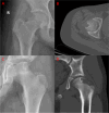
Chondroblastoma of the hip in a 13-year-old male. A A frontal radiograph of the left hip demonstrates a round and lobulated osteolytic lesion in the femoral head, extending up to the subchondral area. B A coronal CT scan demonstrates to better advantage the lytic lesion with the thick sclerotic zone. C A coronal T1 MR image demonstrates a lesion of moderate-signal intensity extending to the subchondral area, surrounded by a low-signal intensity border. D A frontal radiograph of the left hip at the 6-month follow-up shows that the cavity has been filled with autogenous bone
Fig. 3
Fig. 3
X-ray image showing a 2 cm × 9 cm osteolytic lesion with a marginal sclerotic rim
Fig. 4
Fig. 4
Preoperative Imaging: Case 3. A X-ray reveals a well-defined lesion boundary with no sclerotic rim. B CT scan demonstrates local outward protrusion of the lesion, accompanied by surrounding soft tissue swelling. Case 4. C X-ray exhibits a visible sclerotic rim. D CT scan indicates bone hyperplasia and sclerosis at the lesion margin, with the affected area spanning across the epiphyseal plate
Fig. 5
Fig. 5
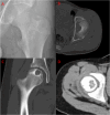
Preoperative Images: Case 5. A The X-ray suggests a lesion with an indistinct boundary and no apparent sclerotic rim. B The CT scan indicates slight discontinuity of the bone cortex on the medial side of the femoral head. Case 6. C The coronal CT scan reveals a prominent sclerotic rim. D The axial CT scan demonstrates streaky bony septations and punctate calcifications within the lesion

Similar articles

Cited by

References

    1. Chen W, DiFrancesco LM. Chondroblastoma: an update. Arch Pathol Lab Med. 2017;141:867–71. - PubMed
    1. Hsu CC, Wang JW, Chen CE, Lin JW. Results of curettage and high-speed burring for chondroblastoma of the bone. Chang Gung Med J. 2003;26:761–7. - PubMed
    1. Suneja R, Grimer RJ, Belthur M, Jeys L, Carter SR, Tillman RM, et al. Chondroblastoma of bone: long-term results and functional outcome after intralesional curettage. J Bone Joint Surg Br. 2005;87:974–8. - PubMed
    1. Laitinen MK, Stevenson JD, Evans S, Abudu A, Sumathi V, Jeys LM, et al. Chondroblastoma in pelvis and extremities- a signle centre study of 177 cases. J Bone Oncol. 2019;17:100248. - PMC - PubMed
    1. Zekry KM, Yamamoto N, Hayashi K, Takeuchi A, Araki Y, Alkhooly AZA, et al. Surgical treatment of chondroblastoma using extended intralesional curettage with phenol as a local adjuvant. J Orthop Surg (Hong Kong). 2019;27:2309499019861031. - PubMed

LinkOut - more resources