. 2024 Nov 18;22(1):720.
doi: 10.1186/s12951-024-02964-w.

Physical, biochemical, and biological characterization of olive-derived lipid nanovesicles for drug delivery applications

Affiliations

Physical, biochemical, and biological characterization of olive-derived lipid nanovesicles for drug delivery applications

Zhu Zhao et al. J Nanobiotechnology. .

Abstract

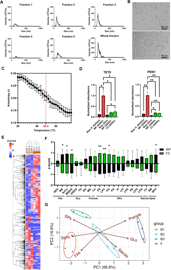
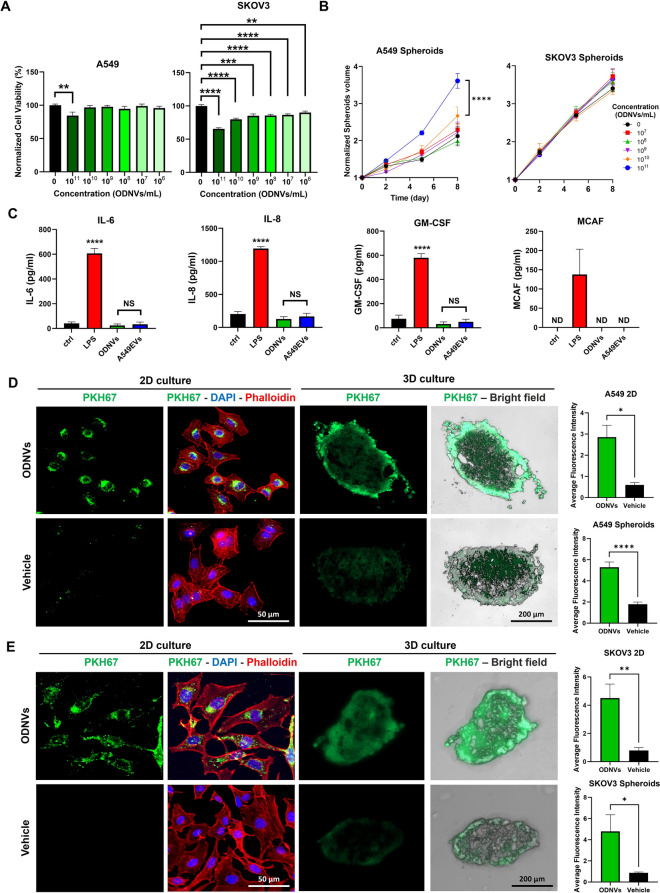
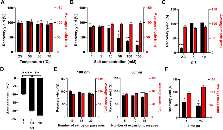
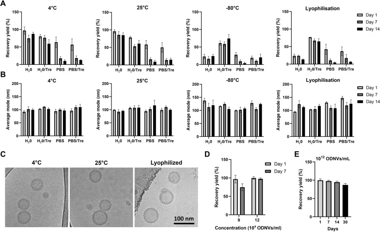
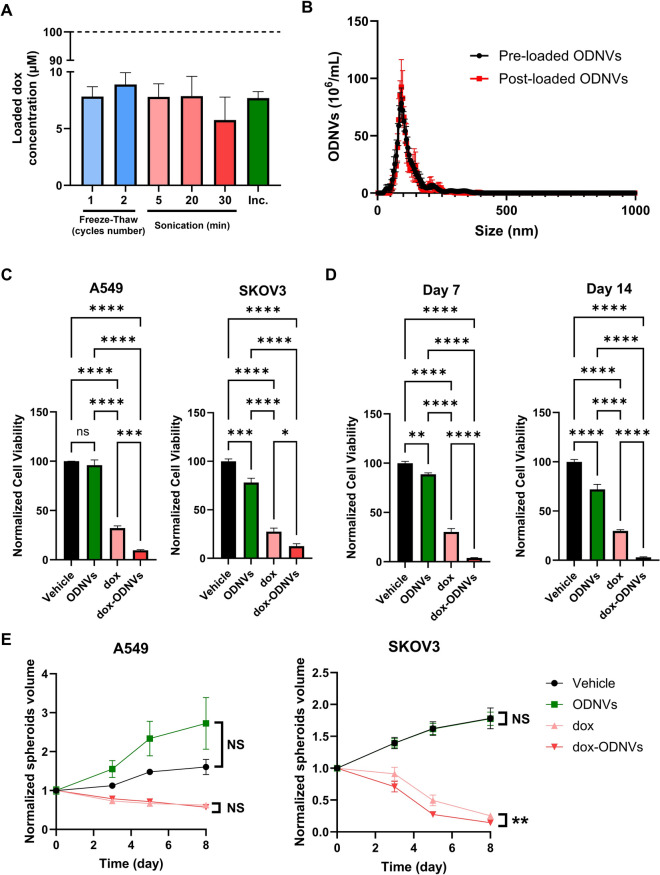
Extracellular vesicles (EVs) have shown great promise as drug delivery system (DDS). However, their complex and costly production limit their development for clinical use. Interestingly, the plant kingdom can also produce EV-like nanovesicles that can easily be isolated and purified from a large quantity of raw material at a high yield. In this study, olive-derived nanovesicles (ODNVs) were isolated from raw fruits using serial centrifugations and their physical and biological features characterized to demonstrate their promising potential to be used as a DDS. Nanotracking particle analysis indicated an average size of 109.5 ± 3.0 nm and yield of 1012 ODNVs/mL for the purest fraction. Microscopy imaging, membrane fluidity assay and lipidomics analysis showed the presence of a rich lipid bilayer that significantly varied between different sources of ODNVs but showed a distinct signature compared to human EVs. Moreover, ODNVs were enriched in PEN1 and TET8 compared to raw fruits, suggesting an extracellular origin. Interestingly, ODNVs size and yield stayed unchanged after exposure to high temperature (70 °C for 1 h), wide pH range (5-10), and 50-100 nm extrusion, demonstrating high resistance to physical and chemical stresses. This high resistance allowed ODNVs to stay stable in water at 4 °C for a month, or with the addition of 25 mM trehalose for long-term freezing storage. Finally, ODNVs were internalized by both 2D and 3D cell culture without triggering significant cytotoxicity and immunogenicity. Importantly, the anticancer drug doxorubicin (dox) could be loaded by passive incubation within ODNVs and dox-loaded ODNVs decreased cell viability by 90% compared to only 70% for free dox at the same concentration, indicating a higher efficiency of drug delivery by ODNVs. In addition, this high cytotoxicity effect of dox-loaded ODNVs was shown to be stable after a 2-week storage at 4 °C. Together, these findings suggested that ODNVs represent a promising candidate as drug nanocarrier for various DDS clinical applications, as demonstrated by their biocompatibility, high resistance to stress, good stability in harsh environment, and improvement of anticancer drug efficacy.


Conflict of interest statement

Declarations Ethics approval and consent to participate Not applicable. Consent for publication All authors agreed to publish this manuscript. Competing interests The authors declare no competing interests.

Figures

Fig. 1
Fig. 1
Characterization of ODNVs size, morphology and biochemical content. A Nanoparticle tracking analysis of size and quantity of the ODNVs collected in the different fractions from canned olives B1. B Representative cryo-EM images of F3 ODNVs. C ODNVs membrane fluidity at various temperatures (n = 3). Red dotted line indicates phase transition temperature. D Expression of TET8 and PEN1 proteins in the tissue and derived nanovesicles of A thaliana (ADNVs) and olive fruits (ODNVs). Data were normalized to protein level in A. Thaliana tissue (n = 3), and differences between each group were calculated using unpaired, two-tailed Student’s t-tests with a significance threshold of α < 0.05. E Hierarchical clustering of the 3240 common lipids identified in WF and F3. F Relative lipid abondance of the main lipid subclasses identified between WF and F3. Differences between each group were calculated using unpaired, two-tailed Student’s t-tests with a significance threshold of α < 0.05. G Principal component analysis showing expression of lipids of the 6 main lipid classes between 3 different brand of canned olives (B1, B2, and B3) and fresh, unprocessed olives (F). All data are represented as mean ± s.e.m. *p < 0.05; **p < 0.01; ***p < 0.001; ****p < 0.0001
Fig. 2
Fig. 2
ODNVs cytotoxicity, immunogenicity and internalization in 2D and 3D culture of lung and ovarian cells. A Viability of A549 and SKOV3 cells cultured in 2D for 3 days and exposed to different concentration of ODNVs assessed by MTT (n = 3). Differences to unexposed sample were evaluated using unpaired, two-tailed Student’s t-tests with a significance threshold of α < 0.05. B Growth curves of A549 and SKOV3 spheroids exposed to different concentration of ODNVs for 8 days (n = 3). Data were analyzed using two-way repeated-measures ANOVA to assess the main effects of treatment. C Level of IL-6, IL-8, GM-CSF and MCAF released in HULEC-5a cells supernatant after exposure to 1.109 A549 EVs/mL, 1.109 ODNVs/mL and 1 µg/mL LPS for 24 h (n = 3). Differences to unexposed sample (ctrl), and between ODNVs and A549 EVs samples, were evaluated using unpaired, two-tailed Student’s t-tests with a significance threshold of α < 0.05. NS = Non-Significant; ND = Not Detected. Representative immunofluorescent pictures of A549 (D) and SKOV3 (E) cells cultured in 2D or 3D (spheroids) and incubated for 24 h with ODNVs (1012 ODNVs/mL) or PBS pre-stained with PKH67 green fluorescent cell membrane labeling. For 2D culture, cells were counterstained for DAPI (blue) and Alexa Fluor 555 Phalloidin (red) to stain DNA and F-actin and visualize nucleus and cytoplasm, respectively. Spheroids were embedded in OCT, sliced and imaged under bright field. Graphs represent the average fluorescence intensity of at least 100 cells or 3 spheroids from three independent experiments. Differences between samples incubated with PKH67-stained ODNVs or PBS were evaluated using unpaired, two-tailed Student’s t-tests with a significance threshold of α < 0.05. All data are represented as mean ± s.e.m. p < 0.05; **p < 0.01; ***p < 0.001; ****p < 0.0001
Fig. 3
Fig. 3
ODNVs resistance to heat, salt, pH, mechanical deformation and blood-like environment. A Relative quantity and mode of ODNVs after 1 h incubation in water at different temperature and compared to ODNVs stored at 4 °C (n = 3). B Relative quantity and mode of ODNVs after 24 h incubation at 4 °C in solutions with different salt concentration and compared to solution with no salt (n = 3). C Relative quantity and mode of ODNVs after 24 h incubation at 4 °C in aqueous solutions of different pH and compared to solution of pH = 7.4 (n = 3). Differences to pH 7.4 were evaluated using unpaired, two-tailed Student’s t-tests with a significance threshold of α < 0.05. D Zeta potential of ODNVs after 24 h incubation at 4 °C in aqueous solutions (1 mM salt) of different pH (n = 3). E Relative quantity and mode of ODNVs subjected to serial extrusion through 100 and 50 nm porous membrane and compared to native ODNVs (n = 4). F Relative quantity and mode of ODNVs after 1 h and 24 h incubation at 37 °C in exosome depleted serum (n = 3). All conditions were tested at 1.109 ODNVs/mL. All data are represented as mean ± s.e.m. Differences were evaluated using one sample t-test with a hypothetical value of 100: *p < 0.05; **p < 0.01; ***p < 0.001; ****p < 0.0001
Fig. 4
Fig. 4
ODNVs size, yield and structure under different storage conditions. Recovery yield (A) and average size (B) of ODNVs (109 ODNVs/mL) under different storage conditions. Samples were resuspended in different storage buffers including DI water (H2O), water + trehalose (H2O/Tre), PBS and PBS + trehalose (PBS/Tre), and kept at refrigerated (4 °C), room (25 °C), freezing (-80 °C) temperature or lyophilized. After 24 h, frozen and lyophilized samples were thawed and reconstituted in water at 4 °C. All samples were then stored for 1, 7 and 14 days. All conditions were compared to fresh ODNVs (n = 3). C Cryo-EM pictures of ODNVs stored for 14 days at 4 °C, 25 °C in H2O and in lyophilized condition after resuspension in H2O/Tre. D Recovery yield of ODNVs stored at different concentrations for 7 days at refrigerated temperature in water and compared to fresh ODNVs (n = 3). E Recovery yield of ODNVs stored at high concentration (1012 ODNVs/mL) up to 1 month at refrigerated temperature in water and compared to fresh ODNVs (n = 3). All data are represented as mean ± s.e.m
Fig. 5
Fig. 5
Chemotherapeutic drug delivery to 2D and 3D lung and ovarian cancer cells using ODNVs. A Concentration of doxorubicin (dox) loaded in ODNVs by 1 or 2 freeze-thaw cycles, sonication for 5, 20 and 30 min and passive incubation (Inc.) for 4 h at 37 °C (n = 3). Initial dox concentration was 100 µM (dotted line). B Nanoparticle tracking analysis of mode and quantity of the ODNVs before and after loading with doxorubicin (100 µM) by passive incubation at 37 °C (4 h). C Viability of A549 and SKOV3 cells exposed to vehicle (water), ODNVs (4.1011 ODNVs/mL), free doxorubicin (dox) at a concentration of 1 µM and ODNVs (4.1011 ODNVs/mL) loaded with dox (dox-ODNVs) at a total concentration of 1 µM for 3 days (n = 3). Differences were evaluated using one-way ANOVA with Tukey’s multiple comparisons test. D Viability of A549 cells exposed to vehicle (water), ODNVs (4.1011 ODNVs/mL), free doxorubicin (dox) at a concentration of 1 µM and ODNVs (4.1011 ODNVs/mL) loaded with dox (dox-ODNVs) at a total concentration of 1 µM for 3 days (n = 3). ODNVs and dox-loaded ODNVs were previously stored at 4 °C in water for 7 and 14 days. Differences were evaluated using one-way ANOVA with Tukey’s multiple comparisons test. E Volume growth of A549 and SKOV3 spheroids exposed to vehicle (water), ODNVs (4.1011 ODNVs/mL), free doxorubicin (dox) at a concentration of 1 µM and ODNVs (4.1011 ODNVs/mL) loaded with dox (dox-ODNVs) at a total concentration of 1 µM for 8 days (n = 3). Dataset were plotted using the second order polynomial model and differences were calculated using extra sum-of-squares F test. All data are represented as mean ± s.e.m: *p < 0.05; **p < 0.01; ***p < 0.001; ****p < 0.0001

References

    1. Gao J, Karp JM, Langer R, Joshi N. The future of drug delivery. Chem Mater. 2023;35:359–63. - PMC - PubMed
    1. Mitchell MJ, Billingsley MM, Haley RM, Wechsler ME, Peppas NA, Langer R. Engineering precision nanoparticles for drug delivery. Nat Rev Drug Discov. 2021;20:101–24. - PMC - PubMed
    1. Vargason AM, Anselmo AC, Mitragotri S. The evolution of commercial drug delivery technologies. Nat Biomed Eng. 2021;5:951–67. - PubMed
    1. Zolnik BS, González-Fernández Á, Sadrieh N, Dobrovolskaia MA. Nanoparticles and the Immune System. Endocrinology. 2010;151:458–65. - PMC - PubMed
    1. Herrmann IK, Wood MJA, Fuhrmann G. Extracellular vesicles as a next-generation drug delivery platform. Nat Nanotechnol. 2021;16:748–59. - PubMed

LinkOut - more resources