

Diffuse pediatric high-grade glioma of methylation-based RTK2A and RTK2B subclasses present distinct radiological and histomolecular features including Gliomatosis cerebri phenotype
- PMID: 39558399
- PMCID: PMC11575044
- DOI: 10.1186/s40478-024-01881-1
Diffuse pediatric high-grade glioma of methylation-based RTK2A and RTK2B subclasses present distinct radiological and histomolecular features including Gliomatosis cerebri phenotype
Abstract
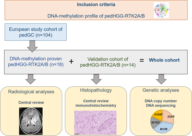
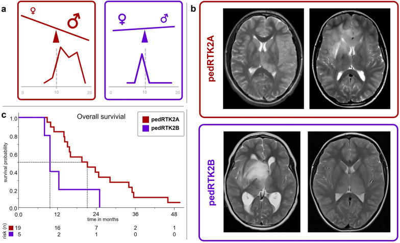
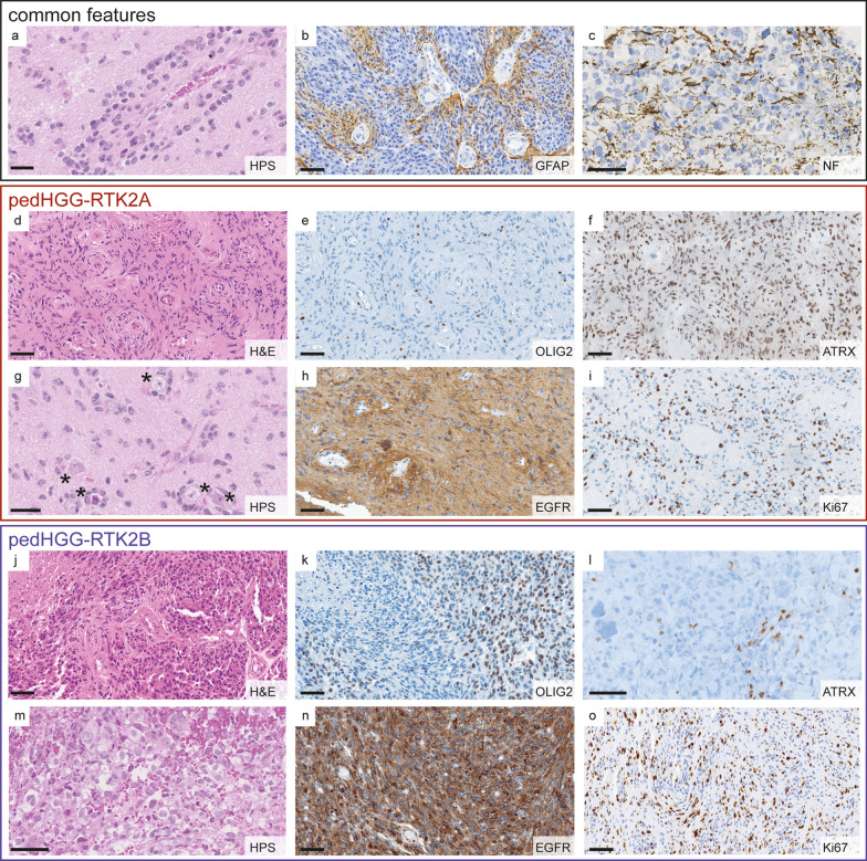
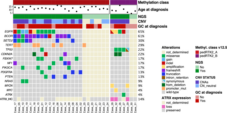
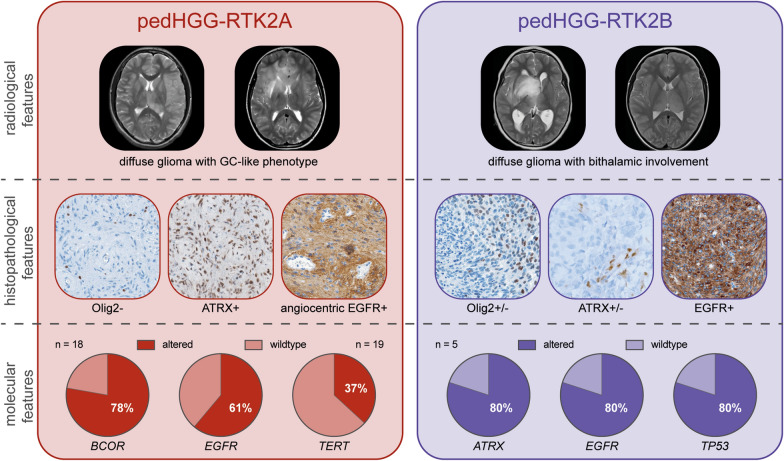
Diffuse pediatric-type high-grade gliomas (pedHGG), H3- and IDH-wildtype, encompass three main DNA-methylation-based subtypes: pedHGG-MYCN, pedHGG-RTK1A/B/C, and pedHGG-RTK2A/B. Since their first description in 2017 tumors of pedHGG-RTK2A/B have not been comprehensively characterized and clinical correlates remain elusive. In a recent series of pedHGG with a Gliomatosis cerebri (GC) growth pattern, an increased incidence of pedHGG-RTK2A/B (n = 18) was observed. We added 14 epigenetically defined pedHGG-RTK2A/B tumors to this GC series and provided centrally reviewed radiological, histological, and molecular characterization. The final cohort of 32 pedHGG-RTK2A/B tumors consisted of 25 pedHGG-RTK2A (78%) and seven pedHGG-RTK2B (22%) cases. The median age was 11.6 years (range, 4-17) with a median overall survival of 16.0 months (range 10.9-28.2). Seven of 11 of the newly added cases with imaging available showed a GC phenotype at diagnosis or follow-up. PedHGG-RTK2B tumors exhibited frequent bithalamic involvement (6/7, 86%). Central neuropathology review confirmed a diffuse glial neoplasm in all tumors with prominent angiocentric features in both subclasses. Most tumors (24/27 with available data, 89%) diffusely expressed EGFR with focal angiocentric enhancement. PedHGG-RTK2A tumors lacked OLIG2 expression, whereas 43% (3/7) of pedHGG-RTK2B expressed this glial transcription factor. ATRX loss occurred in 3/6 pedHGG-RTK2B samples with available data (50%). DNA sequencing (pedHGG-RTK2A: n = 18, pedHGG-RTK2B: n = 5) found EGFR alterations (15/23, 65%; predominantly point mutations) in both subclasses. Mutations in BCOR (14/18, 78%), SETD2 (7/18, 39%), and the hTERT promoter (7/19, 37%) occurred exclusively in pedHGG-RTK2A tumors, while pedHGG-RTK2B tumors were enriched for TP53 alterations (4/5, 80%). In conclusion, pedHGG-RTK2A/B tumors are characterized by highly diffuse-infiltrating growth patterns and specific radiological and histo-molecular features. By comprehensively characterizing methylation-based tumors, the chance to develop specific and effective therapy concepts for these detrimental tumors increases.
Keywords: Gliomatosis cerebri; Methylation; Pediatric high-grade glioma; RTK2A; RTK2B; Receptor tyrosine kinase; pedHGG.
© 2024. The Author(s).
Conflict of interest statement
Figures
References
-
- Capper D, Jones DTW, Sill M, Hovestadt V, Schrimpf D, Sturm D, Koelsche C, Sahm F, Chavez L, Reuss DE, Kratz A, Wefers AK, Huang K, Pajtler KW, Schweizer L, Stichel D, Olar A, Engel NW, Lindenberg K, Harter PN, Braczynski AK, Plate KH, Dohmen H, Garvalov BK, Coras R, Hölsken A, Hewer E, Bewerunge-Hudler M, Schick M, Fischer R, Beschorner R, Schittenhelm J, Staszewski O, Wani K, Varlet P, Pages M, Temming P, Lohmann D, Selt F, Witt H, Milde T, Witt O, Aronica E, Giangaspero F, Rushing E, Scheurlen W, Geisenberger C, Rodriguez FJ, Becker A, Preusser M, Haberler C, Bjerkvig R, Cryan J, Farrell M, Deckert M, Hench J, Frank S, Serrano J, Kannan K, Tsirigos A, Brück W, Hofer S, Brehmer S, Seiz-Rosenhagen M, Hänggi D, Hans V, Rozsnoki S, Hansford JR, Kohlhof P, Kristensen BW, Lechner M, Lopes B, Mawrin C, Ketter R, Kulozik A, Khatib Z, Heppner F, Koch A, Jouvet A, Keohane C, Mühleisen H, Mueller W, Pohl U, Prinz M, Benner A, Zapatka M, Gottardo NG, Driever PH, Kramm CM, Müller HL, Rutkowski S, von Hoff K, Frühwald MC, Gnekow A, Fleischhack G, Tippelt S, Calaminus G, Monoranu C-M, Perry A, Jones C, Jacques TS, Radlwimmer B, Gessi M, Pietsch T, Schramm J, Schackert G, Westphal M, Reifenberger G, Wesseling P, Weller M, Collins VP, Blümcke I, Bendszus M, Debus J, Huang A, Jabado N, Northcott PA, Paulus W, Gajjar A, Robinson GW, Taylor MD, Jaunmuktane Z, Ryzhova M, Platten M, Unterberg A, Wick W, Karajannis MA, Mittelbronn M, Acker T, Hartmann C, Aldape K, Schüller U, Buslei R, Lichter P, Kool M, Herold-Mende C, Ellison DW, Hasselblatt M, Snuderl M, Brandner S, Korshunov A, von Deimling A, Pfister SM (2018) DNA methylation-based classification of central nervous system tumours. Nature 555:469–474. 10.1038/nature26000 - PMC - PubMed
-
- Guerrini-Rousseau L, Tauziède-Espariat A, Castel D, Rouleau E, Sievers P, Saffroy R, Beccaria K, Blauwblomme T, Hasty L, Bourdeaut F, Grill J, Varlet P, Debily M-A (2023) Pediatric high-grade glioma MYCN is frequently associated with Li-Fraumeni syndrome. Acta Neuropathol Commun 11:3. 10.1186/s40478-022-01490-w - PMC - PubMed
-
- IBM Corp. Released (2020) IBM SPSS Statistics for Windows, Version 29.0. Armonk, NY: IBM Corp
-
- International Agency for Research on Cancer, editor. WHO Classification of Tumours Editorial Board. Central nervous system tumours. 5th ed. Lyon (France); 2021
Publication types
MeSH terms
LinkOut - more resources
Full Text Sources
Medical
Research Materials
Miscellaneous
