. 2024 Nov 18;24(1):383.
doi: 10.1186/s12935-024-03552-6.

Cooperative participation of CagA and NFATc1 in the pathogenesis of antibiotics-responsive gastric MALT lymphoma

Affiliations

Cooperative participation of CagA and NFATc1 in the pathogenesis of antibiotics-responsive gastric MALT lymphoma

Hui-Jen Tsai et al. Cancer Cell Int. .

Abstract

Background: This study aimed to explore whether cytotoxin-associated gene A (CagA) can inhibit cell cycle progression by activating nuclear factor of activated T cells (NFAT) in lymphoma B cells and contribute to Helicobacter pylori eradication (HPE) responsiveness (complete remission [CR] after HPE) in gastric mucosa-associated lymphoid tissue (MALT) lymphoma.

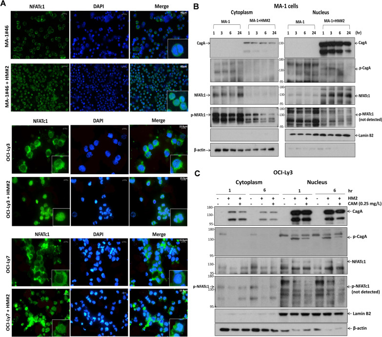
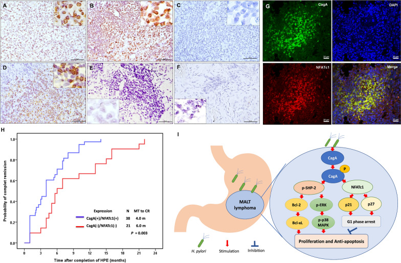
Materials and methods: We co-cultured three B-lymphoma cell lines (MA-1, OCI-Ly3, and OCI-Ly7) with HP strains (derived from HPE-responsive gastric MALT lymphoma) and evaluated the expression patterns of CagA, phosphorylated (p)-CagA (CagAP-Tyr), and CagA-signaling molecules, cell-cycle inhibitors, p-NFATc1 (Ser172), and NFATc1 using western blotting. Furthermore, we evaluated the association between nuclear NFATc1 expression in the tumor cells of 91 patients who received first-line HPE (59 patients with HPE responsiveness and 32 without HPE responsiveness) and HPE responsiveness and CagA expression in tumor cells.

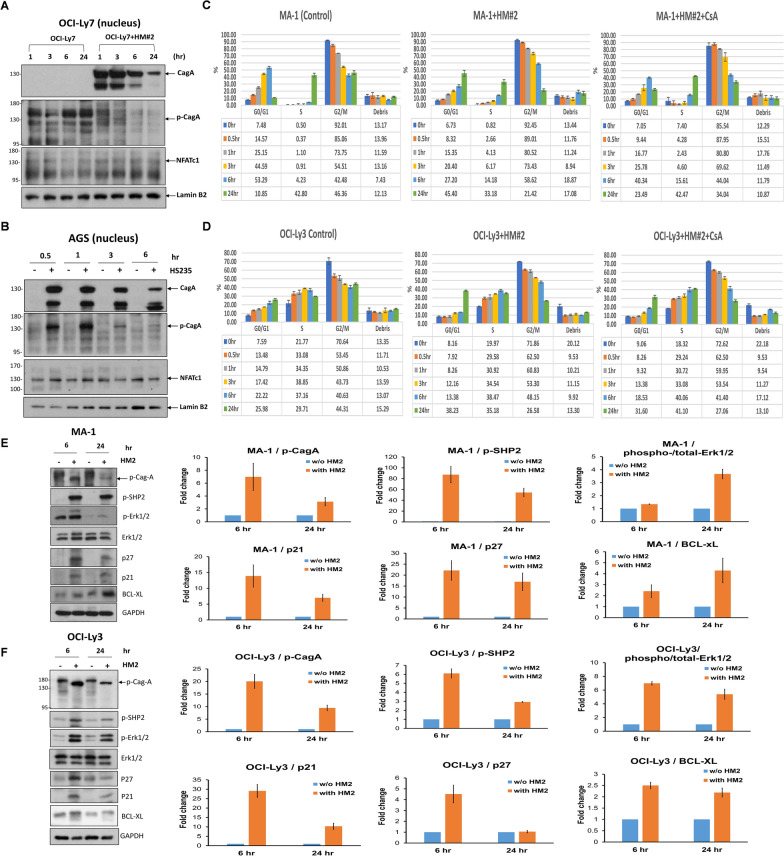
Results: In HP strains co-cultured with B cell lymphoma cell lines, CagA was translocated to the nucleus through tyrosine phosphorylation (CagAP-Tyr) and simultaneously dephosphorylated NFATc1, subsequently causing nuclear NFATc1 translocation and stimulating the expression of p-SHP-2/p-ERK/Bcl-xL. Activated NFATc1 causes G1 cell cycle retardation in both MA-1 and OCI-Ly3 cells by triggering p21 and p27 production. Nuclear NFATc1 localization was significantly associated with the presence of CagA in gastric MALT lymphomas (80% [41/51] vs. 33% [13/40]; p < 0.001) and with HPE responsiveness (73% [43/59] vs. 25% [8/32]; p < 0.001). Patients exhibiting both the presence of CagA and nuclear NFATc1 localization responded more rapidly to HPE than those without (median interval to CR, 4.00 vs. 6.00 months, p = 0.003).

Conclusions: Our findings indicated that CagA and NFATc1 cooperatively participate in the lymphomagenesis of HPE-responsive gastric MALT lymphoma.

Keywords: Helicobacter pylori; CagA; MALT lymphoma; NFATc1; Stomach.


Conflict of interest statement

Declarations Ethics approval and consent to participate The Institutional Review Board of the Research Ethics Committee of National Taiwan University Hospital (approval number: 201801030RINC) approved the study protocol, which included clinical data collection, pathological review, and molecular studies. Consent for publication All listed authors discussed results and commented on the submitted manuscript. Competing interests The authors declare no competing interests.

Figures

Fig. 1
Fig. 1
CagA, tyrosine-phosphorylated CagA, and NFATc1 can be translocated to the nucleus in HP-co-cultured DLBCL cells. A Immunofluorescence showed that NFATc1 translocated to the nucleus in HP (HM#2)-co-cultured MA-1#46 cells (upper panel) (scale bar = 40 μm). Similarly, NFATc1 translocated to the nucleus in HP (HM#2)-co-cultured OCI-Ly3 cells (ABC-DLBCL) (middle panel) (scale bar = 15.9 μm). In HP (HM#2)-co-cultured OCI-Ly7 cells (GCB-DLBCL) (lower panel), NFATc1 also translocated to the nucleus (scale bar = 15.9 μm). B In HP (HM#2)-co-cultured MA-1 cells, nuclear CagA expression was observed at 1, 3, 6, and 24 h, whereas phosphorylated (p)-CagA (CagAP−Tyr) expression was observed at 1 and 3 h after HP stimulation. However, the effect decreased at 6 h and eventually disappeared at 24 h. Simultaneously, nuclear NFATc1 expression was observed at 1, 3, and 6 h after HP stimulation and continued until 24 h in HP (HM#2)-co-cultured MA-1 cells. Compared with that noted in the control MA-1 cells, the expression of cytoplasmic p-NFATc1 decreased at 1 and 3 h, and the decreased effect was significantly noted at 6 and 24 h in HP (HM#2)-co-cultured MA-1 cells. The p-NFATc1 expression was not detected in the nucleus of MA-1 cells. C CagA and p-CagA translocated to the nucleus of the HP (HM#2)-co-cultured OCI-Ly3 cells at 1 and 6 h. Simultaneously, nuclear expression of NFATc1 was detected at 1 and 6 h after HP stimulation. In addition, the cytoplasmic expression of p-NFATc1 was significantly diminished at 1 and 6 h in HP (HM#2)-co-cultured OCI-Ly3 cells. After the treatment of clarithromycin (CAM, 0.25 mg/L) in HP (HM#2)-co-cultured OCI-Ly3 cells, CagA and p-CagA expression decreased at 6 h, and nuclear NFATc1 expression decreased simultaneously at 6 h, whereas cytoplasmic expression of p-NFATc1 was reversed at 1 and 6 h
Fig. 2
Fig. 2
CagA, tyrosine-phosphorylated CagA, and NFATc1 can be translocated to the nucleus in HP-co-cultured GCB-DLBCL and gastric epithelial cells, CagA-related signaling molecules in HP-co-cultured DLBCL cell lines, and HP-co-cultured MA-1 cells and OCI-Ly3 cells exhibit G1 cell-cycle retardation. A In HP (HM#2)-co-cultured OCI-Ly7 cells, nuclear CagA expression was detected 1 and 3 h after HP stimulation; however, the effect decreased gradually at 6 h and decreased predominantly at 24 h. The expression of p-CagA in the nucleus was detected at 1 and 3 h; however, its signal intensity decreased at 6 h and eventually disappeared at 24 h. Simultaneously, the nuclear expression of NFATc1 was detected at 1, 3, 6, and 24 h after HP stimulation. B In HP (HS235)-co-cultured AGS cells, CagA and p-CagA translocated to the nucleus at 0.5 and 1 h. However, the signal intensity of both CagA and p-CagA decreased at 3 and 6 h. Simultaneously, nuclear expression of NFATc1 was detected at 0.5 and 1 h after HP stimulation; however, its signal intensity decreased at 3 and 6 h. C Compared with non-HP-co-cultured MA-1 cells, G1 arrest was predominantly detected at 24 h in HP (HM#2)-co-cultured MA-1 cells. However, G1 phase arrest was reversed after administration of CsA in HP (HM#2)-co-cultured MA-1 cells. The results were expressed in triplicate for each treatment group and measured by flow cytometry analysis (error bar means standard error). D Compared with that noted in non-HP-co-cultured OCI-Ly3 cells, G1 arrest was predominantly detected at 24 h in HP (HM#2)-co-cultured OCI-Ly3 cells. After the administration of CsA to HP (HM#2)-co-cultured OCI-Ly3 cells, the G1 arrest at 24 h was reversed. E Immunoblotting showed that HP induced the expression of CagA-related signaling molecules, including p-CagA, p-SHP2, p-ERK1/2, p21, p27, and Bcl-xL in HP (HM#2)-co-cultured MA-1 cells at 6 and 12 h. Quantification of western blotting in (E) showed that the expression levels of p-CagA, p-SHP2, p21, and p27 were higher at 6 than at 24 h, whereas levels of p-ERK1/2, and Bcl-xL were higher at 24 than at 6 h. F In HP (HM#2)-co-cultured OCI-Ly3 cells, HP provoked the expression of p-CagA, p-SHP2, p-ERK1/2, p21, p27, and Bcl-xL at 6 and at 12 h. Quantification of western blotting in (F) showed that the expression levels of p-CagA, p-SHP2, p-ERK1/2, p21, p27, and Bcl-xL were higher at 6 than at 24 h
Fig. 3
Fig. 3
Nuclear expression pattern of NFATc1 in lymphoma cells and time to CR of patients with HPE-responsive gastric MALT lymphoma A Moderate nuclear localization of NFATc1 was found in the lymphoma cells of gastric mucosa in an HPE-responsive case (time to CR: 6.00 months); right upper inset, × 1000 B Strong nuclear localization of NFATc1 was found in the lymphoma cells of gastric mucosa in an HPE-responsive case (time to CR: 3.00 months); right upper inset, × 1000 C No nuclear localization of NFATc1 was found in lymphoma cells of gastric mucosa in an HPE-irresponsive case; right upper inset, × 1000 D A high baseline expression of nuclear localization of NFATc1 in lymphoma cells of an HPE-responsive case before HPE (case #2); right upper inset, × 1000 E Decreased expression of nuclear localization of NFATc1 in remitting tumor cells (case #2) 1.00 month after completion of HPE; left lower inset, × 1000 F No expression of nuclear localization of NFATc1 in remitting tumor cells (case #2) 4.00 months after completion of HPE (case #2, the time to CR: 7.00 month); left lower inset, × 1000 G Confocal laser-scanning microscopy showing that most CagA-positive cells (green fluorescence) expressed nuclear localization of NFATc1 (red fluorescence); Representative images of nucleus (stained with DAPI, blue) (right upper panel) and merged image of CagA and NFATc1 expression (right lower panel). H Time to CR was calculated from the completion of HPE to first evidence of CR using Kaplan–Meier analysis (CagA( +)/NFATc1( +) [both expression of CagA and nuclear NFATc1 localization in lymphoma cells] vs. CagA(−)/NFATc1(−) [either CagA or nuclear NFATc1localization, or absence expression of CagA and nuclear NFATc1 localization in lymphoma cells]; two-sided log-rank test; p = 0.003). I In the stomach, persistent HP infection can result in the translocation of HP-encoded CagA into B lymphocytes and trigger the tyrosine phosphorylation-dependent signaling pathway, including SHP-2 and its ERK, p38 MAPK, Bcl-2, and Bcl-xL. This CagA-regulated signaling pathway further promotes the proliferation and impedes the apoptosis of lymphoma B cells. Simultaneously, CagA stimulates the production of NFATc1, which further upregulates the expression of p21 and p27, restricting cell cycle progression to the G1 phase and limiting the growth of lymphoma B cells. N number, MT median time, CR complete remission

References

    1. Du MQ, Isaccson PG. Gastric MALT lymphoma: from aetiology to treatment. Lancet Oncol. 2002;3(2):97–104. - PubMed
    1. Zullo A, Hassan C, Cristofari F, et al. Effects of helicobacter pylori eradication on early stage gastric mucosa-associated lymphoid tissuelymphoma. Clin Gastroenterol Hepatol. 2010;8(2):105–10. - PubMed
    1. Kuo SH, Cheng AL. Helicobacter pylori and mucosa-associated lymphoid tissue: what’s new. Hematology Am Soc Hematol Educ Program. 2013;2013:109–17. - PubMed
    1. Farinha P, Gascoyne RD. Helicobacter pylori and MALT lymphoma. Gastroenterology. 2005;128(6):1579–605. - PubMed
    1. Bergman MP, D’Elios MM. Cytotoxic T cells in H pylori-related gastric autoimmunity and gastric lymphoma. J Biomed Biotechnol. 2010. 10.1155/2010/104918. - PMC - PubMed

LinkOut - more resources