Review
. 2024 Nov 18;24(1):156.
doi: 10.1186/s40644-024-00794-5.

Nuclear medicine imaging in non-seminomatous germ cell tumors: lessons learned from the past failures

Affiliations
Review

Nuclear medicine imaging in non-seminomatous germ cell tumors: lessons learned from the past failures

Narjess Ayati et al. Cancer Imaging. .

Abstract

There is an unmet need for a more accurate molecular imaging radiotracer in the field of non-seminomatous germ cell tumors (NSGCT). The clinical problem is that no single imaging modality is able to differentiate teratoma from necrotic tissue in NSGCTs, which the nuclear medicine techniques are no exception. The exponential growth in the list of potentially promising radiotracers may hold promise in the future for imaging of NSGCTs. Here, we have reviewed the past efforts and potential future advances in this field.

Keywords: Imaging; Non-seminomatous germ cell tumor; Radiotracer; Retroperitoneal residual mass; Teratoma.


Conflict of interest statement

Declarations Ethics approval and consent to participate Not applicable. This is a review article. Consent for publication All authors have reviewed the final version and consented for publication of the study. Competing interests The authors declare no competing interests.

Figures

Fig. 1
Fig. 1
A 32-year-old male with non-seminomatous testicular cancer, initially treated with orchidectomy, presented with suspicious para-aortic lymph nodes on CT and underwent an FDG PET scan. The images reveal a subcentimetre left supraclavicular lymph node (SUVmax 3.5; images a-c, crosshairs), bilateral intensely FDG-avid retrocrural lymph nodes (SUVmax 8 on the right and 12 on the left; images d-f, red arrow), and bilateral para-aortic lymphadenopathy extending from the axial level of L1 to L2/L3 on the right (SUVmax 21) and from the axial level of L1/L2 to L2/L3 on the left (SUVmax 25; images g-I). A subsequent biopsy of the left supraclavicular node confirmed metastatic involvement. Retroperitoneal lymph node dissection also confirmed multifocal retroperitoneal nodal metastases
Fig. 2
Fig. 2
A 30-year-old male with a non-seminoma germ cell tumor, previously treated with a left orchidectomy. The PET scan demonstrates multiple low-density nodal lesions in the retroperitoneum, including at the aortocaval and left para-aortic stations (red arrows), with no increased FDG uptake. The patient subsequently underwent retroperitoneal lymph node dissection, which revealed multiple nodal metastases
Fig. 3
Fig. 3
A 33-year-old male with a history of non-seminoma germ cell tumor, previously treated with left orchidectomy, chemotherapy, and retroperitoneal lymph node dissection for bulky para-aortic lymph node metastases, presents for a progress assessment. The PET scan (Image A) demonstrates mild foci of uptake around the aortocaval (SUV max 3.9) and left para-aortic (SUV max 3.3) regions at the level of L3, adjacent to surgical clips (red arrows, a-c), which were reported as indeterminate (either post-surgical inflammatory changes or residual disease). Twelve months later, a follow-up PET scan (Image B) showed an interval reduction in the intensity of retroperitoneal foci of uptake (d-f), consistent with resolving post-operative inflammatory changes
Fig. 4
Fig. 4
A 23-year-old male with non-seminomatous testicular cancer, treated with orchidectomy followed by chemotherapy, presented with suspicious retroperitoneal lymphadenopathy on CT and underwent an FDG PET scan (Image A). The axial (a, d), coronal (b, e), and sagittal (c, f) views show enlarged hypoattenuating retroperitoneal lesions with no metabolic activity (red arrow). Serial CT scans demonstrated ongoing enlargement of these lesions. Ten months later, a follow-up PET scan (Image B) revealed further enlargement of the hypoattenuating retroperitoneal lesions with interval development of peripheral metabolic activity (images g-l, green arow), highly suggestive of nodal metastases. Subsequent nodal dissection confirmed the presence of nodal metastases on histopathology

References

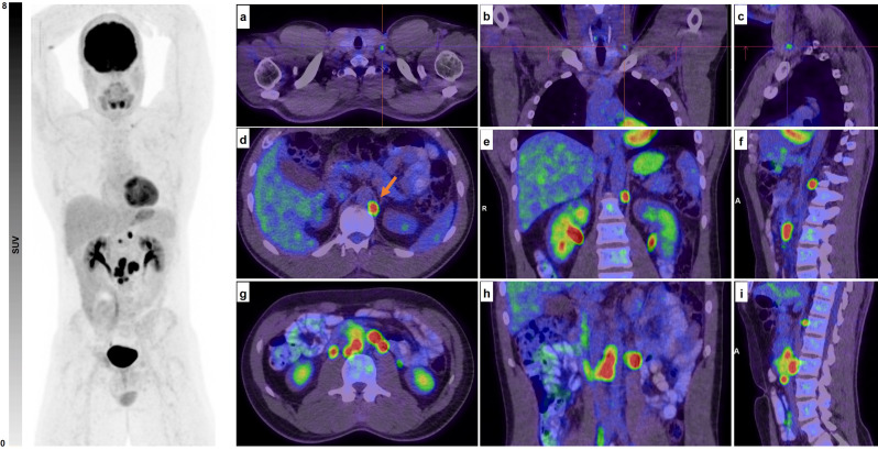
    1. Kaplan İ, Can C, Güzel Y, Alabalik U, Kömek H. 68 GA-FAPI-04 PET/CT Versus 18 F-FDG PET/CT in imaging of malignant mixed germ cell testicular tumor. Clin Nucl Med. 2023;48(4):e195–7. - PubMed
    1. Siegel RL, Miller KD, Wagle NS, Jemal A. Cancer statistics, 2023. CA Cancer J Clin. 2023;73(1):17–48. - PubMed
    1. Nallu A, Mannuel HD, Hussain A. Testicular germ cell tumors: biology and clinical update. Curr Opin Oncol. 2013;25(3):266–72. - PubMed
    1. Daneshmand S, Albers P, Fosså SD, Heidenreich A, Kollmannsberger C, Krege S, et al. Contemporary management of postchemotherapy testis cancer. Eur Urol. 2012;62(5):867–76. - PubMed
    1. Nauman M, Leslie SW. Nonseminomatous Testicular Tumors. StatPearls. Treasure Island (FL): StatPearls Publishing Copyright © 2024. StatPearls Publishing LLC.; 2024. - PubMed

MeSH terms

Substances

Supplementary concepts

LinkOut - more resources