. 2024 Nov 13:19:11819-11846.
doi: 10.2147/IJN.S484277. eCollection 2024.

Locally Acting Budesonide-Loaded Solid Self-Microemulsifying Drug Delivery Systems (SMEDDS) for Distal Ulcerative Colitis

Affiliations

Locally Acting Budesonide-Loaded Solid Self-Microemulsifying Drug Delivery Systems (SMEDDS) for Distal Ulcerative Colitis

Hany S M Ali et al. Int J Nanomedicine. .

Abstract

Background: Budesonide (BUD) is a BCS class II medication with poor water solubility and limited oral bioavailability. In this study, innovative solid self-microemulsifying drug delivery systems (BUD-SMEDDS) were developed for effective local management of distal ulcerative colitis (UC).

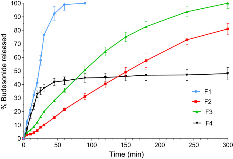
Methods: Based on solubility and emulsification tests, the components of the self-microemulsifying drug delivery system (SMEDDS) were Capryol™ 90, Tween 80, and Transcutol HP. The impacts of BUD-SMEDDS ingredients (as inputs) on the average globule size (AGS), polydispersity index (PDI), and self-emulsification time (SET) as responses were investigated using the Box-Behnken design methodology. Solid rectal systems were then fabricated using the optimized values of SMEDDS components in Lutrol® bases. The developed systems were evaluated for in vitro characteristics and in vivo efficacy using a rat colitis model.

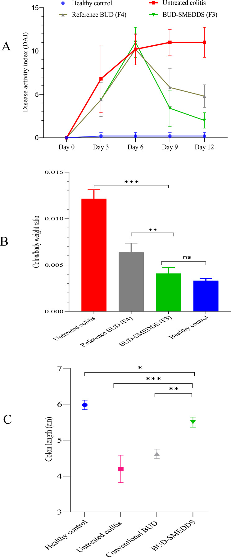
Results: For all responses, the greatest impact was attributed to the oil content of SMEDDS. An optimized BUD-SMEDDS with AGS of 33 ± 2.9 nm, PDI of 0.29 ± 0.03 and SET of 25 ± 2.5 s) was selected for rectal formulations. The developed formulations demonstrated acceptable physical characteristics and mucoadhesive abilities. Differential scanning calorimetric (DSC) analysis revealed the absence of BUD crystallinity in the SMEDDS formulations. The drug release patterns could be regulated by selecting the grade and composition of the incorporated Lutrols. Clinical and histopathological assessments revealed considerable improvements in animals treated with BUD-SMEDDS formulations.

Conclusion: Overall findings confirmed the superior capability of solid SMEDDS as BUD carriers to manage distal colitis in tested animals.

Keywords: Box Behnken design; Budesonide; SMEDDS; Ulcerative colitis; efficacy.


Conflict of interest statement

The authors report no conflicts of interest in this work.

Figures

Figure 1
Figure 1
Illustration of the device used to evaluate mucoadhesive forces.
Figure 2
Figure 2
Results of solubility of budesonide in tested vehicles.
Figure 3
Figure 3
Results of surfactants (A) and co-surfactants (B) screening for oil emulsification.
Figure 4
Figure 4
A pseudo ternary diagram depicting the self-emulsification region (the shaded area).
Figure 5
Figure 5
Contour and response surface plots of interactions between oil–surfactant (a), oil–cosurfactant (b) and surfactant–cosurfactant (c) and the average globule size (AGS).
Figure 6
Figure 6
Contour and response surface plots of interactions between oil–surfactant (a), oil–cosurfactant (b) and surfactant–cosurfactant (c) and polydispersity index (PDI).
Figure 7
Figure 7
Contour and response surface plots of interactions between oil–surfactant (a), oil–cosurfactant (b) and surfactant–cosurfactant (c) and self-emulsification time (SET).
Figure 8
Figure 8
Ramp graph (A) and contour plots (B-D) of desirability for the numerically optimized BUD-SMEDDS formulation.
Figure 9
Figure 9
DSC thermograms of unprocessed budesonide, Lutrol® F68, Lutrol® F127 and formulations F1, F2 and F3.
Figure 10
Figure 10
Release of budesonide from tested formulations F1, F2, F3 and F4.
Figure 11
Figure 11
Disease activity index (A), colon/body weight ratio (B) and colon lengths (C) of tested animal groups.
Figure 12
Figure 12
Photomicrographs of representative stained colonic tissues of tested groups. The first row; Photomicrographs of healthy colons stained by H&E at low power view (A), high power view (B), and trichrome stained (C). The second row; photomicrographs of colitis control colons stained by H&E at low-power view (D), high-power view (E), and trichrome stained (F). The third row; photomicrographs of H&E-stained colons treated by reference budesonide formulation at low-power view (G), and high-power view (H) and by trichrome stain (J). The fourth row; photomicrographs of H&E-stained colons treated by budesonide SMEDDS formulation at low power view (K), and high-power view (L) and by trichrome stain (M).
Figure 13
Figure 13
Total Geboes score of tested animal groups.

References

    1. Rahimi R, Nikfarl S, Rezaie A, Abdollahi M. A meta-analysis of the benefit of probiotics in maintaining remission of human ulcerative colitis: evidence for prevention of disease relapse and maintenance of remission. Arch Med Sci. 2008;4(2):185–190.
    1. Sandborn WJ, Travis S, Moro L, et al. Once-daily budesonide MMX(R) extended-release tablets induce remission in patients with mild to moderate ulcerative colitis: results from the CORE I study. Gastroenterology. 2012;143(5):1218–26e1–2. doi:10.1053/j.gastro.2012.08.003 - DOI - PubMed
    1. Rogler G. Medical management of ulcerative colitis. Dig Dis. 2009;27(4):542–549. doi:10.1159/000233295 - DOI - PubMed
    1. Danese S, Vuitton L, Peyrin-Biroulet L. Biologic agents for IBD: practical insights. Nat Rev Gastroenterol Hepatol. 2015;12(9):537–545. doi:10.1038/nrgastro.2015.135 - DOI - PubMed
    1. Faubion WA, Loftus EV, Harmsen WS, Zinsmeister AR, Sandborn WJ. The natural history of corticosteroid therapy for inflammatory bowel disease: a population-based study. Gastroenterology. 2001;121(2):255–260. doi:10.1053/gast.2001.26279 - DOI - PubMed

MeSH terms