. 2024 Nov 18;2024(11):rjae712.
doi: 10.1093/jscr/rjae712. eCollection 2024 Nov.

Primary division of annular pancreas: a surgical technique

Affiliations

Primary division of annular pancreas: a surgical technique

Kai-Zheong Lim et al. J Surg Case Rep. .

Abstract

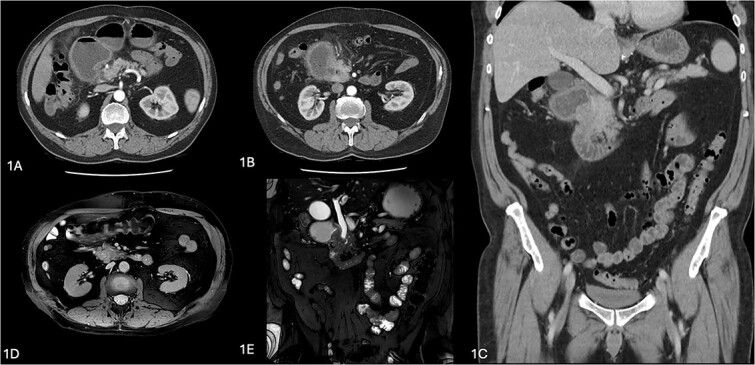
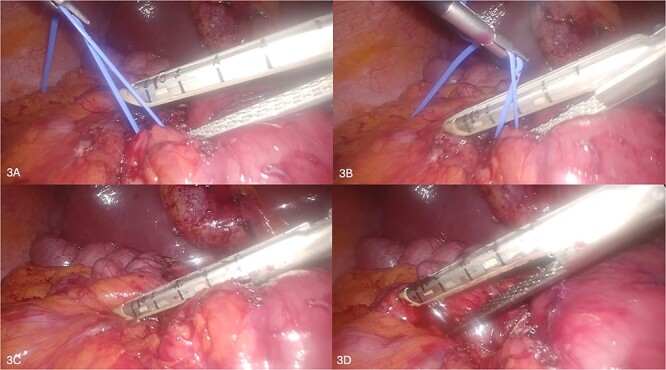
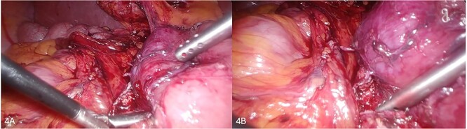
The authors presented a case of duodenal obstruction in a 61-year-old man, resulting from an annular pancreas diagnosed on imaging (computed tomography, magnetic resonance cholangiopancreatography, and endoscopic ultrasound). The patient underwent a diagnostic laparoscopy. Intraoperatively, given a straightforward appearance and anatomy of the annular pancreas overlying the second part of the duodenum, and due to extensive adhesions in the abdomen, a primary division of the annular pancreas was performed, instead of a bypass procedure such as gastrojejunostomy. He had some residual symptoms 1 week postoperatively which was treated with duodenal dilatation endoscopically. On review and follow-up at 1 year, he has remained well with resolution of symptoms, supported by radiological improvement on a computed tomography performed at 4 months post-operatively. We believe this approach has resulted in less morbidity and a shorter period of recovery as compared to a bypass procedure and represents a reasonable therapeutic option for annular pancreas.

Keywords: annular pancreas; duodenal obstruction; gastric outlet obstruction; pancreatic leak; pancreatic mass.


Conflict of interest statement

The authors disclose no conflicts. The authors confirm that the material has not been published previously and is not submitted for publication elsewhere.

Figures

Figure 1
Figure 1
Radiological images of annular pancreas. (A–C) Computerized tomography images showing duodenal obstruction with a transition point at level of pancreatic head with periduodenal fat stranding and duodenal wall thickening, with impression of enhancing pancreatic tissue lateral to duodenum at this site. (C, D) Magnetic resonance cholngiopancreatography images showing an abrupt transition in the caliber of the second part of the duodenum by an obstructive periampullary lesion, the annular pancreas.
Figure 2
Figure 2
(A–D) Sequential depiction of the plane between annular pancreas and duodenum being defined using Maryland grasper (A, B) and silicone vessiloop (B–D).
Figure 3
Figure 3
(A–D) Sequential depiction of the division of annular pancreas using laparoscopic stapler device.
Figure 4
Figure 4
(A, B) Successfully transected annular pancreas above duodenum.
Figure 5
Figure 5
Intraoperative cholangiogram showing pancreaticobiliary malunion with the bile duct entering the main pancreatic duct proximal to the ampulla.
Figure 6
Figure 6
(A, B) A repeat CT at 4 months showing improvement of the gastric and duodenal distension proximal to the previous transition point and resolution of the periduodenal fat stranding and duodenal wall thickening.

References

    1. Yogi Y, Shibue T, Hashimoto S. Annular pancreas detected in adults, diagnosed by endoscopic retrograde cholangiopancreatography: report of four cases. Gastroenterol Jpn 1987;22:92–9. 10.1007/BF02806340. - DOI - PubMed
    1. Zyromski NJ, Sandoval JA, Pitt HA, et al. . Annular pancreas: dramatic differences between children and adults. J Am Coll Surg 2008;206:1019–25, discussion 1025–7. 10.1016/j.jamcollsurg.2007.12.009. - DOI - PubMed
    1. Noh T, Lee SE, Park J. Laparoscopic treatment of annular pancreas in adult: report of a case. Korean J Hepatobiliary Pancreat Surg 2012;16:43–5. 10.14701/kjhbps.2012.16.1.43. - DOI - PMC - PubMed

LinkOut - more resources