

A case of choledochal cyst type IV
- PMID: 39559504
- PMCID: PMC11570900
- DOI: 10.1016/j.radcr.2024.10.033
A case of choledochal cyst type IV
Abstract
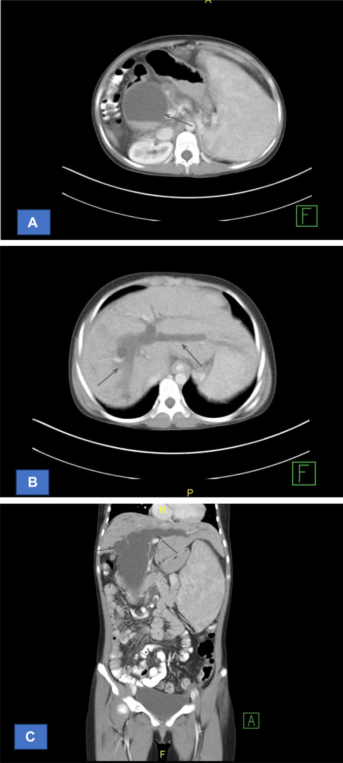
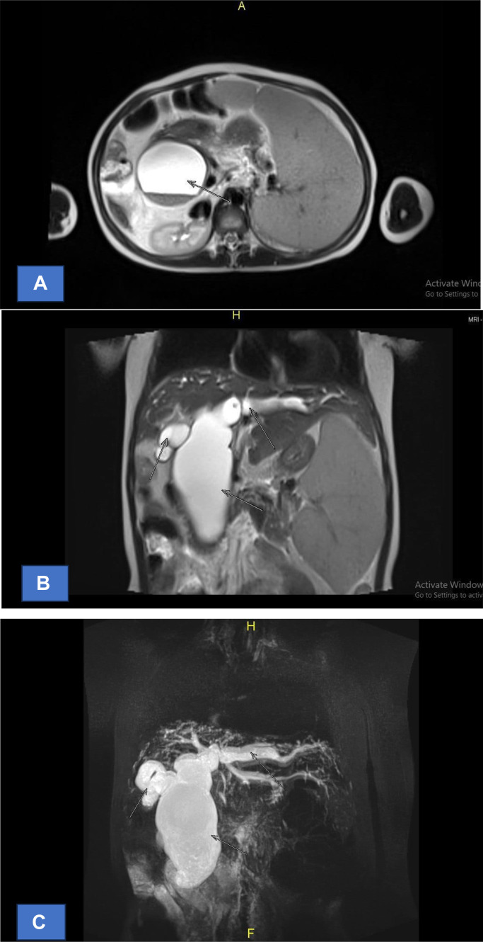
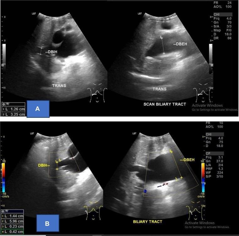
A choledochal cyst (CC) or biliary cyst is a congenital or acquired anomaly affecting the biliary tree. It involves the dilation of the biliary tree that could affect the extrahepatic and/or the intrahepatic segments. A choledochal cyst (CC) has traditionally been considered as a cystic dilation of the extrahepatic bile duct. The incidence of choledochal cysts is high in the Asian population with a female predominance. Choledochal cysts can present at any age, including infancy. However, 80% of choledochal cysts are diagnosed in the first decade of life, with cholestasis being the most common sign in infants, and cholangitis or pancreatitis being less common. Radiological and endoscopic imaging is the cornerstone of CC diagnosis. We report a case of 16 years old patient with choledocal cyst. The case has distinct clinical signs that are easily recognizable.
Keywords: Children; Coledocal cyst; Icteric.
© 2024 The Authors. Published by Elsevier Inc. on behalf of University of Washington.
Figures

References
-
- Juju J, Efa A, Eka W, Fauziah R. Universitas Indonesia Library; Jakarta: 2022. Pengaruh intervensi pemberian posisi semi fowler pada anak post laparatomi kista duktus koledokus = the effect of intervention on giving semi fowler’s position to children post laparatomi of choledochal duct cysts.
-
- Hoilat GJ, John S. StatPearls. StatPearls Publishing; Treasure Island (FL): 2024. Choledochal cyst.
-
- Al-Ba'adani MN, Alammari SA, Jumman NA. Choledochal cysts diagnosis and treatments: case report. OAlib. 2022;09(02):1–6.
Publication types
LinkOut - more resources
Full Text Sources
