. 2024 Oct 30:13:101788.
doi: 10.1016/j.toxrep.2024.101788. eCollection 2024 Dec.

Computational and theoretical insights into the cytotoxic prospects of compounds isolated from Elaeodendron buchananii against Leukemia

Affiliations

Computational and theoretical insights into the cytotoxic prospects of compounds isolated from Elaeodendron buchananii against Leukemia

Ochuko L Erukainure et al. Toxicol Rep. .

Abstract

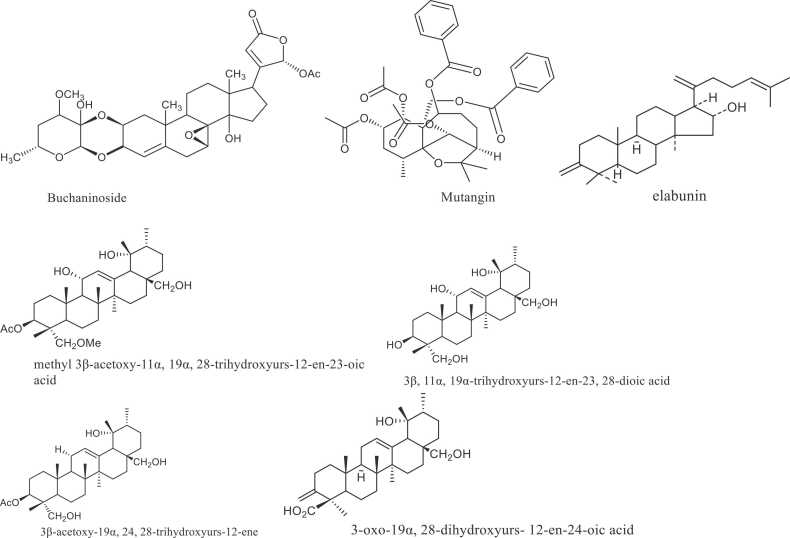
The present study investigated the cytotoxic prospects of isolated compounds from Elaeodendron buchananii against leukemia, using computational tools. Comprehensive literature searches revealed only buchaninoside, mutangin, methyl 3β-acetoxy-11α, 19α, 28-trihydroxyurs-12-en-23-oic acid, 3β, 11α, 19α-trihydroxyurs-12-en-23, 28-dioic acid, 3β-acetoxy-19α, 24, 28-trihydroxyurs-12-ene, 3-oxo-19α,28-dihydroxyurs-12-en-24-oic acid, and elabunin have been isolated from E. buchananii. The compounds were subjected to Density Functional Theory (DFT) and Molecular Dynamics (MD) analyses, with Fms-like tyrosine kinase (FLT3) and catalytic binding sites of Murine Leukemia Virus (MLV) as the target proteins in lukemia. Following DFT analysis, the structures of the compounds were optimized at the PW6B95D3/Def2-TZVP level of theory; their UV-Visible peaks were in the UV region, with mutangin, 3-oxo-19α,28-dihydroxyurs-12-en-24-oic acid and elabunin exhibiting one single peak. The potent Root-Mean-Square Deviation, Root-Mean-Square Fluctuation, solvent-accessible surface area and radius of gyration values indicated a strong and stable molecular interaction between the compounds and the proteins. These were further supported by high ∆G values, with MLV showing the best interaction. Per-residue decomposition plots also revealed high energy contributions in the interactions' binding sites residues. These results indicate that the cytotoxic prospects of the isolated compounds against leukemia as indicated by its molecular interactions with FLT3 and MLV.

Keywords: And Lukemia; Anticancer; Computation; Cytotoxicity; Elaeodendron buchananii.


Conflict of interest statement

The authors declare that they have no known competing financial interests or personal relationships that could have appeared to influence the work reported in this paper.

Figures

None
Graphical abstract
Fig. 1
Fig. 1
Isolated compounds from E. buchananii.
Fig. 2
Fig. 2
Structures of the investigated compounds as optimized at the PW6B95D3/Def2-TZVP level of theory. Detailed names of the compounds are provided in the text.
Fig. 3
Fig. 3
UV-Vis spectra of compounds 2, 4, 6 and 7 computed using TD-DFT at the PW6B95D3/Def2-TZVP level of theory.
Fig. 4
Fig. 4
Infrared spectra of the studied compounds as calculated at the PW6B95D3/Def2-TZVP level of theory.
Fig. 5
Fig. 5
Structural representation of alterations occurring during the binding of the seven compounds bound to FLT3. The (A) conformational stability, C-α atoms RMSD FLT3 to the compounds ran for 100 ns MD simulation. (B) The time evolution RMSF of each residue of the enzyme C-α atom over 100 ns for. (C) Solvent accessible surface area (SASA) backbone atoms relative to the starting minimized structure over 100 ns for FLT3. (D) C-α RoG plots and structural dynamics showing atomic distribution and compactness in various systems during 100 ns simulation period whereas the different colours represent each system atomic distribution structure conformation.
Fig. 6
Fig. 6
Structural representation of alterations occurring during the binding of the compounds bound to MLV. The (A) conformational stability, C-α atoms RMSD MLV to the compounds ran for 100 ns MD simulation. (B) The time evolution RMSF of each residue of the enzyme C-α atom over 100 ns for. (C) Solvent accessible surface area (SASA) backbone atoms relative to the starting minimized structure over 100 ns for MLV. (D) C-α RoG plots and structural dynamics showing atomic distribution and compactness in various systems during 100 ns simulation period whereas the different colours represent each system atomic distribution structure conformation.
Fig. 7
Fig. 7
Per-residue decomposition plots showing individual energy contributions to the binding and stabilization of the compounds at the binding sites of FLT3 (A) 3ß,11a,19a-trihydroxyurs (B) 3ß-acetoxy,28-trihydroxyurs (C) Buchaninoside (D) Elabunin (E) Methyl 3ß-acetoxy-11a (F) Mutangin (G) 3-Oxo9a,28-dihydroxyurs. This reveals that energy contributions were highest in binding sites residues.
Fig. 8
Fig. 8
Per-residue decomposition plots showing individual energy contributions to the binding and stabilization of the compounds at the binding sites of MLV (A) 3ß,11a,19a-trihydroxyurs (B) 3ß-acetoxy,28-trihydroxyurs (C) Buchaninoside (D) Elabunin (E) Methyl 3ß-acetoxy-11a (F) Mutangin (G) 3-Oxo9a,28-dihydroxyurs. This reveals that energy contributions were highest in binding sites residues.

References

    1. Bispo J.A.B., Pinheiro P.S., Kobetz E.K. Epidemiology and etiology of leukemia and lymphoma. Cold Spring Harb. Perspect. Med. 2020;10(6) a034819. - PMC - PubMed
    1. Bray F., Ferlay J., Soerjomataram I., et al. Global cancer statistics 2018: GLOBOCAN estimates of incidence and mortality worldwide for 36 cancers in 185 countries. CA: a Cancer J. Clin. 2018;68(6):394–424. - PubMed
    1. NCI. Cancer Stat Facts: Leukemia 2024 2023 [cited 2024 August 21]. Available from: 〈https://seer.cancer.gov/statfacts/html/leuks.html〉.
    1. Siegel R.L., Giaquinto A.N., Jemal A. Cancer statistics, 2024. CA: a Cancer J. Clin. 2024;74(1) - PubMed
    1. Huang J., Chan S.C., Ngai C.H., et al. Disease burden, risk factors, and trends of leukaemia: a global analysis. Front. Oncol. 2022;12 - PMC - PubMed

LinkOut - more resources