. 2024 Oct 19;27(11):111212.
doi: 10.1016/j.isci.2024.111212. eCollection 2024 Nov 15.

MOTS-c modulates skeletal muscle function by directly binding and activating CK2

Affiliations

MOTS-c modulates skeletal muscle function by directly binding and activating CK2

Hiroshi Kumagai et al. iScience. .

Abstract

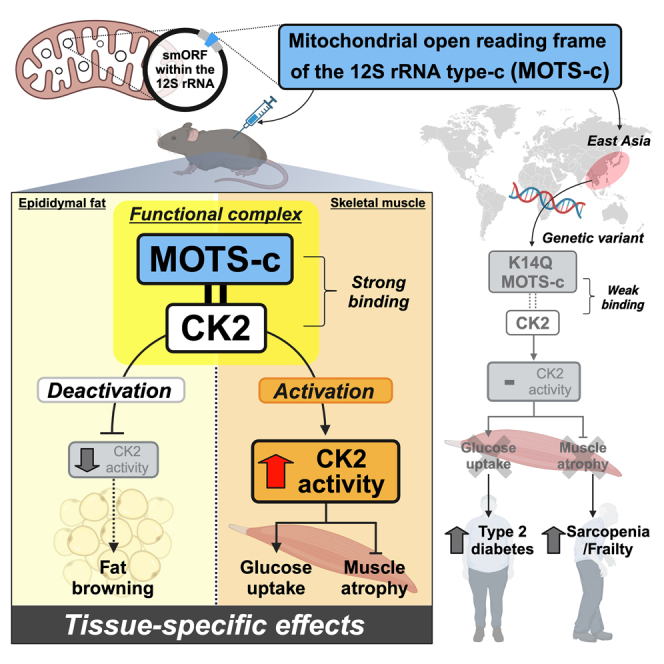
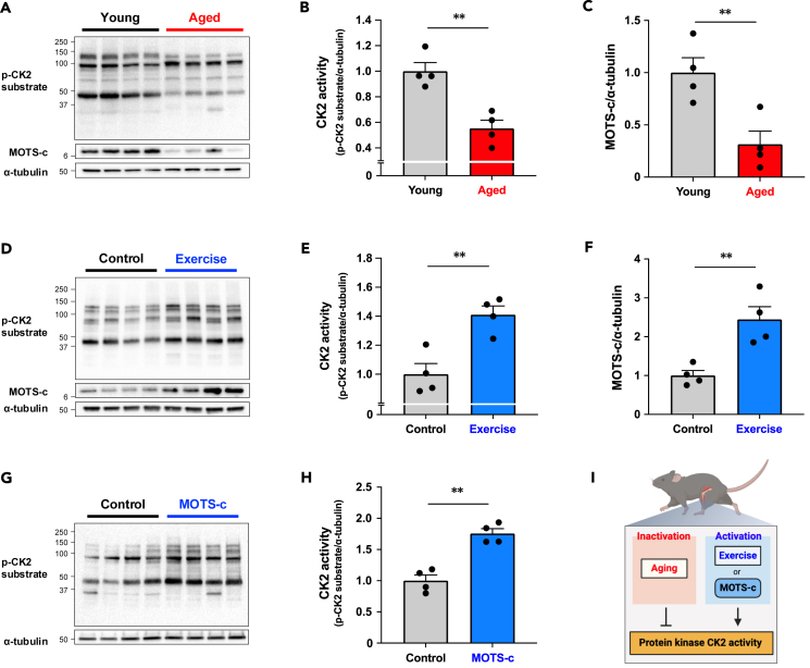
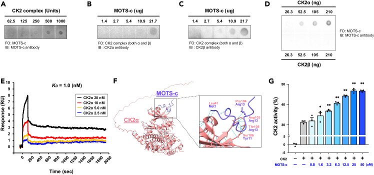
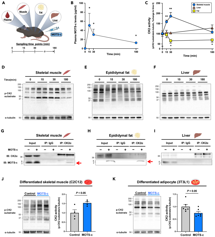
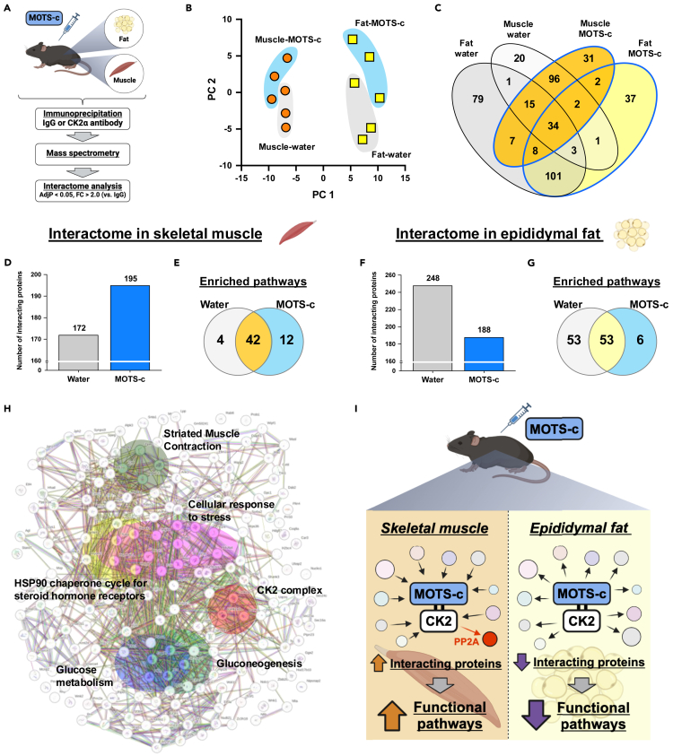
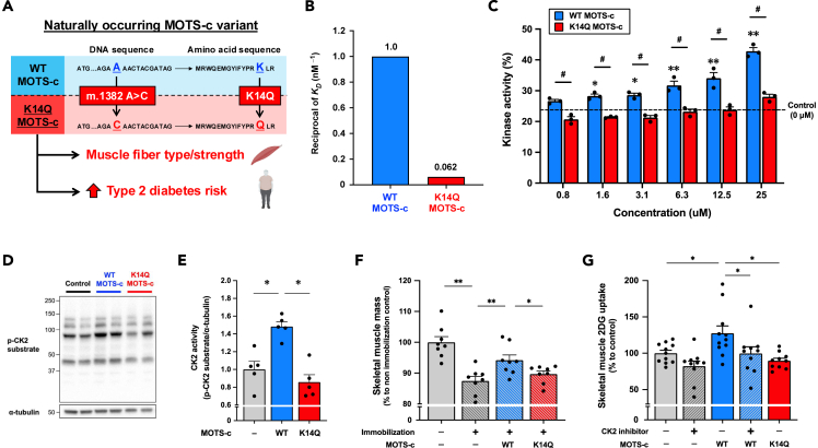
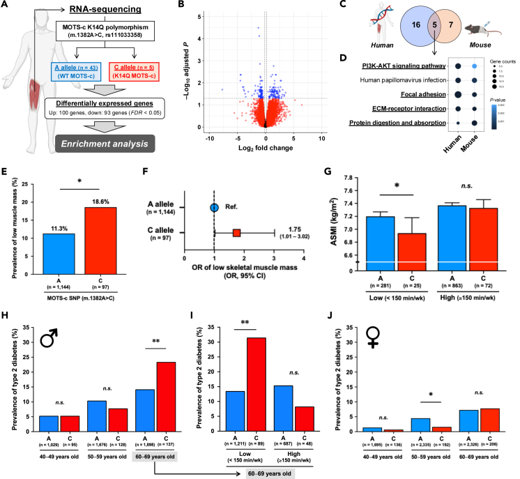
MOTS-c is a mitochondrial microprotein that improves metabolism. Here, we demonstrate CK2 is a direct and functional target of MOTS-c. MOTS-c directly binds to CK2 and activates it in cell-free systems. MOTS-c administration to mice prevented skeletal muscle atrophy and enhanced muscle glucose uptake, which were blunted by suppressing CK2 activity. Interestingly, the effects of MOTS-c are tissue-specific. Systemically administered MOTS-c binds to CK2 in fat and muscle, yet stimulates CK2 activity in muscle while suppressing it in fat by differentially modifying CK2-interacting proteins. Notably, a naturally occurring MOTS-c variant, K14Q MOTS-c, has reduced binding to CK2 and does not activate it or elicit its effects. Male K14Q MOTS-c carriers exhibited a higher risk of sarcopenia and type 2 diabetes (T2D) in an age- and physical-activity-dependent manner, whereas females had an age-specific reduced risk of T2D. Altogether, these findings provide evidence that CK2 is required for MOTS-c effects.

Keywords: Physiology; cell biology.


Conflict of interest statement

Pinchas Cohen is an advisor to and stockholder in CohBar Inc. UCLA has licensed the intellectual property on MOTS-c, on which Pinchas Cohen is listed as an inventor, to CohBar. None of the other authors has any conflicts of interest, financial or otherwise, to disclose.

Figures

None
Graphical abstract
Figure 1
Figure 1
Endogenous and exogenous MOTS-c regulates CK2 activity in the skeletal muscle (A–C) CK2 activity assessed by detecting endogenous proteins containing a pS/pTDXE motif and MOTS-c expression levels in gastrocnemius muscle from young control (2 months) and aged (22 months) mice (n = 4 per group). (D–F) Effect of 4 weeks of voluntary wheel running exercise on CK2 activity and MOTS-c expression levels in gastrocnemius muscle from young mice (n = 4 per group). (G and H) Effect of 8 weeks of MOTS-c administration (5 mg/kg/day) on CK2 activity in quadriceps muscle from high-fat-diet (HFD)-fed mice (n = 4 per group). (I) Summary of MOTS-c and CK2 activity in the skeletal muscle. Data are represented as mean ± SEM for (B, C, E, F, and H). ∗∗p < 0.01.
Figure 2
Figure 2
MOTS-c directly binds and activates CK2 in cell-free systems (A–C) Dot blot assays with (A) CK2 complex (contains both CK2α and CK2β subunits) immobilized, MOTS-c flowed over the membrane, and detected by MOTS-c antibody. (B–C) MOTS-c immobilized, CK2 complex flowed over the membrane, and detected by CK2α (B) and CK2β (C) antibodies. (D) CK2α or CK2β immobilized, MOTS-c flowed over the membrane, and detected by MOTS-c antibody. FO, flow over; IB, immunoblotting. (E): Surface plasmon resonance (Biacore assay) of MOTS-c (10 μg/mL) and CK2α (2.5 nM, 5.0 nM, 10 nM, and 20 nM). MOTS-c was immobilized on the sensor chip and CK2α flowed over the sensor chip. KD, dissociation constant. (F) Molecular docking simulation of the binding between MOTS-c and CK2α by using AlphaFold2. (G) CK2 activity assessed by kinase activity assay with/without MOTS-c in cell-free condition. A different dose of MOTS-c (0–100 μM) was used for the assay. Data are represented as mean ± SEM for (G). ∗p < 0.05, ∗∗p < 0.01 versus CK2 without MOTS-c group.
Figure 3
Figure 3
MOTS-c modulates CK2 activity in a tissue-specific manner (A) Experimental design of a single MOTS-c administration (7.5 mg/kg) experiment in young mice (n = 5 per time point). (B) Plasma MOTS-c levels after MOTS-c administration. ∗: p < 0.05 versus time 0. (C) Quantification of CK2 activity assessed by western blotting in each tissue shown in (E–F). (E–F) CK2 activity assessed by western blotting with p-CK2 substrate antibody in gastrocnemius muscle (D), epididymal fat (E), and liver (F) after MOTS-c administration. ∗p < 0.05, ∗∗p < 0.01 versus time 0 in same tissue. (G–I) MOTS-c detection following CK2α immunoprecipitation (IP) in gastrocnemius muscle (G), epididymal fat (H), and liver (I) 30 min after MOTS-c administration (7.5 mg/kg). The "-" indicates tissues from non-MOTS-c administered mice, while the "+" indicates tissues from MOTS-c administered mice. (J–K) CK2 activity after 10 min MOTS-c treatment (10 μM) in differentiated skeletal muscle (J) and adipocyte (K). Data are represented as mean ± SEM for (B, C, J, and K).
Figure 4
Figure 4
MOTS-c modulates the CK2 interactome in a tissue-specific manner (A) Experimental design for the interactome analyses in young mice (n = 3 per condition). Proteome analysis was performed following CK2α immunoprecipitation in gastrocnemius muscle and epididymal fat with/without MOTS-c administration. (B) Principal components (PCs) of control and MOTS-c-treated mouse gastrocnemius muscle and epididymal fat. (C) Venn diagram of interacting proteins in each condition. (D–E) Numbers of CK2 interacting proteins (D) and enriched Reactome pathways (E) in gastrocnemius muscle. (F–G) Numbers of CK2 interacting proteins (F) and enriched Reactome pathways (G) in epididymal fat. (H) Interacting proteins and significantly enriched Reactome pathways in MOTS-c-administered gastrocnemius muscle compared to the control group. The protein-protein interactions and enrichment analysis were assessed by using the STRING database. (I) Summary of the interactome analysis in gastrocnemius muscle and epididymal fat.
Figure 5
Figure 5
A naturally occurring K14Q MOTS-c is a bio-inactive form of MOTS-c due to its reduced binding to CK2 alpha (A) Nucleotide and amino acid substitutions of naturally occurring MOTS-c variant K14Q MOTS-c, which modulates skeletal muscle function and increases type 2 diabetes risk. (B) Comparison of reciprocal KD between WT and K14Q MOTS-c assessed by the surface plasmon resonance (Biacore) assay. (C) Comparison of CK2 activating effects between WT MOTS-c and K14Q MOTS-c in cell-free condition (n = 3 per group). MOTS-c concentrations are 0, 0.8, 1.6, 3.1, 6.3, 12.5, and 25 μM ∗p < 0.05, ∗∗p < 0.01 versus control. #p < 0.05 versus same concentration of K14Q MOTS-c. (D–E) Comparison of gastrocnemius muscle CK2 activity between WT MOTS-c- and K14Q MOTS-c-administered young mice (2.5 mg/kg, n = 5 per group). ∗p < 0.05 versus control group. (F) Protective effect of MOTS-c administration (WT or K14Q MOTS-c, 15 mg/kg/day, IP injection) against 8 days of immobilization-induced skeletal muscle atrophy (n = 8 per group). Skeletal muscle mass was assessed by a total mass of gastrocnemius, plantaris, and soleus muscles. ∗p < 0.05, ∗∗p < 0.01. (G)Skeletal muscle 2-deoxy-d-glucose (2DG) uptake after MOTS-c administration (WT or K14Q, 7.5 mg/kg) with/without CK2 inhibitor (CX-4945, 25 mg/kg) (n = 10–11 per group). ∗p < 0.05. Data are represented as mean ± SEM for (C, E, F, and G).
Figure 6
Figure 6
The bio-inactive MOTS-c variant increases risks of sarcopenia and type 2 diabetes in an age- and physical-activity-dependent manner (A) A schema of the RNA sequencing analysis in human skeletal muscle from the A allele and C allele carriers of MOTS-c single nucleotide polymorphism (SNP) (m.1382A>C, rs111033358) in 48 Japanese males and females. (B) Differentially regulated genes in human skeletal muscle between the A and C allele carriers. Age and sex were used as covariates. (C–D) The overlapped enriched pathways between human skeletal muscles from the A or C allele carriers and skeletal muscles from WT or K14Q MOTS-c-treated immobilized mice. KEGG pathways were used for the enrichment analyses. (E) Percentage of people with low skeletal muscle mass between the A and C allele carriers of MOTS-c SNP (m.1382A>C, rs111033358) in 1,241 Japanese individuals. Skeletal muscle mass was measured by dual-energy X-ray absorptiometry, and appendicular skeletal muscle mass index (ASMI) was calculated. The cutoff values for low skeletal muscle mass are ASMI <7.0 kg/m2 for men and <5.4 kg/m2 for women. (F) Odds ratio (OR) and 95% confidence interval of low skeletal muscle mass between the A allele and C allele carriers of MOTS-c SNP (m.1382A>C, rs111033358). OR was adjusted for age and sex. (G) Skeletal muscle mass assessed by ASMI between the A and C allele carriers of MOTS-c SNP (m.1382A>C, rs111033358) in low and high physical activity groups. A cutoff value for physical activity is 150 min of moderate-to-vigorous physical activity (MVPA) per week base on the World Health Organization guideline. Data are represented as mean ± SEM. (H) The prevalence of type 2 diabetes in the A and C allele carriers of MOTS-c SNP (m.1382A>C, rs111033358) in 4,966 Japanese males with considering age. (I) The prevalence of type 2 diabetes in the A and C allele carriers in subjects in the 60s with considering physical activity levels. A cutoff value of the physical activity is 150 min of MVPA per week base on the World Health Organization guideline. (J) The prevalence of type 2 diabetes in the A and C allele carriers of MOTS-c SNP (m.1382A>C, rs111033358) in 6,890 Japanese females with considering age.∗p < 0.05, ∗∗p < 0.01.

References

    1. Lee C., Zeng J., Drew B.G., Sallam T., Martin-Montalvo A., Wan J., Kim S.J., Mehta H., Hevener A.L., de Cabo R., Cohen P. The mitochondrial-derived peptide MOTS-c promotes metabolic homeostasis and reduces obesity and insulin resistance. Cell Metabol. 2015;21:443–454. doi: 10.1016/j.cmet.2015.02.009. - DOI - PMC - PubMed
    1. Kumagai H., Miller B., Kim S.-J., Leelaprachakul N., Kikuchi N., Yen K., Cohen P. Novel Insights into Mitochondrial DNA: Mitochondrial Microproteins and mtDNA Variants Modulate Athletic Performance and Age-Related Diseases. Genes. 2023;14:286. doi: 10.3390/genes14020286. - DOI - PMC - PubMed
    1. Kim S.-J., Miller B., Kumagai H., Silverstein A.R., Flores M., Yen K. Mitochondrial-derived peptides in aging and age-related diseases. GeroScience. 2021;43:1113–1121. doi: 10.1007/s11357-020-00262-5. - DOI - PMC - PubMed
    1. Merry T.L., Chan A., Woodhead J.S.T., Reynolds J.C., Kumagai H., Kim S.J., Lee C. Mitochondrial-derived peptides in energy metabolism. Am. J. Physiol. Endocrinol. Metab. 2020;319:E659–E666. doi: 10.1152/ajpendo.00249.2020. - DOI - PMC - PubMed
    1. Miller B., Kim S.J., Kumagai H., Yen K., Cohen P. Mitochondria-derived peptides in aging and healthspan. J. Clin. Invest. 2022;132 doi: 10.1172/JCI158449. - DOI - PMC - PubMed