. 2025 Feb 4;24(2):299-309.
doi: 10.1158/1535-7163.MCT-24-0664.

Modeling the Acute Mucosal Toxicity of Fractionated Radiotherapy Combined with the ATM Inhibitor WSD0628

Affiliations

Modeling the Acute Mucosal Toxicity of Fractionated Radiotherapy Combined with the ATM Inhibitor WSD0628

Darwin A Garcia et al. Mol Cancer Ther. .

Abstract

Ataxia Telangiectasia-mutated (ATM) inhibitors are being developed as radiosensitizers to improve the antitumor effects of radiotherapy, but ATM inhibition can also radiosensitize normal tissues. Therefore, understanding the elevated risk of normal tissue toxicities is critical for radiosensitizer development. This study focused on modeling the relationship between acute mucosal toxicity, radiation dose, fractionation schedule, and radiosensitizer exposure. The ATM inhibitor WSD0628 was combined with single or fractionated doses of radiation delivered to the oral cavity or esophagus of Friend Leukemia virus B (FVB) mice. The potentiation by WSD0628 was quantified by a sensitizer enhancement ratio (SER), which describes the changes in radiation tolerance for radiation combined with WSD0628 relative to radiation-only regimens. WSD0628 profoundly enhanced radiation-induced acute oral and esophageal toxicities. For oral mucosal toxicity, the enhancement by WSD0628 with 3 fractions of radiation resulted in an SER ranging from 1.3 (0.25 mg/kg) to 3.1 (7.5 mg/kg). For the 7.5 mg/kg combination, the SER increased with increasing number of fractions from 2.2 (1 fraction) to 4.3 (7 fractions) for oral toxicity and from 2.2 (1 fraction) to 3.6 (3 fractions) for esophageal toxicity, which reflects a loss of the normal tissue sparing benefit of fractionated radiation. These findings were used to develop a modified biologically effective dose model to determine alternative radiation schedules with or without WSD0628 that result in similar levels of toxicity. Successful radiosensitizer dose escalation to a maximally effective therapeutic dose will require careful deliberation of tumor site and reduction of radiation dose volume limits for organs at risk.


Conflict of interest statement

J.E. Eckel-Passow reports grants from the NIH during the conduct of the study. W. Zhong reports nonfinancial support from Wayshine Biopharm during the conduct of the study and personal fees and nonfinancial support outside the submitted work; in addition, W. Zhong has a patent for US11919899 issued and a patent for US18841286 pending. J.N. Sarkaria reports grants from Wayshine Biopharm during the conduct of the study and grants from Bayer, Black Diamond, Karyopharm, Boston Scientific, Wugen, Rain Therapeutics, Sumitomo Dainippon Pharma Oncology, AbbVie, SK Biopharmaceuticals, Boehringer Ingelheim, AstraZeneca, ABL Bio, Inhibrx, Otomagnetics, grants from Reglagene, and Breakpoint Therapeutics outside the submitted work. No disclosures were reported by the other authors.

Figures
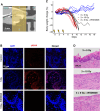
Figure 1.
Figure 1.
WSD0628 profoundly radiosensitizes mouse oral mucosa. A, A single anterior–posterior 10-mm circular x-ray beam was delivered to the oral cavity with mice positioned feet-first prone (FFP) in the irradiator. Green lines represent inferior-superior (I-S) and anterior-posterior (A-P) axes. B, Representative IF images of mouse tongue epithelial cells stained for DAPI (blue) and γH2AX foci (red). WSD0628 (7.5 mg/kg) was administered 1 hour prior to radiation, and tissue was harvested 15 minutes after radiation. DAPI, 4′,6-diamidino-2-phenylindole. C, Relative body weight changes for individual animals during and after 3 fractions of 5 Gy (solid blue), 10 Gy (black), or 5 Gy + 7.5 mg/kg WSD-0628 (red). N = 7 animals per group. Lightning bolts indicate treatment days. Mice were euthanized if they lost ≥20% body weight compared with pretreatment measurements. D, H&E stain of tongue mucosa 10 days after the first dose of treatment.
Figure 2.
Figure 2.
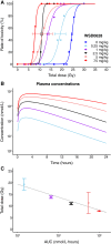
WSD0628 radiosensitization is related to drug exposure. A, Rate of oral mucosal toxicity after 3 fractions of RT combined with 0, 0.25, 1, 2.5, and 7.5 mg/kg WSD0628. N = 5–6 animals per group. B, Simulated total plasma concentration–time profiles after a single dose of WSD0628 used for the calculation of the AUC0–24. C, The resulting D50 estimated from rate of toxicity data as a function of AUC0–24 shown on a linear-log scale. D50 values are plotted with 95% confidence intervals.
Figure 3.
Figure 3.
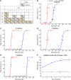
WSD0628 eliminates tissue sparing by fractionated RT in the oral mucosa. A, Fractionation schedules depicting treatment days (gray boxes) and break days (white boxes). *All treatments were delivered within 1 week except for a subset of 7 × RT + WSD0628 groups, which were treated over 8 days due to technical problems with the irradiator on day 4. Rate of oral mucosal toxicity after (B) 1, (C) 3, (D) 5, and (E) 7 fractions of RT-only (blue) and RT + 7.5 mg/kg WSD0628 (red). N = 5–6 animals per group. F, Isoeffect curves for the D50 as a function of number of fractions. D50 values are plotted with 95% confidence intervals.
Figure 4.
Figure 4.
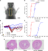
WSD0628 radiosensitization of the esophagus. A, Opposed anterior–posterior and posterior–anterior, 5 × 15 mm, x-ray fields were delivered to the thoracic esophagus. B, Relative body weight changes for individual animals following a single fraction of 12.5 Gy (solid blue), 17.5 Gy (black), or 12.5 Gy + 7.5 mg/kg WSD-0628 (red). N = 7–8 animals per group. C, H&E staining of the esophagus 10 days after the first dose of treatment. Rate of esophageal toxicity after (D) 1 and (E) 3 fractions of RT without (blue) or with 7.5 mg/kg WSD0628 (red). N = 5 animals per group. Total dose resulting in D50 plotted with 95% confidence intervals.

References

    1. García MEG, Kirsch DG, Reitman ZJ. Targeting the ATM kinase to enhance the efficacy of radiotherapy and outcomes for cancer patients. Semin Radiat Oncol 2022;32:3–14. - PMC - PubMed
    1. Durant ST, Zheng L, Wang Y, Chen K, Zhang L, Zhang T, et al. . The brain-penetrant clinical ATM inhibitor AZD1390 radiosensitizes and improves survival of preclinical brain tumor models. Sci Adv 2018;4:eaat1719. - PMC - PubMed
    1. Zimmermann A, Zenke FT, Chiu L-Y, Dahmen H, Pehl U, Fuchss T, et al. . A new class of selective ATM inhibitors as combination partners of DNA double-strand break inducing cancer therapies. Mol Cancer Ther 2022;21:859–70. - PMC - PubMed
    1. Biddlestone-Thorpe L, Sajjad M, Rosenberg E, Beckta JM, Valerie NC, Tokarz M, et al. . ATM kinase inhibition preferentially sensitizes p53-mutant glioma to ionizing radiation. Clin Cancer Res 2013;19:3189–200. - PMC - PubMed
    1. Chen J, Laverty DJ, Talele S, Bale A, Carlson BL, Porath KA, et al. . Aberrant ATM signaling and homology-directed DNA repair as a vulnerability of p53-mutant GBM to AZD1390-mediated radiosensitization. Sci Transl Med 2024;16:eadj5962. - PMC - PubMed

MeSH terms

Substances