. 2024 Nov 26;121(48):e2413018121.
doi: 10.1073/pnas.2413018121. Epub 2024 Nov 19.

The 4EHP-mediated translational repression of cGAS impedes the host immune response against DNA viruses

Affiliations

The 4EHP-mediated translational repression of cGAS impedes the host immune response against DNA viruses

Reese Jalal Ladak et al. Proc Natl Acad Sci U S A. .

Abstract

A critical host response against viral infections entails the activation of innate immune signaling that culminates in the production of antiviral proteins. DNA viruses are sensed by the cytosolic pattern recognition receptor cyclic GMP-AMP synthase (cGAS), which initiates a signaling pathway that results in production of proinflammatory cytokines such as Interferon-β (IFN-β) and activation of the antiviral response. Precise regulation of the antiviral innate immune response is required to avoid deleterious effects of its overactivation. We previously reported that the 4EHP/GIGYF2 translational repressor complex reduces the translation of Ifnb1 mRNA, which encodes IFN-β, upon RNA viral infections. Here, we report a distinct regulatory mechanism by which 4EHP controls replication of DNA viruses by translational repression of the Cgas mRNA, which encodes the DNA viral sensor cGAS. We show that 4EHP is required for effective translational repression of Cgas mRNA triggered by miR-23a. Upon infection, 4EHP deficiency bolsters the elicited innate immune response against the diverse DNA viruses Herpes simplex virus 1 (HSV-1) and Vaccinia Virus (VacV) and concomitantly reduces their rate of replication in vitro and in vivo. This study elucidates an intrinsic regulatory mechanism of the host response to DNA viruses which may provide unique opportunities for countering viral infections.

Keywords: 4EHP; DNA virus; cGAS; innate immunity; microRNA.


Conflict of interest statement

Competing interests statement:The authors declare no competing interest.

Figures
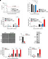
Fig. 1.
Fig. 1.
Compromised HSV-1 infection in 4EHP-depleted mice and MEFs. (A) TMX- or vehicle-treated Eif4e2flox/flox and vehicle-treated Cre+/−:Eif4e2flox/flox (collectively referred to as 4EHP+/+) and TMX-treated Cre+/−:Eif4e2flox/flox (4EHP-/-) mice (n = 8 to 12/each indicated group) were infected intravenously into the retro-orbital sinus with HSV-1 (KOS strain) and survival was monitored for up to 14 d. ***P < 0.001; log-rank test was used. (B) 2 d postinfection, mice were killed and their brain tissue was collected immediately after, homogenized, and used for plaque-forming assay to quantify viral titers. Data are presented as the logarithmic mean ± SD; n = 4, ****P < 0.0001, two-way ANOVA with Śídák’s post hoc test. (C) At 0 h and 12 h postinfection, sera were collected from WT HSV-1-infected 4EHP+/+ and 4EHP-/- littermates and subjected to ELISA to measure IFN-β protein levels. Data are presented as the mean ± SD; n = 3, ns= not significant, ****P < 0.0001, two-way ANOVA with Śídák’s post hoc test. (D) RT-qPCR analysis of Ifnb1 mRNA expression normalized to Gapdh mRNA levels in blood cells isolated from the sera collected in (C). Data are presented as the mean ± SD; n = 3, ns= not significant, ****P < 0.0001, two-way ANOVA with Śídák’s post hoc test. (E and F) WT and 4EHP-KO MEFs were infected with GFP-tagged HSV-dICP0 (KM100 strain) (MOI: 2). 6 h postinfection, viral replication was visualized via fluorescence microscopy (E; Scale bar, 400 µm) and GFP signal was quantified (F). (G) Western blot analysis of expression of the indicated proteins in HSV-dICP0-infected WT and 4EHP-KO MEFs. (H) Measurement of secreted IFN-β levels in supernatant derived from the HSV-dICP0-infected WT and 4EHP-KO MEFs in (E) by ELISA at the indicated time points. Data are presented as the mean ± SD; n = 3, ns= not significant, ****P < 0.0001, two-way ANOVA with Śídák’s post hoc test. (I) RT-qPCR analysis of Ifnb1 mRNA expression normalized to Gapdh mRNA levels in WT and 4EHP-KO MEFs 6 h post HSV-dICP0 infection. Data are presented as the mean ± SD; n = 3, ns= not significant, ***P < 0.001, two-way ANOVA with Śídák’s post hoc test.
Fig. 2.
Fig. 2.
Augmented stimulation of the cGAS-STING pathway in 4EHP-depleted cells induces Ifnb1 gene expression. (A) Western blot analysis of the indicated proteins in vehicle- and cGAMP-treated WT and 4EHP-KO BV-2 cells. (B) ELISA measurement of secreted IFN-β levels in the supernatant of WT and 4EHP-KO BV-2 cells treated with cGAMP at the indicated time points. Data are presented as the mean ± SD; n = 3, ns= not significant, ****P < 0.0001, two-way ANOVA with Śídák’s post hoc test. (C) RT-qPCR analysis of Ifnb1 mRNA expression normalized to Gapdh mRNA levels in vehicle- and cGAMP-treated WT and 4EHP-KO BV-2 cells 12 h posttreatment. Data are presented as the mean ± SD; n = 3, ns= not significant, ****P < 0.0001, two-way ANOVA with Śídák’s post hoc test. (D) shCTRL and sh4EHP THP-1 cells were cotransfected with Ifnb1-Luc (FL) and pRL-TK (RL) vectors, followed by cGAMP treatment for 12 h. Ifnb1-Luc levels were measured and normalized to pRL-TK levels. Data are presented as the mean ± SD; n = 3, *P < 0.05, unpaired student t test with Welch’s correction. (E) Western blot analysis of expression of the total and phosphorylated IRF3 (Ser396) and p65 (Ser536) in vehicle- and cGAMP-treated WT and 4EHP-KO BV-2 cells for 12 h. (F) Quantification of intensity of the indicated bands in (E) using ImageJ. Data are presented as the mean ± SD; n = 3, ns= not significant, ****P < 0.0001, two-way ANOVA with Śídák’s post hoc test. (G and H) WT and 4EHP-KO BV-2 cells were seeded on poly-L-lysine-coated slips and treated with cGAMP. 6 h posttreatment total p65 and IRF3 and phosphorylated p65 (Ser536) and IRF3 (Ser396) and levels were visualized by IF staining (G; Scale bar, 20 µm) and quantified using ImageJ (H). Data are presented as the mean ± SD; n = 3, ***P < 0.001, ****P < 0.0001, two-way ANOVA with Śídák’s post hoc test. (I) Nuclear and cytoplasmic fractions were obtained from cGAMP-treated WT and 4EHP-KO BV-2 cells and probed for total and phosphorylated IRF3 (Ser396) and p65 (Ser536).
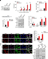
Fig. 3.
Fig. 3.
Translational repression of Cgas mRNA by 4EHP. (A) Western blot analysis of cGAS in vehicle- and cGAMP-treated WT and 4EHP-KO BV-2 cells 12 h posttreatment. (B) Quantification of band intensity in (A) using ImageJ. Data are presented as the mean ± SD; n = 3, **P < 0.01, ****P < 0.0001, two-way ANOVA with Śídák’s post hoc test. (C) RT-qPCR analysis of Cgas mRNA expression normalized to Gapdh mRNA levels in vehicle- and cGAMP-treated WT and 4EHP-KO BV-2 cells 12 h posttreatment. Data are presented as the mean ± SD; n = 3, **P < 0.01, two-way ANOVA with Śídák’s post hoc test. (D) Polysome profiling analysis of untreated WT and 4EHP-KO BV-2 cells. (E) RT-PCR analysis of Cgas mRNA abundance in each polysome fractions collected from (D). (F) Western blot analysis of expression of the indicated proteins in WT and 4EHP-KO MEFs infected with GFP-tagged HSV-dICP0 (MOI: 2) at 6 h postinfection.
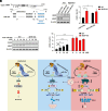
Fig. 4.
Fig. 4.
4EHP-mediated silencing of Cgas mRNA by miR-23a (A) Schematic depicting the interaction between the seed sequence of miR-23a and coding sequence (CDS) of Cgas mRNA (NM_173386.5), along with the structure of the miR-23a:Cgas mRNA complex (60). (B and C) WT and 4EHP-KO BV-2 cells were treated with 20 nM of control (miR-CTRL) and miR-23a (anti-miR-23a) antagomirs. (B) Western blot analysis of expression of the indicated proteins 12 h posttreatment. (C) Quantification of intensity of the indicated bands in (B) using ImageJ. Data are presented as the mean ± SD; n = 3, *P < 0.05, **P < 0.01, two-way ANOVA with Śídák’s post hoc test. (D) Western blot analysis of expression of the indicated proteins in WT and 4EHP-KO BV-2 cells treated with increasing concentrations of anti-miR-23a. (E) Quantification of intensity of the indicated bands in (D) using ImageJ. Data are presented as the mean ± SD; n = 3, ns= not significant, **P < 0.01, ****P < 0.0001, two-way ANOVA with Śídák’s post hoc test. (F) Schematic of the proposed model highlighting that the absence of 4EHP alleviates the miR-23a-mediated Cgas mRNA translation. Upon DNA virus infection, impaired Cgas mRNA silencing results in enhanced activation of the cGAS-STING pathway, increased phosphorylation of the IRF3 and NF-kB transcription factors, augmented Ifnb1 mRNA expression and IFN-β production. The increase in cGAS-STING signaling culminates in greater resistance to DNA virus infection in 4EHP-depleted cells.

References

    1. Li D., Wu M., Pattern recognition receptors in health and diseases. Signal Transduct. Target Ther. 6, 291 (2021). - PMC - PubMed
    1. McNab F., Mayer-Barber K., Sher A., Wack A., O’Garra A., Type I interferons in infectious disease. Nat. Rev. Immunol. 15, 87–103 (2015). - PMC - PubMed
    1. Zevini A., Olagnier D., Hiscott J., Crosstalk between cytoplasmic RIG-I and STING sensing pathways. Trends Immunol. 38, 194–205 (2017). - PMC - PubMed
    1. Rehwinkel J., Gack M. U., RIG-I-like receptors: Their regulation and roles in RNA sensing. Nat. Rev. Immunol. 20, 537–551 (2020). - PMC - PubMed
    1. Seth R. B., Sun L., Ea C. K., Chen Z. J., Identification and characterization of MAVS, a mitochondrial antiviral signaling protein that activates NF-kappaB and IRF 3. Cell 122, 669–682 (2005). - PubMed

MeSH terms

LinkOut - more resources