. 2024 Nov 26;121(48):e2409274121.
doi: 10.1073/pnas.2409274121. Epub 2024 Nov 19.

CD2 expressing innate lymphoid and T cells are critical effectors of immunopathogenesis in hidradenitis suppurativa

Affiliations

CD2 expressing innate lymphoid and T cells are critical effectors of immunopathogenesis in hidradenitis suppurativa

Mahendra Pratap Kashyap et al. Proc Natl Acad Sci U S A. .

Abstract

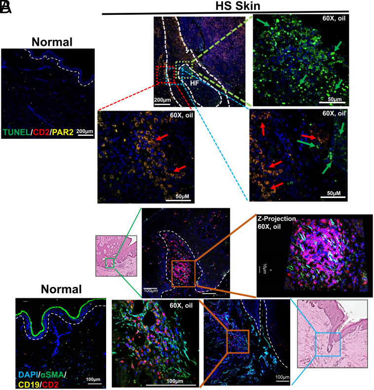
Hidradenitis suppurativa (HS) is a chronic, debilitating inflammatory skin disease with a poorly understood immunopathogenesis. Here, we report that HS lesional skin is characterized by the expansion of innate lymphocytes and T cells expressing CD2, an essential activation receptor and adhesion molecule. Lymphocytes expressing elevated CD2 predominated with unique spatial distribution throughout the epidermis and hypodermis in the HS lesion. CD2+ cells were mainly innate lymphocytes expressing the NK cell marker, CD56, and CD4+ T cells. Importantly, these CD2+ cells interacted with CD58 (LFA3) expressing epidermal keratinocytes and fibroblasts in the hypodermis. Granzyme Abright NKT cells (CD2+CD3+CD56bright) clustered with α-SMA expressing fibroblasts juxtaposed to epithelialized tunnels and fibrotic regions of the hypodermis. Whereas NK cells (CD2+CD56dim) were perforin+, granzymes A+ and B+, and enriched adjacent to hyperplastic follicular epidermis and tunnels of HS showing presence of apoptotic cells. The cytokines IL-12, IL-15, and IL-18, which enhance NK cell maturation and function were significantly elevated in HS. Ex vivo HS skin explant cultures treated with CD2:CD58 interaction-blocking anti-CD2 monoclonal antibody attenuated secretion of inflammatory cytokines/chemokines and suppressed inflammatory gene signature. Additionally, CD2:CD58 blockade altered miRNAs involved in NK/NKT differentiation and/or function. In summary, we show that a cellular network of heterogenous NKT and NK cell populations drives inflammation and is critical in the pathobiology of HS, including tunnel formation and fibrosis. Finally, CD2 blockade is a viable immunotherapeutic approach for the effective management of HS.

Keywords: CD2; CD4 T cell; Hidradenitis suppurativa; NK cell; NKT cell.


Conflict of interest statement

Competing interests statement:E.B. and D.B. are co-founders of ITB-MED. Patent application number: PCT/US2023/022364.

Figures

Fig. 1.
Fig. 1.
Innate and adaptive immune and nonimmune cells in HS lesion. (A) UMAP of cell populations from normal (NS; n = 4) and HS (n = 6) skin. The scRNAseq dataset from 27,442 cells were clustered using Seurat 4.0 and annotated to 19 clusters of immune and nonimmune cell populations. (B) Proportion of annotated cell population in each cluster from NS and HS. (C) Dot plot depicting normalized expression of genes defining selected cell lineage/function in NS and HS defining genes. (D) Feature plot showing cluster localization of selected cell lineage/function-defining genes. (E) Volcano plot of differentially expressed genes in HS (n = 8) relative to NS (n = 6 to 8) determined using qPCR arrays.
Fig. 2.
Fig. 2.
CD2-expressing lymphocytes predominate in HS lesional skin. IHC of serial sections of HS skin stained with antibodies to CD2 (A), CD3 (B), CD4 (C), CD8β (D), CD56 (E), and CD20 (F). The red scale bar in each micrograph is 300 μm. Micrographs for controls are shown in SI Appendix, Fig. S6. Additional details in SI Appendix.
Fig. 3.
Fig. 3.
NKT and NK cells are spatially distributed in different regions of HS Skin. (A) Immunofluorescence staining for CD3, CD56, and CD2 (Upper) and CD2, CD3, and CD16 expressing cells (Lower) in NS and HS. Boxes in H&E “stitched” image (Center) indicate areas of epidermis/hypodermis or tunnel region in HS as depicted in the immunofluorescent images. (Scale bar, 100 µm.) Arrows point to CD2+CD3+CD56bright (NKT cells) and CD2+CD3CD56dim (mature NK cells) in the epidermal/hypodermal and tunnel regions. T cells in the images are CD2+CD3brightCD56. Tunnels show spatially separated NK cells from clusters of NKT cells. E, epidermis and D, dermis/hypodermis (B) Violin plots of absolute numbers of CD56+, CD2+, CD3+, and CD16+ cells in epidermal/hypodermal and tunnel regions. 20× resolution confocal images (minimum 20/section) were acquired and numbers of cells determined in each image using CellSense software. Each dot in each violin plot represents numbers of cells in an image; P values as indicated are from one-way ANOVA nonparametric analysis. (C) Lower panels show CD2+ cells interacting with CD58+ (keratinocyte) cells. Data are representative of HS (n = 3) and NS (n = 3).
Fig. 4.
Fig. 4.
Heterogenous populations of NKT and NK cells in HS. Perforin (A), Granzyme A (B), and CD11b and granzyme B expression (C), in NKT (CD3+CD2+CD56bright) and NK (CD2+CD56dim) cells in NS (Top row) and HS (Middle and Lower), bar is 100 μm. (A) Perforin+ NKT and NK cells are prominent in the epidermis/dermis (E/D), tunnel region (deep-D). (B) Granzyme A is expressed in NKT and NK cells. (C) Some NK cells (CD56dim), but not NKT cells in HS express CD11b and expression of granzyme B is primarily in NK cells. (D) UMAP of 11 immune cell subclusters extracted from scRNA-seq analysis shown in Fig. 1A. (E) Expression of NKT, NK, MAIT, CD4, CD8 associated phenotypic markers and functional genes in each subcluster. (F) Pseudotime trajectory analysis of subclusters with T Naïve-2 as the starting point. Dark purple is initial time and orange is the farthest time in pseudotime scale. (G) The pseudotime trajectory involved gene expression heatmap. (H) The expression pattern of top 10 trajectory-determining genes.
Fig. 5.
Fig. 5.
NKT and NK cells are associated with pathological processes in HS. (A) Localization of NKT/NK cell populations (CD2+) in regions of ongoing apoptosis (TUNEL+). Immunofluorescence staining shows PAR2 expressing CD2+ lymphoid cells adjacent to cells undergoing apoptosis (TUNEL+) within sinus tracts/modified hair follicles of HS. (B) NKT cells interacting with fibroblasts in areas of ongoing fibrosis in HS. Z-projected micrographs show CD2-expressing NKT cells physically interacting with αSMA expressing fibroblasts within modified hair follicles. Very few CD19+ cells are observed in regions of ongoing fibrosis.
Fig. 6.
Fig. 6.
CD2 blockade in HS organotypic skin culture attenuates proinflammatory and fibrotic signaling pathways. Skin tissue from HS skin (n = 3) was cultured in transwell plates with anti-CD2 or control IgG antibodies (A, B, and C) or LPS with anti-CD2 or control IgG (D and E) for 72 h. Cytokine, chemokine, and growth factor levels in supernates of explant cultures (A and D). Student’s t test of IgG vs. anti-CD2 (*P ≤ 0.05, **P ≤ 0.01). Volcano plots (B and E) depicting changes in the expression of inflammatory gene signatures in anti-CD2 treated explants. Disruption of CD2:CD5 interaction in HS skin explant cultures down-modulates the expression of several inflammatory and fibrotic marker genes at the transcriptional level. TUNEL staining of explant tissue cultured with IgG or anti-CD2 from three HS patients (C). Graph is quantification of apoptotic cells (TUNEL+) in explants of each HS patient and change in numbers between IgG and anti-CD2 treatment. Unpaired Student’s t test with Welch’s correction was used to identify statistical significance (P ≤ 0.05) between IgG vs. anti-CD2.

References

    1. Sabat R., et al. , Hidradenitis suppurativa. Nat. Rev. Dis. Primers 6, 18 (2020). - PubMed
    1. von Laffert M., et al. , Hidradenitis suppurativa (acne inversa): Early inflammatory events at terminal follicles and at interfollicular epidermis. Exp. Dermatol. 19, 533–537 (2010). - PubMed
    1. Jfri A., et al. , Prevalence of Hidradenitis suppurativa: A systematic review and meta-regression analysis. JAMA Dermatol. 157, 924–931 (2021). - PMC - PubMed
    1. Kashyap M. P., et al. , Advances in molecular pathogenesis of hidradenitis suppurativa: Dysregulated keratins and ECM signaling. Semin. Cell Dev. Biol. 128, 120–129 (2022). - PMC - PubMed
    1. Byrd A. S., et al. , Neutrophil extracellular traps, B cells, and type I interferons contribute to immune dysregulation in hidradenitis suppurativa. Sci. Transl. Med. 11, eaav5908 (2019). - PMC - PubMed

MeSH terms