. 2024 Nov 19;50(1):21.
doi: 10.1007/s11064-024-04270-1.

The P38MAPK Pathway Mediates the Destruction of the Blood-Brain Barrier in Anti-NMDAR Encephalitis Mice

Affiliations

The P38MAPK Pathway Mediates the Destruction of the Blood-Brain Barrier in Anti-NMDAR Encephalitis Mice

Dayuan Lao et al. Neurochem Res. .

Abstract

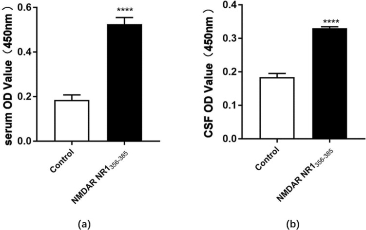
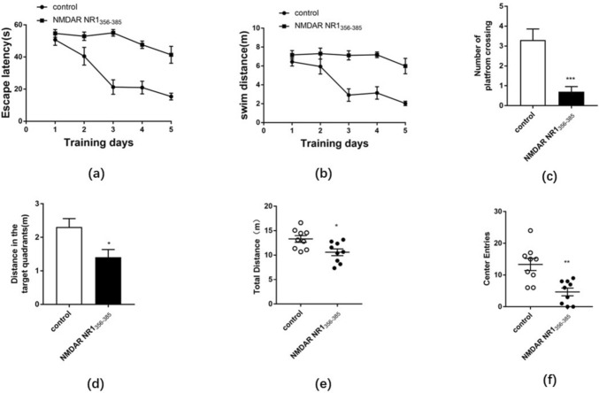
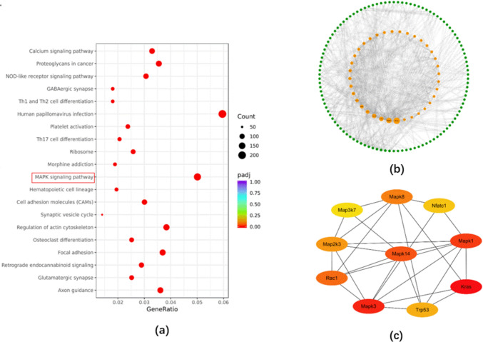
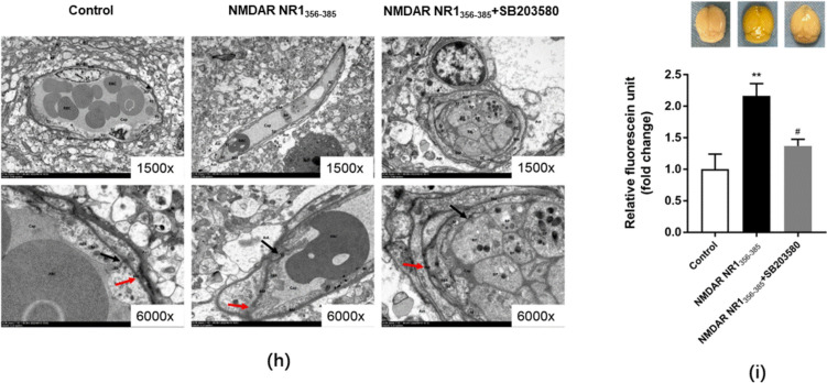
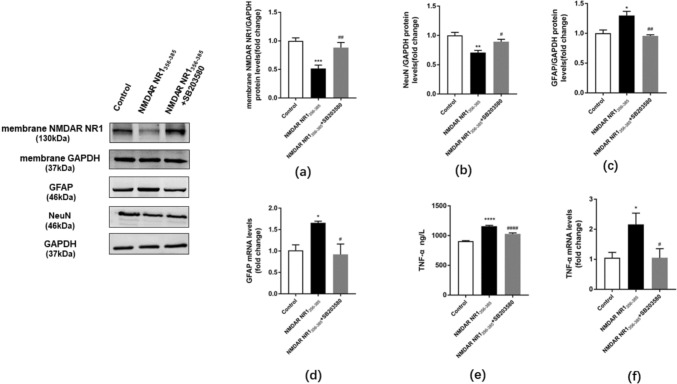
The clinical manifestations of anti-N-methyl-D-aspartate receptor (NMDAR) encephalitis may be closely related to the integrity of the blood-brain barrier (BBB). The P38 mitogen-activated protein kinase (P38MAPK) pathway plays a protective role in neurodegenerative diseases. However, whether the P38MAPK pathway is involved in the underlying mechanism of tight junction (TJ) protein disruption and neuronal damage has not been elucidated. Therefore, in this study, a mouse model of anti-NMDAR encephalitis was established by active immunization with NMDAR NR1356-385 peptides. The critical pathways of P38MAPK were screened by interaction network and co-enrichment analysis. The role of P38MAPK pathways was investigated by the injection of P38MAPK inhibitor SB203580 (10 mg/kg, i.p.). Compared with the control group, the expression of occludin and zonula occludens (ZO)-1 in NMDAR NR1356-385 group mice was downregulated, and the structure and function of BBB were damaged. However, after the intervention of SB203580, the activation of the P38MAPK was inhibited, the expression of matrix metalloproteinase 9 (MMP9) was reduced, and the function of BBB was improved. Meanwhile, inhibiting the P38MAPK pathway reversed the degradation of NMDAR NR1, while reducing the expression of the glial fibrillary acidic protein (GFAP) and pro-inflammatory factor tumor necrosis factor (TNF-α). It also relieved the damage of neuron-specific nucleus (NeuN), thus alleviating psychobehavioral symptoms. In conclusion, our results suggested that the P38MAPK pathway is involved in BBB destruction and neurobehavioral change in mice with anti-NMDAR encephalitis. Targeting the P38MAPK pathway may be a promising option for the treatment of anti-NMDAR encephalitis.

Keywords: Blood–Brain barrier; Matrix metalloproteinase 9; N-Methyl-D-aspartate receptor encephalitis; P38MAPK pathway; Tight junction protein.


Conflict of interest statement

Declarations Conflict of interest The authors report there are no competing interests to declare. Ethical Approval Animal experiments in this study have been approved by the Animal Experimentation Ethics Committee of Guangxi Medical University (Certificate Numbers: 202010031). The experiments were performed and reported in accordance with the ARRIVE guideline.

Figures

Fig. 1
Fig. 1
Diagram of the hypothesis of the P38MAPK pathway mediating blood–brain barrier disruption and neurobehavioral change in the anti-NMDAR encephalitis mouse model
Fig. 2
Fig. 2
Production of anti-NMDAR antibodies in serum and CSF of mice immunized with the NMDAR NR1356-385 peptides. a-b serum and CSF were collected from mice, and anti-NMDAR antibody production was detected by ELISA in the NMDAR NR1356-385 group. Values are expressed as mean ± SEM (n = 6). ****p < 0.0001 versus the control group
Fig. 3
Fig. 3
Behavioral changes in mice immunized with NMDAR NR1356-385 peptide. a-b The escape latency (s) and swimming distance (m) during the acquired training period of MWM. c-d The number of platform crossings and distance in the target quadrants during the training period of MWM exploration. ef Total distance traveled (m) and crossing frequency of the central area of the mice during the 5-min test in the open field. Compared to the control group, the NMDAR NR1356-385 group showed memory impairment and reduced exploration activity. Values are expressed as mean ± SEM (n = 10). * p < 0.05, ** p < 0.01, *** p < 0.001 versus the control group
Fig. 4
Fig. 4
Identification of DEGs and GO enrichment analysis. (n = 3). a DEGs volcano plot of the transcriptome between the control and NMDAR NR1356-385 groups. Upregulated genes were shown in red and downregulated genes were shown in green (padj < 0.01). b Top 10 enriched GO terms for BP, CC, and MF sorted by padj value
Fig. 5
Fig. 5
KEGG enrichment analysis and identification of hub genes (n = 3). a Top 20 of 153 significantly enriched KEGG pathways. GeneRatio represented the ratio of enriched DEGs to total DEGs. The size and color of the dots indicated the count and significance of DEGs enriched in KEGG pathways, respectively. b PPI network analysis of 175 DEGs based on string database. c The top 10 hub genes in the network were arranged using cytoHubba's betweenness method
Fig. 6
Fig. 6
Inhibition of P38MAPK activation affected occludin, ZO-1 expression, and the integrity of BBB in anti-NMDAR encephalitis mice. a-d The relative protein and mRNA expression level of occludin and ZO-1 in brain tissue was detected by western blot and qRT-PCR. e Representative immunofluorescence image of colocalization of occludin (green) and ZO-1 (green) with CD31(red) in brain tissue (Scale bar: 50 μm). f-g Quantitative analysis of immunofluorescence intensity of occludin and ZO-1 relative to CD31. Compared with the control group, the expression levels of occludin and ZO-1 were downregulated in the NMDAR NR1356-385 group. NMDAR NR1356-385 + SB203580 group significantly increased the expression levels of occludin and ZO-1. h The ultrastructural changes of brain microvascular endothelial cells were observed by TEM (original magnification 1500 and 6000). The results showed that the basement membrane of the BBB in the control group was continuous and intact. In the NMDAR NR1356-385 group, the local structure of the basement membrane was blurred and fractured. Compared with the NMDAR NR1356-385 group, SB203580, a P38MAPK inhibitor, could reduce the structural damage of the BBB. i The permeability of the BBB detected with sodium fluorescein. Compared with the control group, the brain tissue of the NMDAR NR1356-385 group was yellow and the amount of sodium fluorescein exudation was significantly increased. Compared with the NMDAR NR1356-385 group, the yellow staining on the brain surface of the NMDAR NR1356-385 group + SB203580 group was lighter and the amount of sodium fluorescein exudation was decreased. Values are expressed as mean ± SEM (n ≥ 3). *p < 0.05, **p < 0.01, ***p < 0.001, ****p < 0.0001 versus control group; #p < 0.05, ##p < 0.01, ####p < 0.0001 versus NMDAR NR1356-385 group
Fig. 6
Fig. 6
Inhibition of P38MAPK activation affected occludin, ZO-1 expression, and the integrity of BBB in anti-NMDAR encephalitis mice. a-d The relative protein and mRNA expression level of occludin and ZO-1 in brain tissue was detected by western blot and qRT-PCR. e Representative immunofluorescence image of colocalization of occludin (green) and ZO-1 (green) with CD31(red) in brain tissue (Scale bar: 50 μm). f-g Quantitative analysis of immunofluorescence intensity of occludin and ZO-1 relative to CD31. Compared with the control group, the expression levels of occludin and ZO-1 were downregulated in the NMDAR NR1356-385 group. NMDAR NR1356-385 + SB203580 group significantly increased the expression levels of occludin and ZO-1. h The ultrastructural changes of brain microvascular endothelial cells were observed by TEM (original magnification 1500 and 6000). The results showed that the basement membrane of the BBB in the control group was continuous and intact. In the NMDAR NR1356-385 group, the local structure of the basement membrane was blurred and fractured. Compared with the NMDAR NR1356-385 group, SB203580, a P38MAPK inhibitor, could reduce the structural damage of the BBB. i The permeability of the BBB detected with sodium fluorescein. Compared with the control group, the brain tissue of the NMDAR NR1356-385 group was yellow and the amount of sodium fluorescein exudation was significantly increased. Compared with the NMDAR NR1356-385 group, the yellow staining on the brain surface of the NMDAR NR1356-385 group + SB203580 group was lighter and the amount of sodium fluorescein exudation was decreased. Values are expressed as mean ± SEM (n ≥ 3). *p < 0.05, **p < 0.01, ***p < 0.001, ****p < 0.0001 versus control group; #p < 0.05, ##p < 0.01, ####p < 0.0001 versus NMDAR NR1356-385 group
Fig. 7
Fig. 7
Inhibition of the P38MAPK pathway affected MMP9 expression in anti-NMDAR encephalitis mice. a The relative protein expression levels of p-P38MAPK/P38MAPK in brain tissue were detected by western blot. b-c The relative protein and mRNA expression level of MMP9 in brain tissue was detected by western blot and qRT-PCR. The relative expressions of p-P38MAPK, MMP9 protein, and mRNA in the brain tissue of mice were increased after immunization. Intervention with SB203580 attenuated this effect. Values are expressed as mean ± SEM (n ≥ 3). *p < 0.05, ***p < 0.001, versus control group; #p < 0.05, ###p < 0.001 versus NMDAR NR1356-385 group
Fig. 8
Fig. 8
Inhibition of P38MAPK activation altered NMDAR NR1/GFAP/TNF-α/NeuN expressions in anti-NMDAR encephalitis mice. a The relative membrane protein expression level of NMDAR NR1 in brain tissue was determined by western blot. Compared with the control group, the relative expression level of NMDAR NR1 membrane protein in the brain tissue of anti-NMDAR encephalitis mice was downregulated, while the relative expression level was upregulated after the SB203580 intervention. b Western blot determined the relative protein expression levels of NeuN in brain tissue. c-d The relative protein and mRNA expression levels of GFAP in brain tissue were detected by western blot and qRT-PCR. e ELISA detected the relative concentration of TNF-α serum in mice. f The relative mRNA expression level of TNF-α in brain tissue was detected by qRT-PCR. Compared with the control group, the protein and mRNA expression levels of GFAP and TNF-α were increased and the protein expression level of NeuN was decreased in anti-NMDAR encephalitis mice. However, inhibition of P38MAPK activation reversed these changes. Values are expressed as mean ± SEM (n ≥ 3). *p < 0.05, **p < 0.01, ***p < 0.001,****p < 0.0001 versus control group; #p < 0.05, ##p < 0.01, ####p < .0001 versus NMDAR NR1356-385 group
Fig. 9
Fig. 9
Inhibition of P38MAPK neurological dysfunction in anti-NMDAR encephalitis mice. a-b The escape latency (s) and swimming distance (m) during the acquired training period of MWM. c-d The number of platform crossings and distance in the target quadrants during the training period of MWM exploration. ef Total distance traveled (m) and crossing frequency of the central area of the mice during the 5-min test in the open field. The NMDAR NR1356-385 group showed memory impairment and decreased exploratory activity compared to the control group. Compared with the NMDAR NR1356-385 group, the NMDAR NR1356-385 + SB203580 group showed increased exploratory activity time in mice. Values are expressed as mean ± SEM (n = 6). ***p < 0.001, ****p < 0.0001 versus control group; ###p < 0.001, ####p < 0.0001 versus NMDAR NR1356-385 group

References

    1. Moscato EH, Peng XY, Jain A et al (2014) Acute mechanisms underlying antibody effects in anti-N-methyl-D-aspartate receptor encephalitis. Ann Neurol 76:108–119 - PMC - PubMed
    1. Dalmau J, Tüzün E, Wu H-Y et al (2007) Paraneoplastic anti-N-methyl-D-aspartate receptor encephalitis associated with ovarian teratoma. Ann Neurol 61:25–36 - PMC - PubMed
    1. Franchini L, Carrano N, Di Luca M et al (2020) Synaptic GluN2A-containing NMDA receptors: from physiology to pathological synaptic plasticity. Int J Mol Sci 21:1538 - PMC - PubMed
    1. Kadry H, Noorani B, Cucullo L (2020) A blood–brain barrier overview on structure, function, impairment, and biomarkers of integrity. Fluids and Barriers of the CNS 17:69 - PMC - PubMed
    1. Sweeney MD, Zhao Z, Montagne A et al (2019) Blood-brain barrier: from physiology to disease and back. Physiol Rev 99:21–78 - PMC - PubMed

MeSH terms

LinkOut - more resources