. 2024 Nov 19;20(11):e1011461.
doi: 10.1371/journal.pgen.1011461. eCollection 2024 Nov.

Cell non-autonomous signaling through the conserved C. elegans glycoprotein hormone receptor FSHR-1 regulates cholinergic neurotransmission

Affiliations

Cell non-autonomous signaling through the conserved C. elegans glycoprotein hormone receptor FSHR-1 regulates cholinergic neurotransmission

Morgan Buckley et al. PLoS Genet. .

Abstract

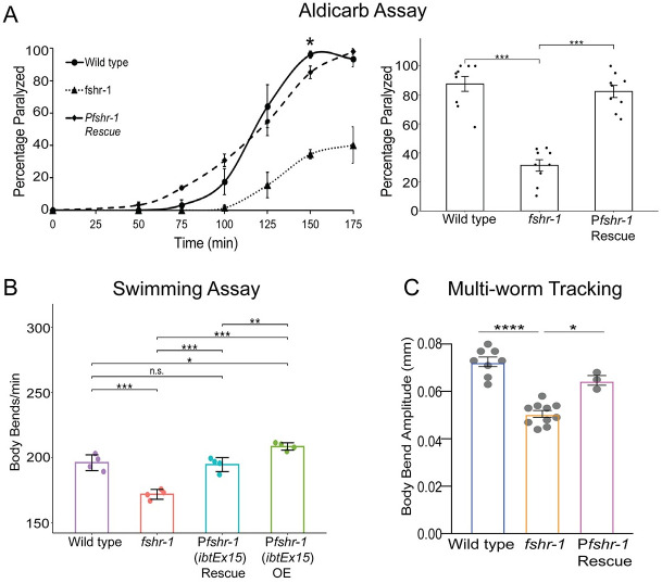
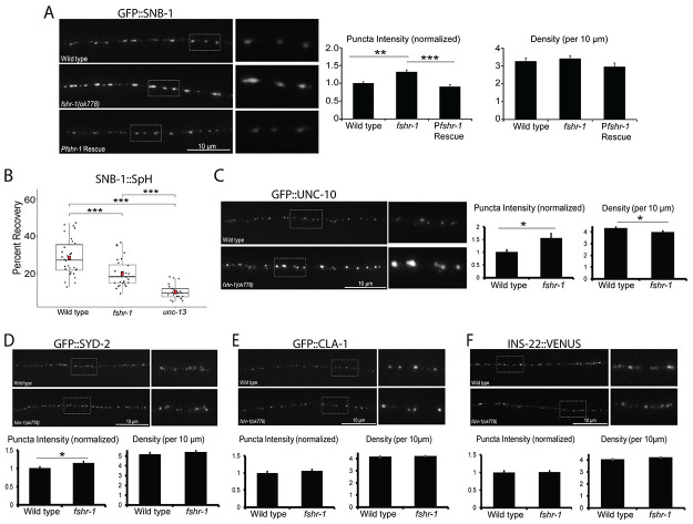
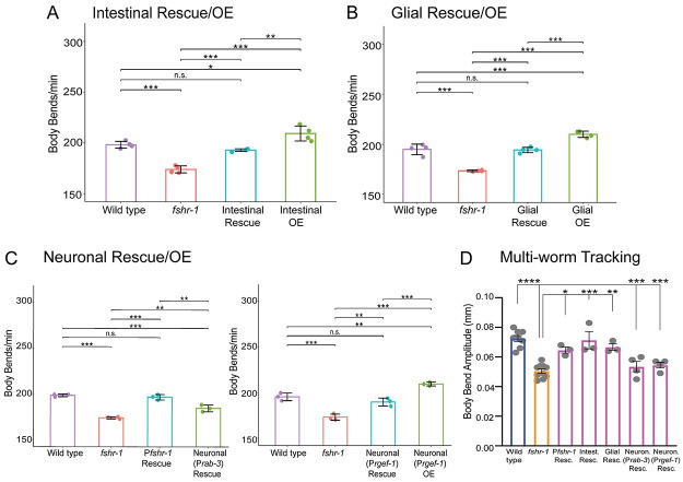
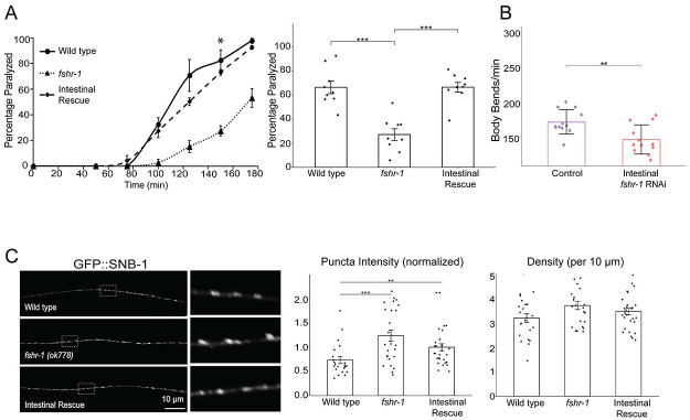
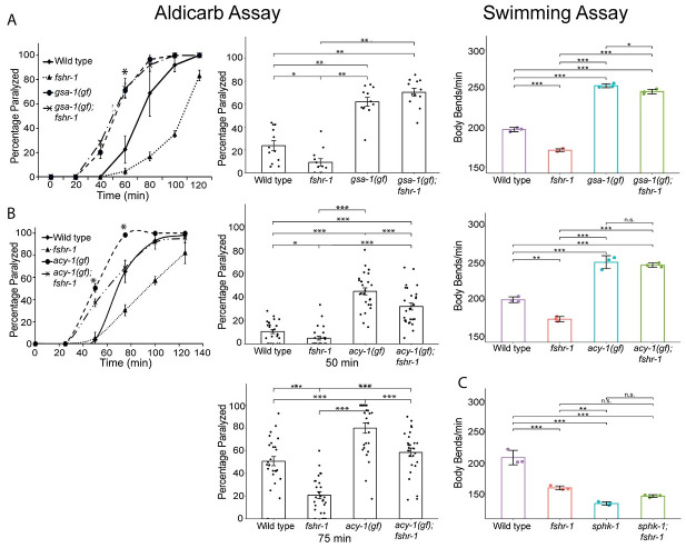
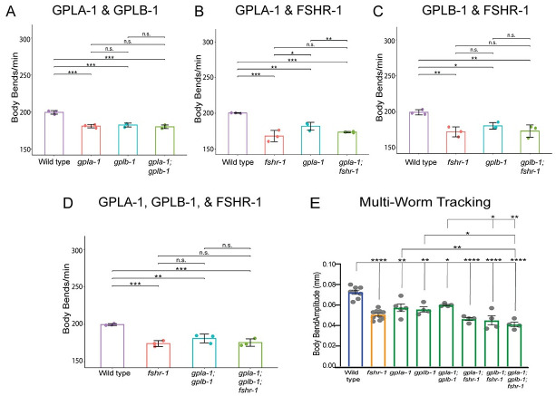
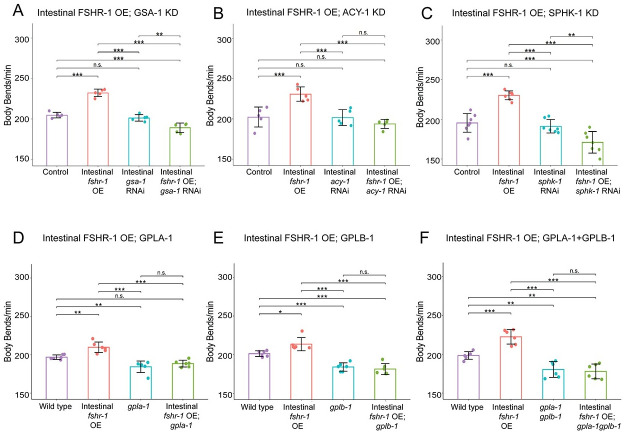
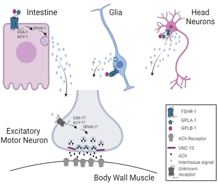
Modulation of neurotransmission is key for organismal responses to varying physiological contexts such as during infection, injury, or other stresses, as well as in learning and memory and for sensory adaptation. Roles for cell autonomous neuromodulatory mechanisms in these processes have been well described. The importance of cell non-autonomous pathways for inter-tissue signaling, such as gut-to-brain or glia-to-neuron, has emerged more recently, but the cellular mechanisms mediating such regulation remain comparatively unexplored. Glycoproteins and their G protein-coupled receptors (GPCRs) are well-established orchestrators of multi-tissue signaling events that govern diverse physiological processes through both cell-autonomous and cell non-autonomous regulation. Here, we show that follicle stimulating hormone receptor, FSHR-1, the sole Caenorhabditis elegans ortholog of mammalian glycoprotein hormone GPCRs, is important for cell non-autonomous modulation of synaptic transmission. Inhibition of fshr-1 expression reduces muscle contraction and leads to synaptic vesicle accumulation in cholinergic motor neurons. The neuromuscular and locomotor defects in fshr-1 loss-of-function mutants are associated with an underlying accumulation of synaptic vesicles, build-up of the synaptic vesicle priming factor UNC-10/RIM, and decreased synaptic vesicle release from cholinergic motor neurons. Restoration of FSHR-1 to the intestine is sufficient to restore neuromuscular activity and synaptic vesicle localization to fshr-1-deficient animals. Intestine-specific knockdown of FSHR-1 reduces neuromuscular function, indicating FSHR-1 is both necessary and sufficient in the intestine for its neuromuscular effects. Re-expression of FSHR-1 in other sites of endogenous expression, including glial cells and neurons, also restored some neuromuscular deficits, indicating potential cross-tissue regulation from these tissues as well. Genetic interaction studies provide evidence that downstream effectors gsa-1/GαS, acy-1/adenylyl cyclase and sphk-1/sphingosine kinase and glycoprotein hormone subunit orthologs, GPLA-1/GPA2 and GPLB-1/GPB5, are important for intestinal FSHR-1 modulation of the NMJ. Together, our results demonstrate that FSHR-1 modulation directs inter-tissue signaling systems, which promote synaptic vesicle release at neuromuscular synapses.


Conflict of interest statement

The authors have declared that no competing interests exist.

Figures

Fig 1
Fig 1. fshr-1 is required for neuromuscular function in multiple assays.
(A) Aldicarb paralysis assays, (B) swimming assays, and (C) multi-worm tracking assays were performed on wild type worms, fshr-1(ok778) mutants, rescued animals (Rescue) re-expressing fshr-1 under the endogenous fshr-1 promoter (Pfshr-1, ibtEx15) in the fshr-1 mutant background, and over-expression (OE) animals expressing fshr-1 under the endogenous fshr-1 promoter (Pfshr-1, ibtEx15). (A) (Left panel) Representative aldicarb assays showing the mean percentage of worms paralyzed on 1mM aldicarb ± s.e.m. for n = 3 plates of approximately 20 young adult animals each per strain. (Right panels) Bar graphs showing cumulative mean data ± s.e.m. pooled from 3–4 independent experiments for worms paralyzed at the timepoint indicated by an asterisk (*) in the left panel. Scatter points show individual plate averages. Statistical significance of the data was analyzed using a one-way ANOVA and Tukey’s post hoc test or a Wilcoxon Rank Sum test followed by a Steel-Dwass multiple comparison analysis, as appropriate. (B) Mean body bends per minute ± S.D. obtained in swimming experiments. Each scatter point represents an independent experiment testing 30 animals per genotype. (C) Mean body bend amplitude ± S.D. obtained from multi-worm tracking experiments. Each scatter point represents an independent experiment from a population average of 20 animals. Statistical significance of the data was analyzed using a one-way ANOVA with Tukey’s multiple comparison. For A-C, results of analyses for which p ≤ 0.05 are indicated by horizontal lines above the bars. *p ≤ 0.05, **p ≤ 0.01, ***p ≤ 0.001, **** p ≤ 0.0001, n.s., not significant.
Fig 2
Fig 2. fshr-1 mutants have decreased synaptic vesicle release accompanied by accumulation of some synaptic vesicle and active zone proteins.
(A) Wild type worms, fshr-1(ok778) mutants, and fshr-1 mutants re-expressing fshr-1 genomic DNA under the endogenous fshr-1 promoter (Pfshr-1, agEx43) that also expressed GFP::SNB-1 in cholinergic (ACh) neurons were imaged using a 100x objective. (Left panel) Representative images of the dorsal nerve cords halfway between the vulva and the tail of young adult animals. Boxed areas are shown in higher resolution to the right of the main images. (Right panels) Quantification of puncta (synaptic) intensity and puncta density (per 10 μm) ± s.e.m. Puncta intensity is shown normalized to wild type. For (A), n = 24 animals imaged for wild type, n = 31 for fshr-1, and n = 26 for Pfshr-1 rescue. (B) Percent recovery of SNB-1::Superecliptic pHluorin (SpH) fluorescence at ACh motor neuron presynapses following photobleaching in wild type, fshr-1(ok778), and fusion-defective unc-13(se69) animals. For wild type, n = 30 animals; for fshr-1, n = 28; for unc-13, n = 21. (C-F) Wild type or fshr-1(ok778) mutant animals that also expressed (C) GFP::UNC-10, (D) GFP::SYD-2, (E) GFP::CLA-1, or (F) INS-22::VENUS in ACh neurons were imaged using a 100x objective. (Upper panels) Representative images of the dorsal nerve cords halfway between the vulva and the tail of young adult animals. (Lower panels) Quantification of normalized puncta (synaptic) intensity and puncta density (per 10 μm) ± s.e.m. For (C), n = 25 animals imaged for wild type, n = 24 for fshr-1. For (D), n = 31 for wild type, n = 35 for fshr-1. For (E), n = 30 for wild type, n = 31 for fshr-1. For (F), n = 31 for wild type, n = 35 for fshr-1. One-way ANOVA followed by Tukey’s post hoc tests were used to compare the means of the datasets in A and B; Student’s t tests were used to compare datasets in C-F. *p ≤ 0.05, **p ≤ 0.01, ***p ≤ 0.001. Upper two images and numeric data in (A) originally published in modified format in Hulsey-Vincent et al., 2023a [63].
Fig 3
Fig 3. fshr-1 re-expression in multiple distal tissues is sufficient to restore neuromuscular signaling to fshr-1(lf) mutants.
(A-C) Swimming assays and (D) multi-worm tracking experiments were performed on wild type worms, fshr-1(ok778) mutants, and animals re-expressing fshr-1 under either an intestinal promoter (A, D; Pges-1, ibtEx35), a pan-glial promoter (B, D; Pmir-228, ibtEx51) or a pan-neuronal promoter (C, D; Prab-3, ibtEx34 or Prgef-1, ibtEx67) in either a wild type (OE, overexpression) or fshr-1 mutant background (Rescue/Resc.). (A-C) Mean body bends per minute ± S.D. obtained in swimming experiments. Each scatter point represents an independent experiment testing 30 animals per genotype. (D) Mean body bend amplitude ± S.D. obtained from multi-worm tracking experiments. Each scatter point represents an independent experiment from a population average of 20 animals. Statistical significance of the data was analyzed using a one-way ANOVA with Tukey’s multiple comparison. Colors on the bars indicate strain groups as follows: Blue = wild type, yellow = fshr-1 mutants; magenta = fshr-1 rescue strains. For all experiments, one-way ANOVA and Tukey’s post hoc tests were used to compare the means of the datasets (*p ≤ 0.05, ** p ≤ 0.01, ***p 0.001; **** p ≤ 0.0001, n.s., not significant; in (D), asterisks above the two Neuron Rescue strains indicate statistically significant differences from wild type worms, whereas asterisks above the Pfshr-1, Intestinal Rescue, and Glial Rescue bars indicate differences from fshr-1 mutants).
Fig 4
Fig 4. fshr-1 expression in the intestine is necessary and sufficient for neuromuscular function and structure.
(A, C) Intestinal rescue (Pges-1, ibtEx35) and (B) Intestine-specific RNAi [Pnhx-2::rde-1; rde-1(ne219)] effects on fshr-1 neuromuscular phenotypes. (A) (Left panel) Representative aldicarb assays showing the mean percentage of wild type, fshr-1(ok778) mutant, and intestinal rescue worms paralyzed on 1mM aldicarb ± s.e.m. for n = 3 plates of approximately 20 young adult animals per strain. (Right panel) Bar graphs showing cumulative data ± s.e.m.pooled from 3 independent aldicarb experiments for worms paralyzed at the timepoint indicated by an asterisk (*) in the left panel. Scatter points show individual plate averages. (B) Mean body bends per minute ± S.D. obtained in swimming experiments done on L4440 vector only-treated (Control) or fshr-1 RNAi-treated worms (Intestinal fshr-1 RNAi). (C) Wild type, fshr-1(ok778) mutant, and intestinal rescue worms that also expressed GFP::SNB-1 in ACh neurons were imaged using a 100x objective. (Left panels) Representative images of the dorsal nerve cords halfway between the vulva and the tail of young adult animals. Boxed areas are shown in higher resolution to the right of the main images. (Right panels) Quantification of normalized puncta (synaptic) intensity and puncta density (per 10 μm) ± s.e.m. Scatter points show individual worm means (n = 22–29 animals per genotype). One-way ANOVA and Tukey’s post hoc were used to compare the means of the datasets; *p ≤ 0.05, **p ≤ 0.01, ***p ≤ 0.001, **** p ≤ 0.0001, n.s., not significant.
Fig 5
Fig 5. gsa-1(gf), acy-1(gf), and sphk-1(lf) mutations suppress fshr-1(lf) aldicarb phenotypes consistent with a downstream function.
Aldicarb paralysis and swimming assays were performed on wild type worms, fshr-1(ok778) mutants, and (A) gsa-1(ce81) or (B) acy-1(md1756) gain-of-function (gf) mutants, or (C) sphk-1(ok1097) loss-of-function mutants, along with their respective double mutants (gsa-1;fshr-1, acy-1;fshr-1, or sphk-1;fshr-1). (Left panels) Representative aldicarb assays showing the mean percentage of worms paralyzed on 1mM aldicarb ± s.e.m. for n = 3 plates of approximately 20 young adult animals each per strain. (Center panels) Bar graphs show cumulative data ± s.e.m. pooled from (A) 4 or (B) 8–9 independent aldicarb experiments for worms paralyzed at the timepoint indicated by an asterisk (*) in the upper panels. Scatter points show individual plate averages. Statistical significance of the data was analyzed using a Wilcoxon Rank Sum test followed by a Steel-Dwass multiple comparison analysis, as appropriate. Results of analyses for which p ≤ 0.05 are indicated by horizontal lines above the bars. (Right panels) Mean body bends per minute ± S.D. obtained in swimming experiments. Each scatter point represents an independent experiment testing 30 animals per genotype, analyzed by one-way ANOVA and Tukey’s post hoc test, *p ≤ 0.05, **p ≤ 0.01, ***p ≤ 0.0001, n.s., not significant.
Fig 6
Fig 6. GLPA-1/FLR-2/GPA2 and GPLB/GB5 glycoprotein act in a common genetic pathway with FSHR-1 at the NMJ.
(A-D) Mean body bends per minute ± S.D. obtained in swimming experiments testing fshr-1 and α and β glycoprotein ligand mutants [fshr-1(ok778), gpla-1(ibt1) α, gplb-1 (ibt4) β worms] or combinations of double and triple mutants in these genes. Each scatter point represents an independent experiment testing 30 animals per genotype. (E) Mean body bend amplitude obtained from multi-worm tracking experiments. Each scatter point represents an independent experiment with a population average of 20 animals. Colors indicate groups of mutants as follows: blue = wild type, yellow = fshr-1; green = glycoprotein mutants. For all experiments, one-way ANOVA and Tukey’s post hoc tests were used to compare the means of the datasets (*p ≤ 0.05, **p ≤ 0.01, ***p ≤ 0.001; ****p ≤ 0.0001; n.s., not significant).
Fig 7
Fig 7. Regulation of neuromuscular function by intestinal FSHR-1 requires intestinally expressed GSA-1, ACY-1, AND SPHK-1, as well as the GLPA-1 and GPLB glycoproteins.
(A-C) Mean body bends per minute ± S.D. obtained in swimming experiments testing intestinal RNAi sensitized [Pnhx-2::rde-1; rde-1(ne219)] control-treated animals overexpressing fshr-1 in the intestine (Pges-1, ibtEx35, Intestinal fshr-1 OE), treated with feeding RNAi targeting (A) gsa-1, (B) acy-1, or (C) sphk-1, or animals with intestinal FSHR-1 overexpression and gsa-1, acy-1, or sphk-1 RNAi compared to L4440 empty vector-treated worms (Control). Each scatter point represents an independent experiment testing 10 animals per genotype. (D-F) Mean body bends per minute ± S.D. obtained in swimming experiments testing animals overexpressing fshr-1 in the intestine (Pges-1, ibtEx35), (D) gpla-1(ibt1), (E) gplb-1(itb4), or (F) gpla-1gplb-1 mutations compared to animals with both intestinal fshr-1 overexpression and glycoprotein mutation, and wild type controls. Each scatter point represents an independent experiment testing 30 animals per genotype. For all experiments, one-way ANOVA and Tukey’s post hoc tests were used to compare the means of the datasets (*p ≤ 0.05, **p ≤ 0.01, ***p ≤ 0.001; n.s., not significant).
Fig 8
Fig 8. Hypothesized mechanism for FSHR-1 cross-tissue regulation of neuromuscular function.
Our data support a model in which FSHR-1 acts in distal tissues, including the intestine, and possibly glia or head neurons, to promote neurotransmitter release from cholinergic body wall motor neurons leading to body wall muscle excitation. This cell non-autonomous regulation of neuromuscular function likely requires secretion of currently unknown molecules from the intestine or other distal tissues in response to FSHR-1 activation that, in turn, act on unknown receptors on the cholinergic motor neurons to promote synaptic vesicle release through effects on UNC-10/RIM. Results of our epistasis experiments further suggest that FSHR-1 is activated by the glycoprotein ligands, GPLA-1/GPA2 and GPLB-1/GPB5. Known effectors of FSHR-1 in other contexts, GSA-1, ACY-1, and SPHK-1, act downstream of FSHR-1 in the intestine during neuromuscular junction regulation; however, further studies will be required to determine whether these molecules also act in motor neurons themselves or other distal tissues following FSHR-1 activation. Created in BioRender. BioRender.com/u21o416.

Update of

References

    1. Ben Achour S, Pascual O. Glia: The many ways to modulate synaptic plasticity. Neurochemistry International. 2010. Nov 1;57(4):440–5. doi: 10.1016/j.neuint.2010.02.013 - DOI - PubMed
    1. Foster JA, Rinaman L, Cryan JF. Stress & the gut-brain axis: Regulation by the microbiome. Neurobiology of Stress. 2017. Dec 1;7:124–36. - PMC - PubMed
    1. Halliwell B. Oxidative stress and neurodegeneration: where are we now? J Neurochem. 2006. Jun;97(6):1634–58. doi: 10.1111/j.1471-4159.2006.03907.x - DOI - PubMed
    1. Kim KW, Jin Y. Neuronal responses to stress and injury in C. elegans. FEBS Letters. 2015. Jun 22;589(14):1644–52. - PMC - PubMed
    1. Westfall S, Lomis N, Kahouli I, Dia SY, Singh SP, Prakash S. Microbiome, probiotics and neurodegenerative diseases: deciphering the gut brain axis. Cell Mol Life Sci. 2017. Oct;74(20):3769–87. doi: 10.1007/s00018-017-2550-9 - DOI - PMC - PubMed

MeSH terms

Substances

LinkOut - more resources