Review
. 2024 Nov 19;56(1):23.
doi: 10.1007/s12029-024-01143-2.

Initial Application of Fluorescence Imaging for Intraoperative Localization of Small Neuroendocrine Tumors in the Pancreas: Case Report and Review of the Literature

Affiliations
Review

Initial Application of Fluorescence Imaging for Intraoperative Localization of Small Neuroendocrine Tumors in the Pancreas: Case Report and Review of the Literature

Shihang Xi et al. J Gastrointest Cancer. .

Abstract

Background: Indocyanine green is commonly used for laparoscopic hepatectomy but remains uncommon in pancreatic surgery. Given the increasing number of small neuroendocrine tumors found in the pancreas and the heavy reliance on laparoscopic ultrasound for intraoperative localization, we attempted to use indocyanine green for these tumors. Our results show good localization and have the potential to provide a valuable clinical aid.

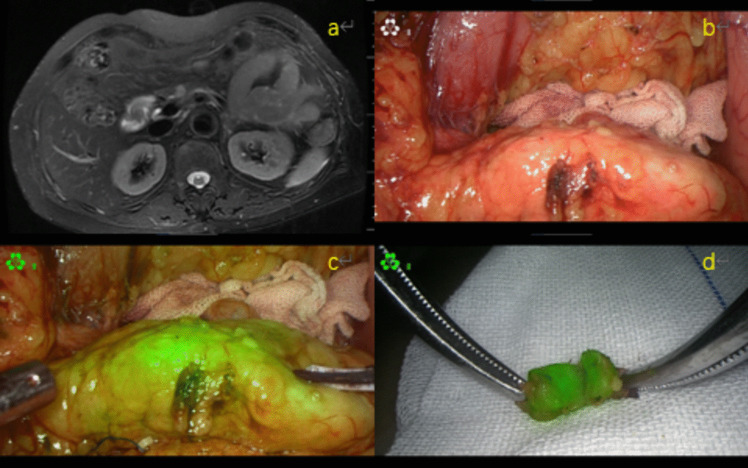
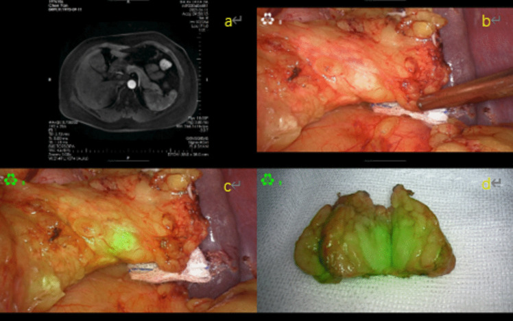
Case presentation: This case report details five patients with preoperative diagnosis of pancreatic neuroendocrine tumors of small endocrine tumors, intraoperative successful localization, and successful completion of laparoscopic partial resection of pancreatic tumors by indocyanine green fluorescence staining; none of the patients experienced serious complications after surgery and were discharged from the hospital, and routine pathology confirmed that four cases were pancreatic neuroendocrine tumors of G1 stage, and one case was pancreatic neuroendocrine cell hyperplasia.

Conclusion: Fluorescence imaging technology safely aids in the intraoperative localization of small pancreatic neuroendocrine tumors.

Keywords: Fluorescence imaging; Intraoperative localization; Small neuroendocrine tumor of the pancreas.


Conflict of interest statement

Declarations Ethics Approval The procedures that human research followed in this study were in accordance with the Declaration of Helsinki. The retrospective study protocol has been approved by the Scientific Research and New Technology of Wannan Medical College Yijishan Hospital IRB (2024–30). Informed Consent Written informed consent was obtained from the patient for publication of this report and the accompanying images, and the patient’s anonymity was upheld.Competing Interests The authors declare no competing interests. Competing Interests The authors declare no competing interests.

Figures

Fig. 1
Fig. 1
Patient 1: a abdominal-enhanced MRI indicates potential pancreatic neck occupancy; b laparoscopic white light mode reveals no visible lesion in the pancreatic neck; c laparoscopic fluorescence mode featuring green fluorescence imaging of the tumor; d laparoscopic fluorescence mode dissects the tumor, allowing visualization of the parenchyma while the margins of the incision remain no visualization
Fig. 2
Fig. 2
Patient 2: a abdominal enhanced MRI indicating potential pancreatic tail occupancy. b The laparoscopic white light mode seemingly reveals a nodular lesion located in the tail of the pancreas. c Laparoscopic fluorescence mode reveals the tumor, imaged in green fluorescence. d Laparoscopic fluorescence mode dissects the tumor, allowing visualization of the parenchyma, while the margins of the incision remain no visualization
Fig. 3
Fig. 3
Patient 4: a Abdominal enhanced MRI indicating possible pancreatic body occupancy. b Multiple nodular lesions appeared to be visible in the body of the pancreas under the white light mode of laparoscopy. c The tumor was fluorescently visualized in green color in the fluorescence mode of laparoscopy. d Fluorescence laparoscopy dissection revealed no visualization within the parenchyma, but fluorescent visualization was observed on the surface

References

    1. Kurita Y, Hara K, Kobayashi N, et al. The detection rate of endoscopic ultrasound and computed tomography in diagnosing pancreatic neuroendocrine neoplasms including small lesions: a multicenter study. J Hepatobiliary Pancreat Sci. 2022;29(8):950–9. 10.1002/jhbp.1144. - PubMed
    1. Rindi G, Mete O, Uccella S, et al. Overview of the 2022 WHO classification of neuroendocrine neoplasms. Endocr Pathol. 2022;33(1):115–54. 10.1007/s12022-022-09708-2. - PubMed
    1. Magi L, Marasco M, Rinzivillo M, et al. Management of functional pancreatic neuroendocrine neoplasms. Curr Treat Options Oncol. 2023;24(7):725–41. 10.1007/s11864-023-01085-0. - PMC - PubMed
    1. Scott AT, Howe JR. Evaluation and management of neuroendocrine tumors of the pancreas. Surg Clin North Am. 2019;99(4):793–814. 10.1016/j.suc.2019.04.014. - PMC - PubMed
    1. Lubner MG, MankowskiGettle L, Kim DH, et al. Diagnostic and procedural intraoperative ultrasound: technique, tips and tricks for optimizing results. Br J Radiol. 2021;94(1121):20201406. 10.1259/bjr.20201406. - PMC - PubMed

Substances

LinkOut - more resources