. 2024 Nov 19;15(1):10012.
doi: 10.1038/s41467-024-54374-z.

Dual receptor-binding, infectivity, and transmissibility of an emerging H2N2 low pathogenicity avian influenza virus

Affiliations

Dual receptor-binding, infectivity, and transmissibility of an emerging H2N2 low pathogenicity avian influenza virus

Ju Sun et al. Nat Commun. .

Abstract

The 1957 H2N2 influenza pandemic virus [A(H2N2)pdm1957] has disappeared from humans since 1968, while H2N2 avian influenza viruses (AIVs) are still circulating in birds. It is necessary to reveal the recurrence risk and potential cross-species infection of these AIVs from avian to mammals. We find that H2 AIVs circulating in domestic poultry in China have genetic and antigenic differences compared to the A(H2N2)pdm1957. One H2N2 AIV has a dual receptor-binding property similar to that of the A(H2N2)pdm1957. Molecular and structural studies reveal that the N144S, and N144E or R137M substitutions in hemagglutinin (HA) enable H2N2 avian or human viruses to bind or preferentially bind human-type receptor. The H2N2 AIV rapidly adapts to mice (female) and acquires mammalian-adapted mutations that facilitated transmission in guinea pigs and ferrets (female). These findings on the receptor-binding, infectivity, transmission, and mammalian-adaptation characteristics of H2N2 AIVs provide a reference for early-warning and prevention for this subtype.


Conflict of interest statement

Competing interests The authors declare no competing interests.

Figures

Fig. 1
Fig. 1. Phylogenetic and antigenic characteristics of the H2Ny avian influenza viruses identified in this study.
a Phylogenetic tree of the HA gene of H2 influenza viruses. Pink and orange grids represent the H2 human virus lineage and the current circulating H2 avian influenza virus (AIV) lineage, respectively. The dotted viruses in the AIV and human virus lineages are the sequenced H2Ny AIVs and reference human viruses used in this study. b Antigenic cartograph representation of the hemagglutination inhibition (HI) data was generated using an antiserum panel. One unit (grid) represents a 2-fold change in the HI assay. H2N2 human viruses are encircled in red oval, while H2 AIVs are encircled in black oval. c Individual sera from humans (n = 419) born prior to 1968 were examined for antibodies against the H2N2 pandemic strain A/SG/1/1957 and the AIV strain Dk/FJ/2021 by hemagglutination inhibition (HI) and microneutralization (MN) assays. Sera with HI or MN titers ≥ 1:40 are considered positive. Data are represented as the mean ± SD. d Sequence alignment of the key residues in the receptor-binding site (RBS) of the HAs from representative H2 viruses.
Fig. 2
Fig. 2. Receptor-binding properties of the wild-type H2N2 avian and human viruses.
ac Receptor-binding properties at the virus level. Results of the solid-phase binding assays for Dk/FJ/2021 (a), A/SG/1/1957 (b), and A/KR/426/1968 (c) to both α2,3-linked (3′-SLNLN) and α2,6-linked (6′-SLNLN) sialylglycan receptors. Blue and red lines represent 3′-SLNLN and 6′-SLNLN, respectively. Vaccine strains (A/SG/1/1957 and A/KR/426/1968) were rescued by reverse genetics, in which HA and NA genes from the wild-type A/SG/1/1957 or A/KR/426/1968 strains and the internal genes from the vaccine backbone A/PR/8/34. A/CA/04/2009 and A/VN/1194/2004 were used as controls. df Receptor-binding properties at the protein level. BIAcore diagram of HAs from Dk/FJ/2021 (d), A/SG/1/1957 (e), and A/KR/426/1968 (f) binding to 3′-SLNLN or 6′-SLNLN. The binding affinity (KD) values were calculated using the BIAcore 3000 analysis software (BIAcore version 4.1). The binding curve to the 3′-SLNLN is shown in blue, and that for the 6′-SLNLN is shown in red.
Fig. 3
Fig. 3. Receptor-binding properties of the H2N2 HA mutants.
Receptor-binding properties at the protein level. BIAcore diagram of A/SG/1/1957-R137M mutant HA (a), A/SG/1/1957-N144E mutant HA (b), and Dk/FJ/2021-S144N mutant HA (c) binding to 3′-SLNLN or 6′-SLNLN. The binding affinity (KD) values were calculated using BIAcore 3000 analysis software (BIAcore version 4.1). The binding curve of the 3′-SLNLN is shown in blue, and that for the 6′-SLNLN is shown in red.
Fig. 4
Fig. 4. Structure basis of the interaction between H2N2 HAs and avian and human receptor analogs.
The secondary structural elements of the RBS (130-loop, 140-loop, 190-helix, and 220-loop; H3 number) are labeled in the ribbon representation, together with selected residues in the stick representation. Hydrogen bonds are indicated by the dashed lines. ab Molecular interactions of Dk/FJ/2021 wild-type HA with human (a) and avian (b) receptor analogs. c Comparison of the RBSs between the Dk/FJ/2021 HA-avian receptor complex (light pink) and the Dk/FJ/2021-S144N HA mutant-avian receptor complex (orange). A similar trans conformation for glycan binding showed no observable drift between the two complexes. d, e Molecular interactions of A/SG/1/1957 HA with human (d) and avian (e) receptor analogs. f Comparison of the RBSs between the A/SG/1/1957 HA-human receptor complex (cyan) and A/SG/1/1957-R137M HA mutant-human receptor complex (lavender). The human receptor analog shifts towards the 130-loop by 0.8 Å in the A/SG/1/1957-R137M complex. g Comparison of the RBSs between the A/SG/1/1957 HA-human receptor complex (cyan) and A/SG/1/1957-N144E HA mutant-human receptor complex (green). The human receptor analog shifts towards the 220-loop by 1.3 Å in the A/SG/1/1957-N144E complex. h Comparison of the RBSs between the A/SG/1/1957 HA-human receptor complex (cyan) and A/KR/426/1968 HA-human receptor complex (pale green). The human receptor analog shifts towards the 130-loop by 0.8 Å in the A/KR/426/1968 complex.
Fig. 5
Fig. 5. Dynamic adaptation of H2N2 avian influenza virus in mice.
a 7-week-old BALB/c mice were intranasally (i.n.) inoculated with 106 EID50 of the Dk/FJ/2021 strain, and then the virus was passaged in mice by lung-to-lung, and the viruses in each passage were named MA1-MA15. The changes in body weight (n = 3) and lung viral RNA loads (n = 3) of infected mice are shown. The dashed line indicates 75% of the initial body weight or the limit of detection. b Dynamic changes of two full mutation sites, PB2-E627K and NS1-G183S, were analyzed using the Sanger sequencing chromatogram and the proportions of amino acids are shown according to the Next-generation sequencing results. The red-dashed boxes show the peaks of nucleotide G and A in the PB2 and NS1 genes.
Fig. 6
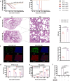
Fig. 6. Pathogenicity of the H2N2 avian influenza virus.
a Groups of five mice were i.n. inoculated with the Dk/FJ/2021 wild-type (WT) and mouse-adapted (MA) mutant viruses at doses from 102 to 106 EID50, and the body weight changes and survival rates were monitored for 14 days. The dashed line indicates 75% of the initial body weight. b The lungs of mice infected with the indicated viruses were collected at 5 dpi. Lung sections were stained with H&E, and one representative result was shown. The histopathological scores (n = 3) were measured. Scale bar, 100 μm. c Three-dimensional image of the NHBE cells infected by WT and MA mutant strains with 106 EID50 at 12 hpi. The viral NP protein (green) was detected by indirect immunofluorescence, ciliated epithelial cells were detected with anti-β-tubulin IV antibodies (red) and cell nucleus were immunostained with DAPI (blue). Percentages of influenza NP-positive cells per DAPI-positive cells were calculated. Data are expressed as mean ± SEM of three randomly selected fields. d Mice (n = 3) in each group were euthanized at 3, 5, and 7 dpi, and the lung suspensions were used for IFN-β detection. Data are shown as mean ± SEM. e Supernatants of A549 cells infected with indicated viruses were collected to determine IFN-β at 6, 12, 18, 24, and 30 hpi. Data are shown as mean ± SEM of three replicates. f The NS1 (G183 or S183) expression vector together with the plasmids of IFN-β with luciferase reporter and the internal reference of Renilla luciferase reporter were co-transfected into HEK293T cells. At 24 h post-transfection, the cells were stimulated with Sendai virus for 12 h. The cell lysates were then collected and luciferase activity was measured. Data are shown as mean ± SEM of three replicates. Statistical significance was based on Student’s t test and one-way or two-way ANOVA. p < 0.05 was considered to indicate statistical significance, and p values are shown on the graphs.
Fig. 7
Fig. 7. Transmission of the H2N2 avian influenza virus.
a, b Groups of three guinea pigs (a) and two ferrets (b) were i.n. inoculated with the WT and MA mutant viruses at a dose of 107 EID50. The direct contact (DC) and respiratory droplet exposure (RD) animals were housed in the corresponding cages at 1 dpi. Nasal washes were collected every two days from all animals to detect viral RNA loads. c The body weight changes of ferrets (n = 2) in the inoculated, DC, and RD groups were monitored for 14 days. The dashed line indicates the limit of detection.

References

    1. Liu, W. J. et al. Emerging HxNy influenza A viruses. Cold Spring Harb. Perspect. Med.12, a038406 (2022). - PMC - PubMed
    1. Shortridge, K. F. et al. Interspecies transmission of influenza viruses: H5N1 virus and a Hong Kong SAR perspective. Vet. Microbiol.74, 141–147 (2000). - PubMed
    1. Gao, G. F. From “A“IV to “Z“IKV: attacks from emerging and re-emerging pathogens. Cell172, 1157–1159 (2018). - PMC - PubMed
    1. Shi, W., Shi, Y., Wu, Y., Liu, D. & Gao, G. F. Origin and molecular characterization of the human-infecting H6N1 influenza virus in Taiwan. Protein Cell4, 846–853 (2013). - PMC - PubMed
    1. Chen, H. et al. Clinical and epidemiological characteristics of a fatal case of avian influenza A H10N8 virus infection: a descriptive study. Lancet383, 714–721 (2014). - PubMed

Publication types

MeSH terms

Substances

Associated data

LinkOut - more resources