. 2024 Nov 19;14(1):28649.
doi: 10.1038/s41598-024-79226-0.

House dust mites stimulate thymic stromal lymphopoietin production in human bronchial epithelial cells and promote airway remodeling through activation of PAR2 and ERK signaling pathway

Affiliations

House dust mites stimulate thymic stromal lymphopoietin production in human bronchial epithelial cells and promote airway remodeling through activation of PAR2 and ERK signaling pathway

Yi-An Hsieh et al. Sci Rep. .

Abstract

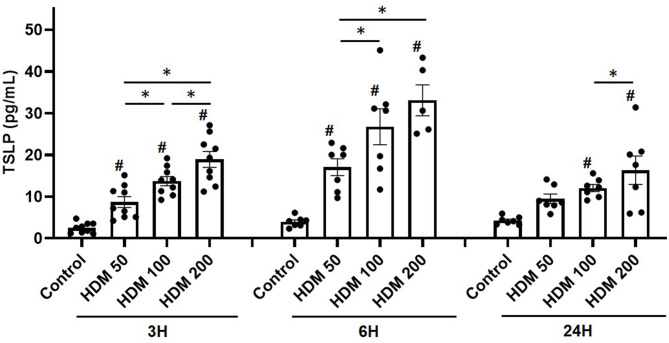
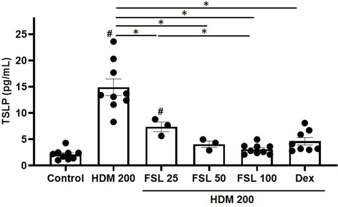
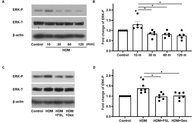
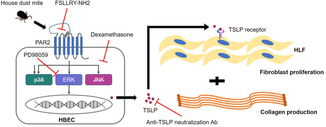
House dust mites (HDM) are common aeroallergens linked to airway inflammation and remodeling in asthma. Protease-activated receptor 2 (PAR2) and thymic stromal lymphopoietin (TSLP) may mediate these immune responses. However, how the epithelium influences fibroblasts toward airway remodeling remains unclear. We hypothesize that HDM stimulates human bronchial epithelial cells (HBECs) to produce TSLP via PAR2 activation, driving fibroblasts toward remodeling processes. HBECs were treated with HDM, with or without the PAR2 antagonist FSLLRY-NH2 (FSL), and TSLP expression was measured by qPCR and ELISA. Phosphorylation of MAPKs was assessed by western blotting. Human lung fibroblasts (HLFs) were exposed to recombinant TSLP or conditioned medium (CM) from HDM-stimulated HBECs, with or without anti-TSLP antibodies. Fibroblast proliferation and collagen production were assessed as remodeling markers. HDM increased ERK phosphorylation (not p38 or JNK) and TSLP expression at mRNA and protein levels. FSL preincubation significantly reduced ERK phosphorylation and TSLP production: HDM-stimulated CM induced fibroblast proliferation and collagen production, effects suppressed by anti-TSLP or FSL. Direct treatment with recombinant TSLP also promoted fibroblast proliferation and collagen synthesis. These findings suggest that HDM promotes HBEC-to-HLF paracrine interactions via PAR2-ERK-TSLP axis, participating in airway remodeling. PAR2 antagonists may represent potential therapeutic targets for HDM-induced remodeling processes.

Keywords: Airway inflammation; House dust mite; Protease-activated receptor 2; Remodeling; Thymic stromal lymphopoietin.


Conflict of interest statement

Declarations Conflict of interest All authors declare that they have no conflict of interest. Ethics approval This study was approved by the Institutional Review Board of Taipei Veterans General Hospital (ID 2019-08-004B).

Figures

Fig. 1
Fig. 1
Effects of HDM on TSLP production. HBECs were treated with various concentrations of HDM (50, 100, and 200 µM) and incubated for different periods (3, 6, and 24 h), followed by collection of the supernatants for measurements of TSLP. The bar charts with dot plots indicate mean values with SEM in error bars. #, P < 0.05, vs. control. * P < 0.05, Mann-Whitney U test. TSLP levels (pg/mL) were generated in duplicate from 5 to 9 experiments. HDM, house dust mite. TSLP, thymic stromal lymphopoietin.
Fig. 2
Fig. 2
Effects of HDM on TSLP mRNA expression. HBECs were treated with various concentrations of HDM (50, 100, and 200 µM) and incubated for 3 h, followed by cell lysis for measurements of TSLP mRNA expression. The bar chart with dot plots indicates mean values and SEM in error bars. #, P < 0.05, vs. control. * P < 0.05, Mann-Whitney U test. The mRNA expression was determined by quantitative real-time PCR in 4 to 8 separate experiments.
Fig. 3
Fig. 3
Effects of PAR2 antagonist on HDM-induced TSLP production. HBECs were treated with HDM 200 µM in the absence or presence of 2-h preincubation of various concentrations of PAR2 antagonist (FSL, 25, 50, and 100 µM) or dexamethasone (100 µM) for 6 h. At the end of the experiments, the supernatant was collected for TSLP measurements using the ELISA method. The bar charts with dot plots indicate mean values and SEM in error bars. #, P < 0.05, vs. control. * P < 0.05, Mann-Whitney U test. TSLP levels (pg/mL) were generated in duplicate from 4 to 9 separate experiments. FSL, FSLLRY-NH2; Dex, dexamethasone.
Fig. 4
Fig. 4
Effects of PAR2 on HDM-induced ERK phosphorylation. HBECs were treated with HDM 200 µM for various periods (10, 30, 60, and 120 min) or treated with HDM 200 µM in the absence or presence of 2-h preincubation of PAR2 antagonist (FSL 100 µM) or dexamethasone (100 µM) for 10 min, then harvested for Western blotting (A) or (C) and comparisons of protein bands (B) or (D). Protein bands were analyzed by densitometry. The bar charts with dot plots indicate mean values with SEM in error bars. #, P < 0.05, vs. control. * P < 0.05, Mann-Whitney U test. Blots are representative of 5 to 6 separate experiments. ERK-P, phosphorylation of extracellular signal-regulated kinases; ERK-T, total ERK. Refer to Fig. 3 for other abbreviations.
Fig. 5
Fig. 5
Effect of ERK inhibitor on HDM-induced ERK phosphorylation and TSLP production. HBECs were treated with HDM 200 µM in the absence or presence of 2-h preincubation of ERK inhibitor (PD 98059 10 µM) for 10 min, then harvested for Western blotting (A) and comparisons of protein bands (B). Protein bands were analyzed by densitometry. HBECs were also treated with HDM 200 µM in the absence or presence of 2-h preincubation of PD 98,059 (10 µM) and incubated for 6 h, followed by measurements of TSLP in the supernatants (C). The bar charts with dot plots indicate mean values with SEM in error bars. #, P < 0.05, vs. control. * P < 0.05, Mann-Whitney U test. Blots are representative of 3 separate experiments. TSLP levels (pg/mL) were generated in duplicate from 6 separate experiments. PD, PD 98,059. Refer to Figure for other abbreviations.
Fig. 6
Fig. 6
Effects of PAR2 on fibroblast proliferation and collagen production in HLFs through CM from HBECs. Conditioned medium (CM) was prepared from HBECs treated with HDM 200 µM in the absence or presence of 2-h preincubation of FSL 100 µM or dexamethasone 100 µM for 6 h. HLFs were incubated in 500 µL of a medium mixture (CM: fibroblast medium = 1:4) for 24 h, followed by fibroblast counting (A) and measurement of collagens in the supernatants (B). Bar charts with dot plots indicate mean values and SEM in error bars. #, P < 0.05, vs. control. * P < 0.05, Mann-Whitney U test. The fibroblast counts and collagen levels were generated in duplicate from 3 to 7 separate experiments.
Fig. 7
Fig. 7
Effects of TSLP stimulation or suppression on fibroblast proliferation and collagen production in HLFs. Recombinant human TSLP with various concentrations (0.1, 1, and 10 µg/mL) were added to HLFs and incubated for 24 h, followed by calculating fibroblast counts (A) and measuring collagen in the supernatants (B). CM was prepared from HBECs treated with HDM 200 µM for 6 h. HLFs were treated in 500 µL of a medium mixture (CM: fibroblast medium = 1:4) containing anti-TSLP neutralization antibody (50 or 100 ng/mL) for 24 h, followed by fibroblast counting (C) and measurement of collagens in the supernatants (D). Bar charts with dot plots indicate mean values and SEM in error bars. #, P < 0.05, vs. control. * P < 0.05, Mann-Whitney U test. The fibroblast counts and collagen levels were calculated from 3 to 7 experiments.
Fig. 8
Fig. 8
A proposed mechanism in this study. HDM stimulates PAR2 activation on HBECs, followed by a downstream signaling pathway through ERK phosphorylation and TSLP production, which can be suppressed by preincubation of PAR2 antagonist or ERK inhibitor. HBECs-generated TSLP in a conditioned medium contributes to fibroblast proliferation and collagen production in human fibroblasts. Based on the findings, this concept can be proved that fibroblast proliferation and collagen production in HLFs can be enhanced by recombined TSLP or suppressed by anti-TSLP neutralization antibodies. HBECs, human bronchial epithelial cells. HLFs, human lung fibroblasts.

References

    1. Hough, K. P. et al. Airway Remodeling in Asthma. Front. Med. (Lausanne). 7, 191. 10.3389/fmed.2020.00191 (2020). - PMC - PubMed
    1. Joseph, C. & Tatler, A. L. Pathobiology of Airway Remodeling in Asthma: the emerging role of Integrins. J. Asthma Allergy. 15, 595–610. 10.2147/Jaa.S267222 (2022). - PMC - PubMed
    1. Varricchi, G. et al. Biologics and airway remodeling in severe asthma. Allergy. 77, 3538–3552. 10.1111/all.15473 (2022). - PMC - PubMed
    1. Pearce, N., Pekkanen, J. & Beasley, R. How much asthma is really attributable to atopy? Thorax. 54, 268–272. 10.1136/thx.54.3.268 (1999). - PMC - PubMed
    1. Abu Khweek, A., Kim, E., Joldrichsen, M. R., Amer, A. O. & Boyaka, P. N. Insights into Mucosal Innate Immune responses in House Dust mite-mediated allergic asthma. Front. Immunol.11, 534501. 10.3389/fimmu.2020.534501 (2020). - PMC - PubMed

LinkOut - more resources