. 2025 Jan;21(1):59-89.
doi: 10.1038/s44320-024-00076-z. Epub 2024 Nov 19.

Global atlas of predicted functional domains in Legionella pneumophila Dot/Icm translocated effectors

Affiliations

Global atlas of predicted functional domains in Legionella pneumophila Dot/Icm translocated effectors

Deepak T Patel et al. Mol Syst Biol. 2025 Jan.

Abstract

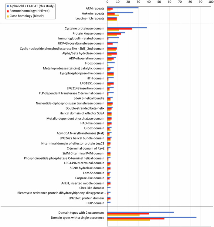
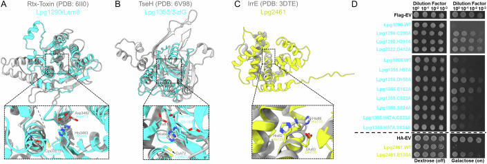
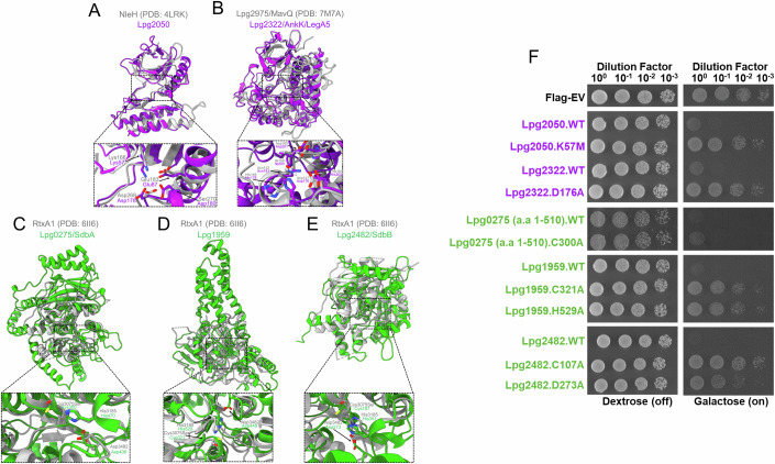
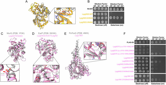
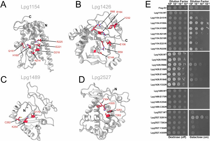
Legionella pneumophila utilizes the Dot/Icm type IVB secretion system to deliver hundreds of effector proteins inside eukaryotic cells to ensure intracellular replication. Our understanding of the molecular functions of the largest pathogenic arsenal known to the bacterial world remains incomplete. By leveraging advancements in 3D protein structure prediction, we provide a comprehensive structural analysis of 368 L. pneumophila effectors, representing a global atlas of predicted functional domains summarized in a database ( https://pathogens3d.org/legionella-pneumophila ). Our analysis identified 157 types of diverse functional domains in 287 effectors, including 159 effectors with no prior functional annotations. Furthermore, we identified 35 cryptic domains in 30 effector models that have no similarity with experimentally structurally characterized proteins, thus, hinting at novel functionalities. Using this analysis, we demonstrate the activity of thirteen functional domains, including three cryptic domains, predicted in L. pneumophila effectors to cause growth defects in the Saccharomyces cerevisiae model system. This illustrates an emerging strategy of exploring synergies between predictions and targeted experimental approaches in elucidating novel effector activities involved in infection.

Keywords: Legionella pneumophila; Bacterial Effectors; Cryptic Domains; Protein Modeling; Yeast Toxicity.


Conflict of interest statement

Disclosure and competing interests statement. The authors declare no competing interests.

Figures

Figure 1
Figure 1. Functional domain occurrences from 368 L. pneumophila effector models.
The number of occurrences of different ECOD domain types: blue bars—identified using FATCAT in 3D models built with AlphaFold (this study), red bars—identified with remote homology recognition program HHPred, yellow bars—identified by close homology detected with BlastP. As expected, domain-type occurrences identified by homology are a subset of those identified in 3D models.
Figure 2
Figure 2. Revelation of two previously unrecognized cysteine protease and metalloprotease effectors that are known to cause yeast toxicity.
(A, B) Structural alignment of the cysteine protease domain of Lpg1290/Lem8 (cyan, residues 244–457) with Rtx-toxin (gray, residues 3309–3562) (Lee et al, 2019), followed by the alignment of the cysteine protease domain model of Lpg1355/SidG (cyan, residues 16–195 + 563–665) with TseH effector (gray, residues 21–223) from Vibrio cholera (Hersch et al, 2020). Furthermore, in the case of Lpg1355/SidG, an adjacent ɑ-helix in the predicted catalytic pocket harbors two residues, Asp158 and Glu162, orientated in a position that suggests an involvement in the biochemical activity of this effector. (C) Model of Lpg2461 (yellow, residues 1–212) structurally aligned with IrrE from Deinococcus derserti (residues 1–281) (Vujicic-Zagar et al, 2009). Below is a close view of the potential catalytic residues (cyan or yellow sticks) of each effector model determined by their top structural hit (gray sticks) from our FATCAT analysis. (D) Yeast toxicity panel of FLAG-tagged constructs of Lpg1290/Lem8, Lpg1355/SidG, and their respective mutants, followed by the HA-tagged constructs of Lpg2461 and its mutant. Expression of these constructs are found in Appendix Figs. S10, 11. In the case of Lpg1290/Lem8, a Cys280Ala mutation was tested instead of the Cys280Ser mutation used in a previous study. Serial dilutions were spotted on SD media containing either dextrose (repressing) or galactose (inducing). A representative experiment of three independent replicates is shown.
Figure 3
Figure 3. Mutagenesis analysis of predicted kinase and α/β hydrolase domains in L. pneumophila effector repertoire.
(A, B) Model of Lpg2050 (purple, residues 1–205) aligned with the structure of the T3SS effector NleH (gray, residues 128-293) (Grishin et al, 2014a). Structural overlay of Lpg2322/AnkK/LegA5’s (purple) predicted kinase domain (residues 1–314) onto the lipid kinase domain of Lpg2975/MavQ (gray, residues 1–375) (Hsieh et al, 2021). Putative catalytic residues are shown in the zoomed view. (CE) Alignments of the effectors possessing α/β hydrolase domains (green) to the top structural hit (gray) from the FATCAT server, followed by the inset view of the putative catalytic residues. (F) The corresponding yeast panel pinpointing potential residues important for the function of these domains in causing yeast toxicity. Effector proteins and their mutants were expressed with an N-terminal FLAG tag (Appendix Fig. S10).
Figure 4
Figure 4. The predicted ADP-ribosyltransferase domain of Lpg2523/Lem26 and glycosyltransferase domains of Lpg0275/SdbA, Lpg0402/LegA9, and Lpg1961 are linked to yeast toxicity.
(A, B) Model of Lpg2523/Lem26’s ADP-ribosyltransferase domain (residues 1–324) (gold) overlayed onto the structurally characterized ADP-ribosyltransferase effector Tre1 from S. proteamaculans (residues 8–192, gray) (Ting et al, 2018). Residues potentially involved in the catalysis of this predicted ADP-ribosyltransferase domain are also shown in the zoomed-in panel. In the following panel, Glu294 of the ExE motif and His545 of the predicted phosphodiesterase domain were tested for their roles in causing yeast toxicity. The expression of Lpg2523/Lem26 and its mutants are found in Appendix Fig. S11. (CF) Representation of the glycosyltransferase domain models (pink) from Lpg0275/SdbA (residues 511–1050), Lpg0402 (residues 1–399), and Lpg1961 (residues 27–328) aligned with their top structural hit from the FATCAT analysis. Predicted residues involved in catalysis were assessed in the yeast toxicity panel. The expression level of each construct is found in Appendix Fig. S10.
Figure 5
Figure 5. Cryptic domains in L. pneumophila effectors contribute to the yeast toxicity phenotype.
(AD) Structural models of the cryptic domains from L. pneumophila effectors that are toxic to yeast when expressed ectopically (gray). Residue boundaries of each cryptic domain are described in Table 8. Potentially important residues in the cryptic domains that were mutated and tested in (E) were shown in sticks (red). (E) Yeast spot dilution assay of cryptic domain containing effectors and their respective mutants. The protein expression levels of the mutants tested were analyzed using western blot (Appendix Fig. S12).
Figure EV1
Figure EV1. Both domains of Lpg0275/SdbA are involved in yeast toxicity.
(A) Schematic of the domain organization of Lpg0275/SdbA. The N-terminal domain shown in green corresponds to the hydrolase domain, whereas the pink indicates the glycosyltransferase domain. (B) Alphafold2 model of Lpg0275/SdbA. The green represents the hydrolase domain, and the pink represents the glycosyltransferase domain. Below is a zoomed-in view of the predicted catalytic residues of each predicted enzymatic domain. (C) Yeast toxicity panel of strains expressing FLAG-tagged constructs of full-length wildtype Lpg0275 and its variants.
Figure EV2
Figure EV2. Lpg1154/RavQ forms a unique “T” shape containing a highly conserved groove that may serve as an active site.
(A) The Lpg1154/RavQ model (residues 59–389, red) shows the base and stalk that form the “T” shape. (B) An electrostatic potential surface representation of a potential active site cavity of Lpg1154/RavQ, followed by a zoom-in of the conserved residues identified in Lpg1154/RavQ and its orthologs from the Legionella genus, which are arranged in a potential active site.
Figure EV3
Figure EV3. Lpg1426/VpdC has a cryptic domain on the N-terminus that has structural similarity to the Ntox11 putative toxin found in human pathogenic amoeba.
(A) Cartoon representation of the Lpg1426/VpdC Alphafold2 model. The cryptic domain is found on the N-terminus (red), followed by a central phospholipase domain (white) and the C-terminal helical bundle involved in interactions with ubiquitin. (B) Structural alignment of the Lpg1426/VpdC cryptic domain (residues 1–298, red) onto the Ntox11 Alphafold2 model (residues 285–446, salmon) from N. fowleri. (C) Surface representation of the electrostatic potential of the Lpg1426/VpdC model that also shows a conserved negatively-charged pocket (dotted circle) is present in the cryptic domain. (D) Zoom in on the positively charged region where highly conserved residues (salmon sticks), which are present in the Legionella orthologs of Lpg1426/VpdC, form a pocket.
Figure EV4
Figure EV4. Lpg1489/RavX has a central globular cryptic domain surrounded by disordered loops.
(A) Structural alignment of the full-length Lpg1489/RavX models generated by Alphafold2 (red) and ESMFold (salmon). (B) ESMFold model of the Lpg1489/RavX cryptic domain (residues 83–263, red) onto the E. coli enterotoxin (PDB: 1LTA, residues 1–181, gray).
Figure EV5
Figure EV5. The cryptic domain of Lpg2527/LnaB is present in other L. pneumophila effectors, harboring a conserved set of residues that resemble a potential active site.
(A) Alphafold2 model of Lpg2527/LnaB which highlights the cryptic domain (red) and the helical bundle that was previously shown to be important in the activation of the NF-κB pathway (dark gray) (Losick et al, 2010). (B) Representation of the Lpg2527/LnaB cryptic domain (shown in red) that is present in other L. pneumophila effectors (Lpg0208/LegK4, Lpg0209, and Lpg0437/Ceg14/SidL). (C) Zoomed-in image of the putative active site of Lpg2527/LnaB that is also conserved in its Legionella orthologs and other L. pneumophila effectors containing this domain.

References

    1. Abu Kwaik Y, Gao LY, Stone BJ, Venkataraman C, Harb OS (1998) Invasion of protozoa by Legionella pneumophila and its role in bacterial ecology and pathogenesis. Appl Environ Microbiol 64:3127–3133 - PMC - PubMed
    1. Adams M, Sharma R, Colby T, Weis F, Matic I, Bhogaraju S (2021) Structural basis for protein glutamylation by the Legionella pseudokinase SidJ. Nat Commun 12:6174 - PMC - PubMed
    1. Alexander JAN, Locher KP (2023) Emerging structural insights into C-type glycosyltransferases. Curr Opin Struct Biol 79:102547 - PubMed
    1. Altindis E, Dong T, Catalano C, Mekalanos J (2015) Secretome analysis of Vibrio cholerae type VI secretion system reveals a new effector-immunity pair. mBio 6:e00075 - PMC - PubMed
    1. Altschul SF, Madden TL, Schaffer AA, Zhang J, Zhang Z, Miller W, Lipman DJ (1997) Gapped BLAST and PSI-BLAST: a new generation of protein database search programs. Nucleic Acids Res 25:3389–3402 - PMC - PubMed

MeSH terms

Substances