. 2024 Nov 19;30(1):221.
doi: 10.1186/s10020-024-00982-w.

Post-sepsis chronic muscle weakness can be prevented by pharmacological protection of mitochondria

Affiliations

Post-sepsis chronic muscle weakness can be prevented by pharmacological protection of mitochondria

Meagan S Kingren et al. Mol Med. .

Abstract

Background: Sepsis, mainly caused by bacterial infections, is the leading cause of in-patient hospitalizations. After discharge, most sepsis survivors suffer from long-term medical complications, particularly chronic skeletal muscle weakness. To investigate this medical condition in detail, we previously developed a murine severe sepsis-survival model that exhibits long-term post-sepsis skeletal muscle weakness. While mitochondrial abnormalities were present in the skeletal muscle of the sepsis surviving mice, the relationship between abnormal mitochondria and muscle weakness remained unclear. Herein, we aimed to investigate whether mitochondrial abnormalities have a causal role in chronic post-sepsis muscle weakness and could thereby serve as a therapeutic target.

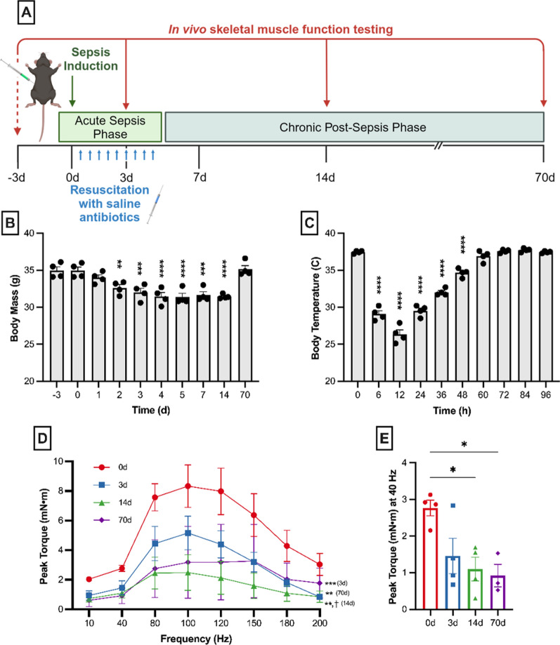
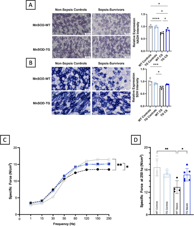
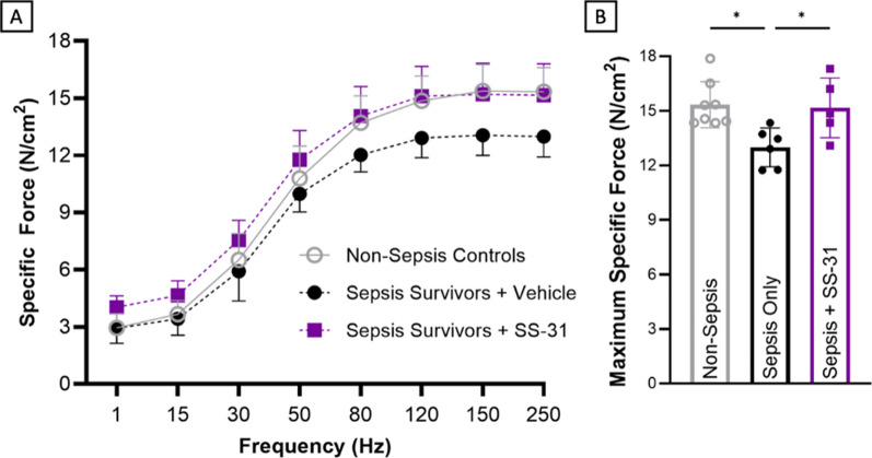
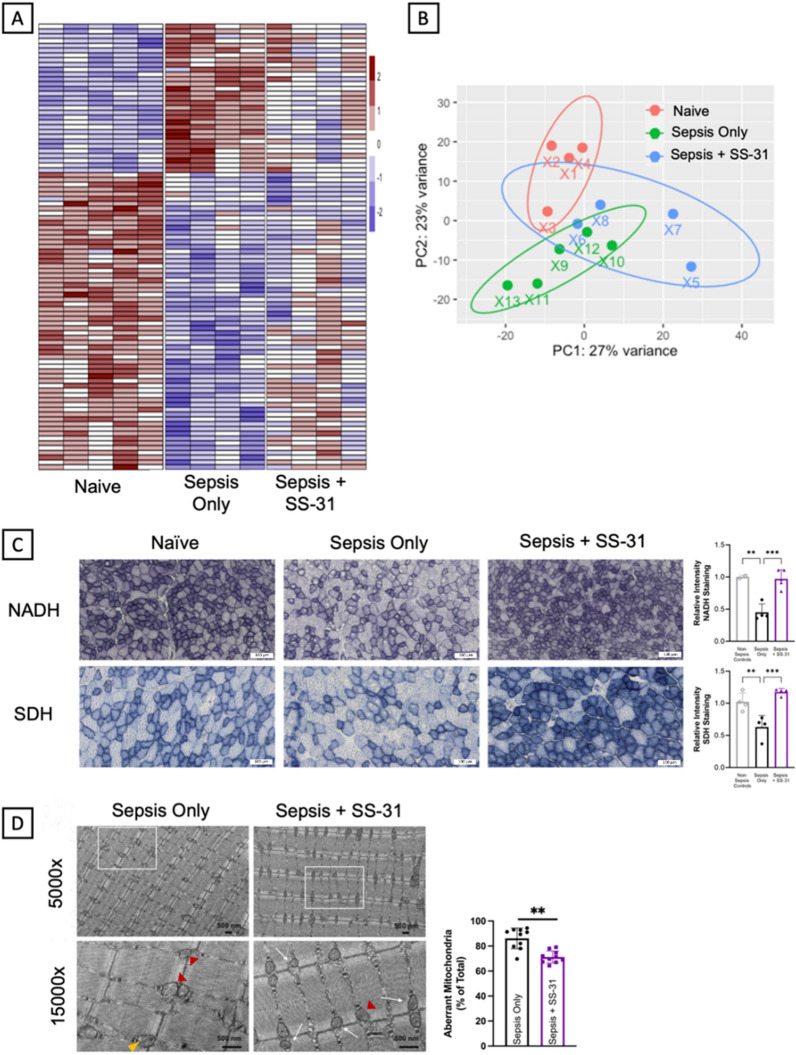
Methods: Experimental polymicrobial abdominal sepsis was induced in 16-18 months old male and female mice using cecal slurry injection with subsequent antibiotic and fluid resuscitation. To evaluate the pathological roles of mitochondrial abnormalities in post-sepsis skeletal muscle weakness, we utilized a transgenic mouse strain overexpressing the mitochondria-specific antioxidant enzyme manganese superoxide dismutase (MnSOD). Following sepsis development in C57BL/6 mice, we evaluated the effect of the mitochondria-targeting synthetic tetrapeptide SS-31 in protecting mitochondria from sepsis-induced damage and preventing skeletal muscle weakness development. In vivo and in vitro techniques were leveraged to assess muscle function at multiple timepoints throughout sepsis development and resolution. Histological and biochemical analyses including bulk mRNA sequencing were used to detect molecular changes in the muscle during and after sepsis RESULTS: Our time course study revealed that post sepsis skeletal muscle weakness develops progressively after the resolution of acute sepsis and in parallel with the accumulation of mitochondrial abnormalities and changes in the mitochondria-related gene expression profile. Transgenic mice overexpressing MnSOD were protected from mitochondrial abnormalities and muscle weakness following sepsis. Further, pharmacological protection of mitochondria utilizing SS-31 during sepsis effectively prevented the later development of muscle weakness.

Conclusions: Our study revealed that the accumulation of mitochondrial abnormalities is the major cause of post-sepsis skeletal muscle weakness. Pharmacological protection of mitochondria during acute sepsis is a potential clinical treatment strategy to prevent post-sepsis muscle weakness.

Keywords: Critical care illness; Mitochondrial myopathy; Muscle weakness; Post-sepsis syndrome.


Conflict of interest statement

Declarations Ethics approval and consent to participate All animal experiments were conducted according to procedures described in our Animal Use Protocol #2009-0541 which is approved by the Institutional Animal Care and Use Committee at the University of Kentucky. Consent for publication Not Applicable. Competing interests The authors declare no competing interests.

Figures

Fig. 1
Fig. 1
Skeletal muscle weakness develops progressively after sepsis resolution. A Late middle-aged C57BL/6 mice were subjected to severe sepsis followed by resuscitation with antibiotics and saline beginning 12 h after sepsis induction. Sepsis surviving mice (n = 4) suffered severe acute sepsis illness with significant decreases in body mass (B) and temperature (C). D Effect of sepsis on muscle function was examined by testing in vivo plantar flexion on the same four mice prior to sepsis induction, during acute illness (3d), and following recovery from sepsis (14d and 70d). Post-sepsis skeletal muscle weakness was evident by day 3 and persisted through at least day 70. Notably, skeletal muscle weakness worsened from day 3 to day 14, did not recover by day 70, and was still significantly weaker than pre-sepsis levels. E Analysis of plantar flexion at a physiologically relevant stimulus frequency (40 Hz) showed significantly reduced muscle function in the chronic post-sepsis phase. Data was expressed as mean ± SEM. Difference from baseline was expressed as *P < 0.05, **P < 0.01, ***P < 0.001, and ****P < 0.0001 (B-E), while difference from day 3 was denoted with ꝉ P < 0.05 and ꝉꝉ P < 0.01 (D-E only)
Fig. 2
Fig. 2
Skeletal muscle mitochondrial abnormalities develop progressively. Bulk mRNA sequencing was conducted on TA muscles from mice prior to sepsis (0d), during acute sepsis (4d) and after recovery during the chronic sepsis phase (14d). A Samples clustered based on sepsis timepoint (n = 6–7) as shown by PCA. B Out of the 2,278 DEGs at day 4, 213 of these remained altered on day 14, at which point an additional 514 DEGs emerged. Venn diagram depicts the number of DEGs only altered at day 4 (2065; 74%) and day 14 (514; 18.4%) on the left and right, respectively, with the DEGs altered at both timepoints shown in the middle (213; 7.6%). C A heatmap of the DEGs is shown. D Cross sectional area was reduced at day 4 and recovered by day 14, demonstrating that atrophy in the acute phase is recovered by the chronic post-sepsis phase (E) Western blot analyses demonstrated decreased expression of complexes I, IV, and V by day 4 that remained depressed at day 14. F In situ enzyme staining for complex I (NADH) and complex II (SDH) on TA muscle sections revealed intensity was significantly decreased by day 14, but not as early as day 4 during acute sepsis. G TEM was used to visualize changes in mitochondrial ultrastructure on in EDL samples from days 0, 3, and 14 (n = 3 each). Yellow and red arrow heads indicate examples of mitochondria with ruptured mitochondrial membranes and organelles with disrupted cristae/centralization into a vacuole-like structure, respectively. White squares in 5,000 × images indicate the area of 15,000 × images. Aberrant mitochondria accumulated progressively after sepsis, with a majority of the organelles demonstrating altered structure by day 14. Data is presented as mean ± SEM. Significant differences are compared to baseline where *P < 0.05, **P < 0.01, and ***P < 0.001
Fig. 3
Fig. 3
Overexpression of MnSOD protects against sepsis induces mitochondrial and muscular abnormalities. Late middle-aged MnSOD-overexpressing transgenic mice (MnSOD-TG) and littermate wildtype mice (MnSOD-WT) were subjected to experimental sepsis. Sepsis survivor mice were euthanized 21-days post-sepsis induction for in vitro muscle function testing and tissue analyses. A NADH staining for complex I activity in TA muscle sections was decreased as a result of sepsis, though MnSOD overexpression provided significant protection when compared to WT sepsis survivors. B SDH staining for complex II activity revealed MnSOD overexpression completely protected against typical sepsis reductions in staining intensity. Scale bars (A and B) represent 100 μm. C-D Function was assessed utilizing in vitro function testing 21 days after sepsis induction. MnSOD overexpression completely protected against the muscle weakness found in WT animals, indicating mitochondrial protection prevents chronic muscle weakness. *P < 0.05, **P < 0.01, ***P < 0.001, and ****P < 0.0001
Fig. 4
Fig. 4
SS-31 administration after sepsis development prevents post-sepsis muscle weakness. Following the development of severe sepsis, mice repeatedly received SS-31 or control vehicle in resuscitation saline starting from 12 h after sepsis induction. In vitro function testing of the EDL on day 21 post-sepsis induction demonstrated that SS-31 protects against muscle weakness development as indicated in (A) and (B). *P < 0.05
Fig. 5
Fig. 5
SS-31 administration after sepsis development prevents mitochondrial abnormalities from occurring. A Heatmap of DEGs reveal the patterns of SS-31-treated sepsis survivors shift towards naïve controls from vehicle-treated sepsis survivors. B PCA indicates clear differences between non-sepsis controls (red) and sepsis survivors, while SS-31 treated sepsis survivors (blue) cluster in between the two other groups. C NADH and SDH activity staining in TA muscle sections indicates SS-31 protects against sepsis-induced decreases as shown in the representative images and quantification. Scale bars represent 100 μm. D TEM analyses to assess mitochondrial morphology in EDL muscle sections. Representative images of vehicle- and SS-31- treated sepsis survivors (n = 3 each) are shown. Yellow and red arrow heads indicate examples of mitochondria with ruptured mitochondrial membranes and organelles with disrupted cristae/centralization into a vacuole-like structure, respectively. White squares in 5,000x images indicate the area of 15,000x images. Black scale bars indicate 500 nm. There are clear trends indicating that SS-31 confers protection against sepsis induced mitochondrial damage. **P < 0.01 and ***P < 0.001
Fig. 6
Fig. 6
Diagram depicting the development of chronic post-sepsis skeletal muscle weakness. Sepsis induces significant inflammation that resolves early during sepsis pathogenesis. This inflammation is accompanied by acute muscle weakness that mostly resolves within two weeks and progressive muscle weakness. While mitochondrial abnormalities occur early, they accumulate as inflammation and muscle atrophy resolve, leaving altered mitochondrial function and morphology as major causes of chronic skeletal muscle weakness

Similar articles

Cited by

References

    1. Angus DC, et al. Epidemiology of severe sepsis in the United States: analysis of incidence, outcome, and associated costs of care. Crit Care Med. 2001;29:1303–10. - PubMed
    1. Azevedo LCP. Mitochondrial dysfunction during sepsis. Endocr Metab Immune Disord Drug Targets. 2010;10:214–23. - PubMed
    1. Birk AV, Chao WM, Bracken C, Warren JD, Szeto HH. Targeting mitochondrial cardiolipin and the cytochrome c/cardiolipin complex to promote electron transport and optimize mitochondrial ATP synthesis. Br J Pharmacol. 2014;171:2017–28. - PMC - PubMed
    1. Borrelli A, et al. The functional role of MnSOD as a biomarker of human diseases and therapeutic potential of a new isoform of a human recombinant MnSOD. BioMed Res Int. 2014;2014: 476789. - PMC - PubMed
    1. Calaf GM. Cancer : oxidative stress and dietary antioxidants. Amsterdam Boston: Elsevier; 2014.

MeSH terms

Substances

LinkOut - more resources