. 2024 Nov 20;23(1):413.
doi: 10.1186/s12933-024-02501-x.

Lipoxin A4 improves cardiac remodeling and function in diabetes-associated cardiac dysfunction

Affiliations

Lipoxin A4 improves cardiac remodeling and function in diabetes-associated cardiac dysfunction

Ting Fu et al. Cardiovasc Diabetol. .

Abstract

Background: Diabetic heart disease may eventually lead to heart failure, a leading cause of mortality in diabetic individuals. The lack of effective treatments for diabetes-induced heart failure may result from a failure to address the underlying pathological processes, including chronic, low-grade inflammation. Previous studies have reported that lipoxin A4 (LXA4), known to promote resolution of inflammation, attenuates diabetes-induced atherosclerosis, but its impact on diabetic hearts has not been sought. Thus, we aimed to determine whether LXA4 therapeutic treatment attenuates diabetes-induced cardiac pathology.

Methods: Six-week-old male apolipoprotein E-deficient (ApoE-/-) mice were followed for 16 weeks after injection of streptozotocin (STZ, 55 mg/kg/day, i.p. for 5 days) to induce type-1 diabetes (T1DM). Treatment with LXA4 (5 μg/kg, i.p.) or vehicle (0.02% ethanol, i.p.) was administered twice weekly for the final 6 weeks. One week before endpoint, echocardiography was performed within a subset of mice from each group. At the end of the study, mice were euthanized with sodium pentobarbital (100 mg/kg i.p.) and hearts were collected for ex vivo analysis, including histological assessment, gene expression profiling by real-time PCR and protein level measurement by western blot.

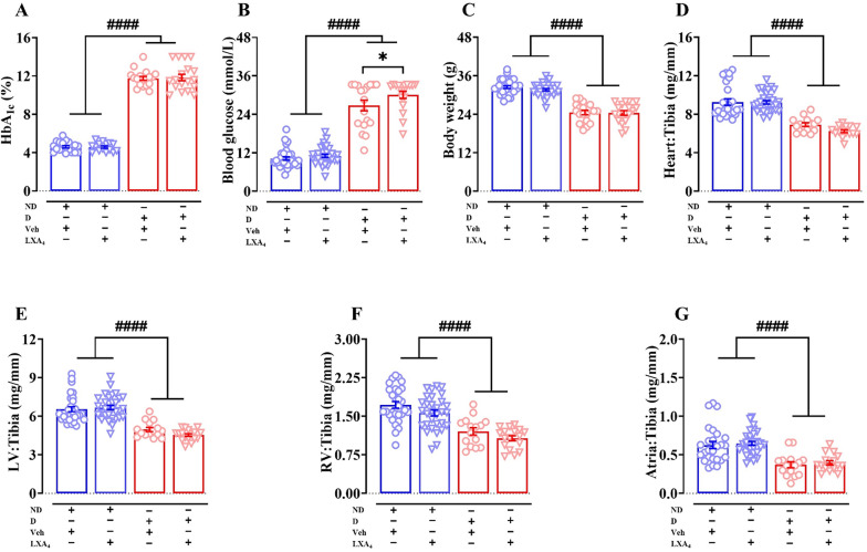
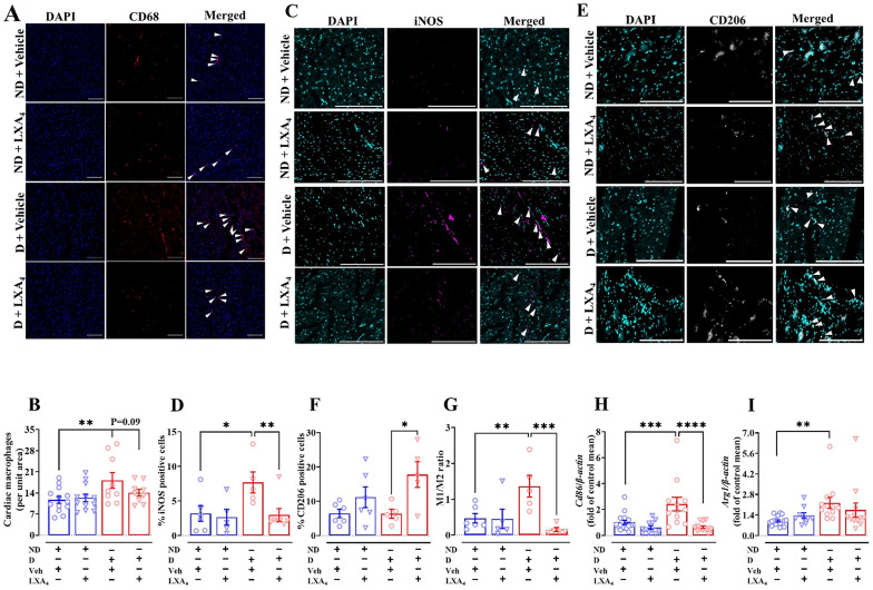
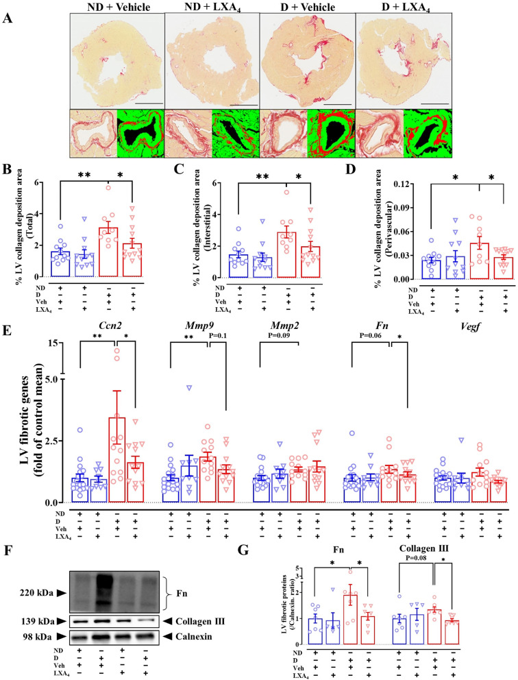
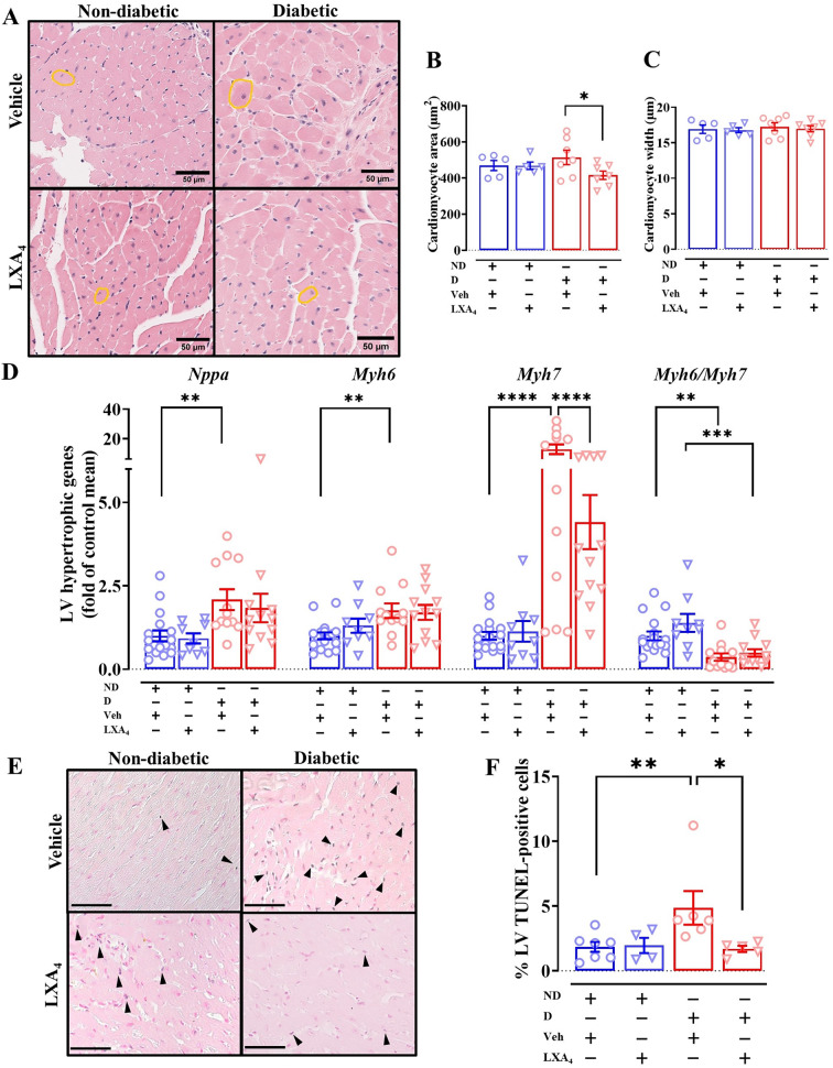
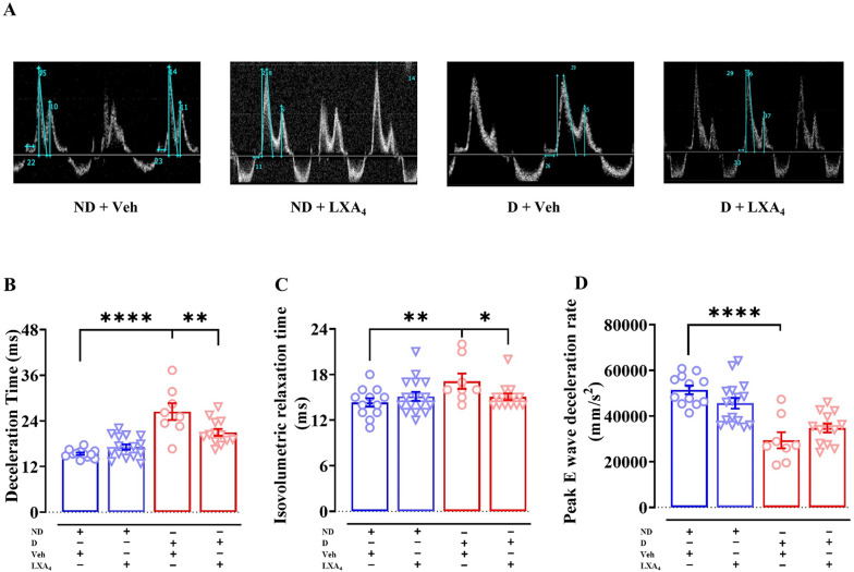
Results: As expected diabetic mice showed a significant elevation in plasma glycated hemoglobin (HbA1c) and glucose levels, along with reduced body weight. Vehicle-treated diabetic mice exhibited increased cardiac inflammation, macrophage content, and an elevated ratio of M1-like to M2-like macrophage markers. In addition, myocardial fibrosis, cardiomyocytes apoptosis and hypertrophy (at the genetic level) were evident, with echocardiography revealing early signs of left ventricular (LV) diastolic dysfunction. Treatment with LXA4 ameliorated diabetes-induced cardiac inflammation, pro-inflammatory macrophage polarization and cardiac remodeling (especially myocardial fibrosis and cardiomyocytes apoptosis), with ultimate improvement in cardiac function. Of note, this improvement was independent of glucose control.

Conclusions: These findings demonstrated that LXA4 treatment attenuated the extent of cardiac inflammation in diabetic hearts, resulting in limited cardiac remodeling and improved LV diastolic function. This supports further exploration of LXA4-based therapy for the management of diabetic heart disease. The recent development of stable LXA4 mimetics holds potential as a novel strategy to treat cardiac dysfunction in diabetes, paving the way for innovative and more effective therapeutic strategies.

Keywords: Cardiac fibrosis; Cardiac inflammation; Diabetic cardiomyopathy; Lipoxin A4; Resolution of inflammation; Specialized pro-resolving lipid mediators.


Conflict of interest statement

Declarations Competing interests The authors declare no competing interests.

Figures

Fig. 1
Fig. 1
Metabolic parameters at study endpoint. A Endpoint HbA1c levels. B Endpoint blood glucose levels. C Endpoint body weight. DG Cardiac weight relative to tibia length. Data are presented as means ± SEM. Two-way ANOVA followed by Fisher’s LSD post hoc test was used to compare the effects of phenotype and treatment. *p < 0.05, ####p < 0.0001. HbA1c, glycated haemoglobin; LV, left ventricle; RV, right ventricle; ND, non-diabetic; D, diabetic; Veh, vehicle
Fig. 2
Fig. 2
Effects of lipoxin A4 on macrophage polarization in LV. A Representative images of macrophages in the left ventricle, co-stained with nuclei (blue, DAPI) and macrophage (red, CD68). Scale bar = 200 μm. B Quantitative data of CD68-positive macrophages. C Representative images of M1-like macrophages in the left ventricle, co-stained with nuclei (cyan, DAPI) and M1-like macrophage (magenta, iNOS). Scale bar = 200 μm. D Quantitative data of iNOS-positive macrophages. E Representative images of M2-like macrophages in the left ventricle, co-stained with nuclei (cyan, DAPI) and M2-like macrophage (grey, CD206). Scale bar = 200 μm. F Quantitative data of CD206-positive macrophages. G M1/M2 macrophage ratio H mRNA expression of mCd86 in mouse left ventricle. I mRNA expression of mArg1 in mouse left ventricle. Data are presented as mean ± SEM, Two-way ANOVA followed by Fisher’s LSD post hoc test was used to compare the effects of phenotype and treatment. *P < 0.05, **P < 0.01, ***P < 0.001 and ****P < 0.0001. Arg1, arginase-1
Fig. 3
Fig. 3
Effects of lipoxin A4 on cardiac inflammation in diabetic mice. A mRNA levels of inflammatory markers. B mRNA levels of pro-resolving markers. C Representative LV immunoblot of Alox-5, Alox-15 and calnexin. D LV protein expression of Alox-5 and Alox-15. Data are presented as mean ± SEM, Two-way ANOVA followed by Fisher’s LSD post hoc test was used to compare the effects of phenotype and treatment. *P < 0.05, **P < 0.01, ***P < 0.001 and ****P < 0.0001. Il, interleukin; Cox, cyclooxygenase; Saa1, serum amyloid A1; Alox, arachidonate lipoxygenase; Fpr, formylpeptide receptor
Fig. 4
Fig. 4
Effects of lipoxin A4 on cardiac fibrosis in diabetic mice. A Representative image of the left ventricle with picrosirius red staining. Scale bar = 1 mm. BD Quantification of the entire left ventricle, interstitial and perivascular collagen deposition in the indicated groups. E mRNA levels of fibrotic markers. F Representative LV immunoblot of fibronectin, collagen III and calnexin. G LV protein expression of fibronectin and collagen III. Data are presented as mean ± SEM. Two-way ANOVA followed by Fisher’s LSD post hoc test was used to compare the effects of phenotype and treatment. *P < 0.05, **P < 0.01. Ccn2, connective tissue growth factor; Mmp9, matrix metallopeptidase 9; Mmp2, metallopeptidase 2; Fn, fibronectin; Vegf, vascular endothelial growth factor
Fig. 5
Fig. 5
Effects of lipoxin A4 on cardiac hypertrophy and cardiomyocyte apoptosis in diabetic mice. A mRNA levels of hypertrophic markers. B Representative images of the left ventricle with H&E staining. Scale bar = 50 µm. C and D Quantification of cardiomyocyte area and width in the indicated groups. E Representative images of the left ventricle with TUNEL staining, scale bar = 100 µm. F Quantification of the percentage of apoptotic cardiomyocytes (blue and pink co-stained nuclei) to total cells (pink-stained nuclei). Data are presented as mean ± SEM. Two-way ANOVA followed by Fisher’s LSD post hoc test was used to compare the effects of phenotype and treatment. *P < 0.05, **P < 0.01, ***P < 0.001 and ****P < 0.0001. Nppa, natriuretic peptide A; Myh7, β-myosin heavy chain; Myh6, α-myosin heavy chain
Fig. 6
Fig. 6
Effects of Lipoxin A4 on diastolic function in diabetic mice. A Representative Doppler image from each group B Deceleration time. C Isovolumetric relaxation time. D Rate of E-wave deceleration. Data are presented as mean ± SEM, n = 8–16. Two-way ANOVA followed by Fisher’s LSD post hoc test was used to compare the effects of phenotype and treatment; *P < 0.05, **P < 0.01, ***P < 0.001, ****P < 0.0001

References

    1. Marx N, et al. 2023 ESC guidelines for the management of cardiovascular disease in patients with diabetes. Eur Heart J. 2023;44(39):4043–140. - PubMed
    1. Ritchie RH, Abel ED. Basic mechanisms of diabetic heart disease. Circ Res. 2020;126(11):1501–25. - PMC - PubMed
    1. American Diabetes, A., 10. Cardiovascular disease and risk management: standards of medical care in diabetes-2021. Diabetes Care. 2021;44(Suppl 1):S125–S150. - PubMed
    1. Marwick TH, et al. Implications of underlying mechanisms for the recognition and management of diabetic cardiomyopathy. J Am Coll Cardiol. 2018;71(3):339–51. - PubMed
    1. Packer M. Differential pathophysiological mechanisms in heart failure with a reduced or preserved ejection fraction in diabetes. JACC Heart Fail. 2021;9(8):535–49. - PubMed

Publication types

MeSH terms