. 2024 Nov 19;22(1):554.
doi: 10.1186/s12964-024-01911-4.

IL-10 mediates pleural remodeling in systemic lupus erythematosus

Affiliations

IL-10 mediates pleural remodeling in systemic lupus erythematosus

Qian Niu et al. Cell Commun Signal. .

Abstract

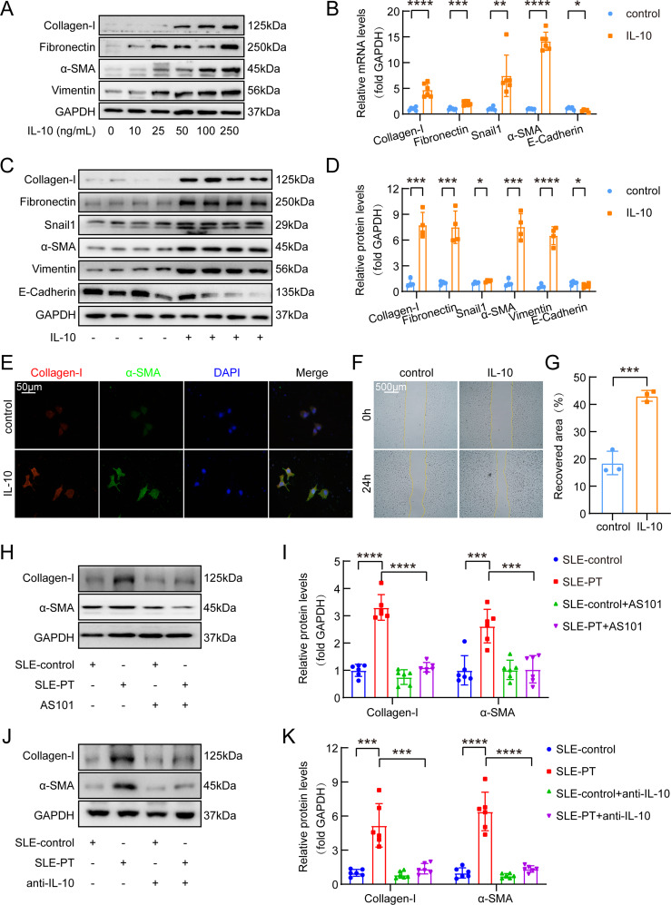
Background: Interleukin-10 (IL-10), a pivotal anti-inflammatory cytokine, has gotten attention for its involvement in tissue remodeling and organ fibrosis. Pleurisy and subsequent pleural remodeling are recognized as quantifiable indicators of systemic lupus erythematosus (SLE) activity. However, the role of IL-10 in SLE-associated pleural remodeling remains unknown. In this study, we investigated role of IL-10 in SLE-associated pleural remodeling and the underlying mechanism.

Methods: Clinical data and serum specimens were obtained from SLE patients, while pleural mesothelial cells and mouse models served as primary experimental subjects. The protein expression-related technologies, histopathological staining, and other experimental methods were used in the study.

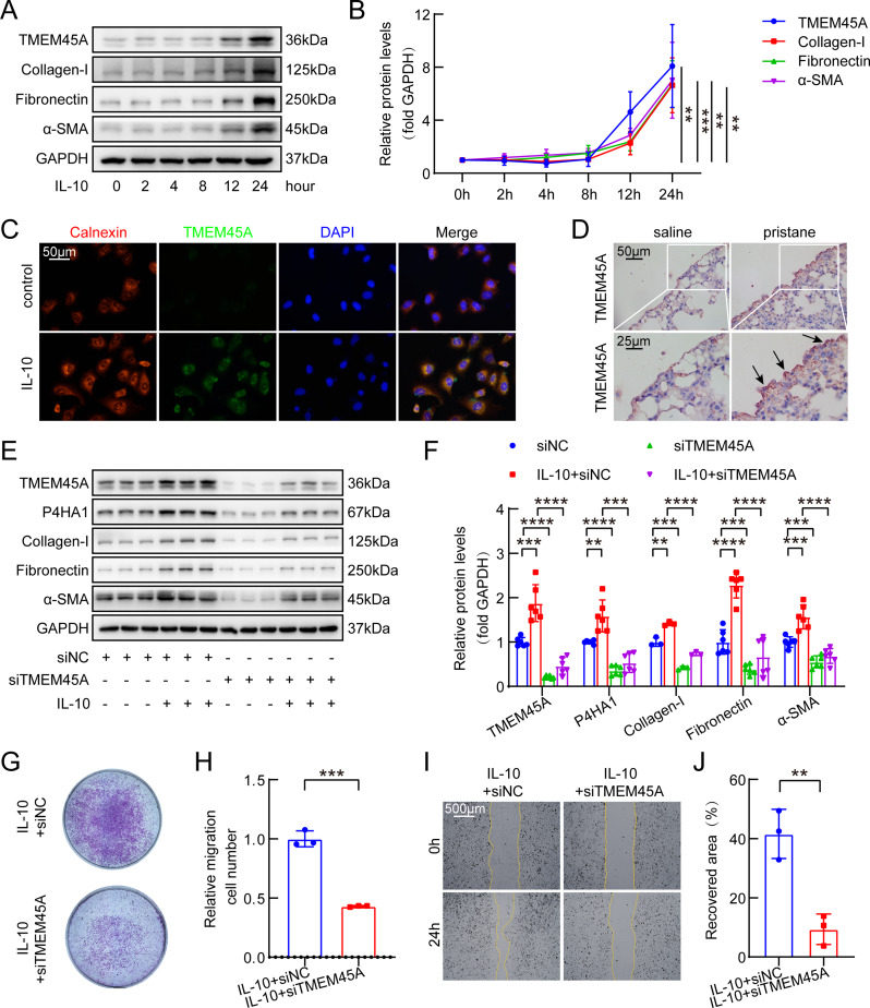
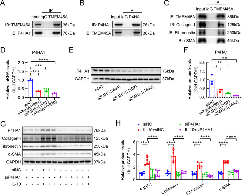
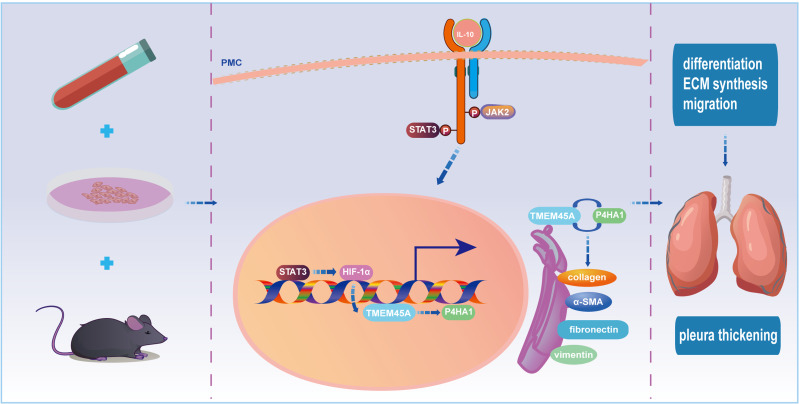
Results: Our investigation got several key findings. Firstly, serum obtained from SLE patients with pleural thickening was found to induce pleural mesothelial cell remodeling. Subsequently, heightened levels of IL-10 were found in serum from SLE patients with pleural thickening compared to that of SLE patients without pleural thickening. Secondly, administration of recombinant IL-10 was confirmed its ability to induce pleural mesothelial cell remodeling, on the contrary, this remodeling was effectively mitigated by IL-10 inhibition. Notably, blockade of IL-10 significantly prevented collagen deposition and prevented thickening in pleura of SLE mouse models. Lastly, the IL-10/JAK2/STAT3/HIF1α/TMEM45A/P4HA1 signaling axis was elucidated to mediate pleural remodeling and thickening.

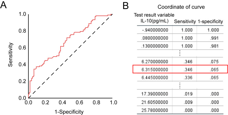
Conclusions: Our study uncovered that IL-10 mediated pleural remodeling in SLE. We suggested that serum IL-10 level exceeding 6.32 pg/mL was a potential reference threshold for predicting pleural thickening in SLE patients.

Keywords: IL-10; Pleural remodeling; Systemic lupus erythematosus (SLE).


Conflict of interest statement

Declarations Ethics approval and consent for participants The clinical data and patient serum samples used in experiments were from inpatients in the Union Hospital, Tongji Medical College, Huazhong University of Science and Technology. The study was approved by the Medical Ethics Committee of Tongji Medical College, Huazhong University of Science and Technology (Approval number: S1220) and all patients signed informed consent. The study involving animals were carried out according to the ethical policies and procedures approved by the Medical Ethics Committee of Tongji Medical College, Huazhong University of Science and Technology (Approval number: 20200411). Consent for publication All authors have read and agreed with the submission of the manuscript. This manuscript has not been published or presented elsewhere in part or in entirety. Competing interests The authors declare no competing interests.

Figures

Fig. 1
Fig. 1
Serum derived from SLE-PT patients induced PMC remodeling. (A-B) Primary rat PMCs were incubated with serum (5%) obtained from SLE-control or SLE-PT patients for 48 h, following which intracellular levels of collagen-I and α-SMA were measured by western blotting. n = 12. (C-F) Primary rat PMCs were treated with serum (5%) from either SLE-control or SLE-PT patients for 24 h, whereupon cell proliferation was evaluated using CCK-8 (C, n = 10) and EdU (D, n = 4) assays. Scale bar, 50 μm. Migration capability was assessed through wound healing assay (E-F). n = 3. Scale bar, 500 μm. Data are shown as mean ± SEM of n individual experiments. Statistical significance was determined using unpaired Student’s t-test. *P < 0.05, ***P < 0.001, ****P < 0.0001
Fig. 2
Fig. 2
Elevated levels of IL-10 were found in the serum of SLE-PT patients. Serum samples were obtained from SLE-control and SLE-PT patients. (A) IL-10 levels were quantified utilizing a combination cytokine detection kit based on immunofluorescence technology. n = 220 (SLE-control) and n = 150 (SLE-PT). (B) The correlation between IL-10 levels and pleural thickness in SLE-PT patients was analyzed. n = 150. Data are shown as mean ± SEM of n individual experiments. Statistical significance was determined using unpaired Student’s t-test (A) and Pearson correlation test (B). ****P < 0.0001
Fig. 3
Fig. 3
IL-10 induced remodeling and migration of PMCs. (A) Human PMCs were incubated with recombinant human IL-10 across a concentration gradient (0, 10, 25, 50, 100 and 250 ng/mL) for 24 h, followed by quantification of intracellular protein levels of collagen-I, fibronectin, α-SMA and vimentin using western blot analysis. (B-G) Human PMCs were treated with recombinant human IL-10 (100 ng/mL). After 24 h, quantitative real-time PCR (qRT-PCR) was employed to assess relative mRNA expression levels of collagen-I, fibronectin, snail1, α-SMA and E-Cadherin (B). n = 5~6. Western blot analysis was performed to determine relative protein levels of collagen-I, fibronectin, snail1, α-SMA, vimentin and E-Cadherin (C-D). n = 4~5. Immunofluorescence staining was conducted to visualize Collagen-I (red) and α-SMA (green) proteins (E). Scale bar, 50 μm. Migration capability was evaluated through wound healing assay (F-G). n = 3. Scale bar, 500 μm. (H-I) Primary rat PMCs were pretreated with small molecule inhibitor of IL-10 (AS101, 1 µg/mL) for 1 h before exposure to patient sera for 24 h. Western blot analysis was conducted to assess protein levels of collagen-I and α-SMA. n = 6. (J-K) Primary rat PMCs were pretreated with neutralizing antibody of IL-10 (anti-IL-10, 1 µg/mL) for 1 h prior to incubation with patient sera for 24 h. Protein levels of collagen-I and α-SMA were then measured by western blot analysis. n = 6. Data are shown as mean ± SEM of n individual experiments. Statistical significance was determined using unpaired Student’s t-test. *P < 0.05, **P < 0.01, ***P < 0.001, ****P < 0.0001
Fig. 4
Fig. 4
Anti-IL-10 and IL-10 receptor blocker attenuated pleural fibrosis in pristane-induced SLE model. Eight-week-old female BALB/c mice received intraperitoneal injections of pristane (0.5 mL/mouse) to induce SLE model. Neutralizing antibody targeting IL-10 (anti-IL-10, 10 µg/kg) and IL-10 receptor blocker (anti-IL-10R, 10 µg/kg) were intrapleurally administered at days 5, 9, 14, 18, 22 and 27. All mice were euthanized on day 90. (A-C) Peripheral blood collected at day 60 was subjected to ELISA for the quantification of ANA, anti-dsDNA and anti-Sm levels. n = 8 (saline), n = 5 (anti-IL-10), n = 6 (anti-IL-10R), n = 7 (pristane), n = 6 (pristane + anti-IL-10), n = 6 (pristane + anti-IL-10R). (D) Serum IL-10 levels were detected by ELISA. n = 8 (saline), n = 5 (anti-IL-10), n = 6 (anti-IL-10R), n = 7 (pristane), n = 6 (pristane + anti-IL-10), n = 6 (pristane + anti-IL-10R). (E) Lung tissues were harvested for Masson’s trichrome staining to visualize collagen deposition in visceral, parietal, and diaphragmatic pleura. Scale bar, 50 μm. (F-H) Analysis of pleural thickness was performed based on Masson’s trichrome staining. n = 8 (saline), n = 5 (anti-IL-10), n = 5 (anti-IL-10R), n = 4 (pristane), n = 6 (pristane + anti-IL-10), n = 5 (pristane + anti-IL-10R). Data are shown as mean ± SEM of n individual experiments. Statistical significance was determined using unpaired Student’s t-test. *P < 0.05, **P < 0.01, ***P < 0.001, ****P < 0.0001
Fig. 5
Fig. 5
TMEM45A was a mediator of IL-10-induced PMC remodeling and migration. (A-B) Human PMCs were exposed to recombinant human IL-10 (100 ng/mL) over a time gradient (0, 2, 4, 8, 12 and 24 h), followed by measurement of intracellular protein levels of TMEM45A, collagen-I, fibronectin and α-SMA by western blot. n = 3. (C) Following incubation with recombinant human IL-10 (100 ng/mL) for 24 h, calnexin (a marker of endoplasmic reticulum, red) and TMEM45A (green) proteins were detected by immunofluorescence staining in human PMCs. Scale bar, 50 μm. (D) Immunohistochemical staining was conducted to assess TMEM45A protein expression in lung tissue from pristane-induced SLE model. Brown, TMEM45A protein positive. Scale bars, 50 μm and 25 μm. (E-J) After transfection with TMEM45A siRNA (siTMEM45A), cells were treated with IL-10 (100 ng/mL) for 24 h, and relative protein levels of TMEM45A, P4HA1, collagen-I, fibronectin and α-SMA were measured by western blot (E-F). n = 6 (TMEM45A, P4HA1, fibronectin and α-SMA), n = 3 (collagen-I). Migration capability was detected by transwell migration assay (G-H), and wound healing assay (I-J). n = 3. Scale bar, 500 μm. Data are shown as mean ± SEM of n individual experiments. Statistical significance was determined using unpaired Student’s t-test. **P < 0.01, ***P < 0.001, ****P < 0.0001
Fig. 6
Fig. 6
TMEM45A interacted with P4HA1 and ECM, TMEM45A/P4HA1 mediated IL-10-induced PMC remodeling. (A) Co-IP assays were conducted in human PMCs using anti-TMEM45A and anti-IgG antibodies, followed by detection of protein precipitated complexes using anti-TMEM45A and anti-P4HA1 antibodies via western blot analysis. (B) Co-IP assays were performed in human PMCs using anti-P4HA1 and anti-IgG antibodies, followed by detection of protein precipitated complexes using anti-P4HA1 and anti-TMEM45A antibodies via western blot analysis. (C) Co-IP assays were conducted using anti-TMEM45A and anti-IgG antibodies in human PMCs, followed by detection of protein precipitated complexes using anti-TMEM45A, anti-collagen-I, anti-fibronectin, and anti-α-SMA antibodies via western blot analysis. (D) Human PMCs were transfected with siRNAs specific for P4HA1 (site 694, 1107 and 1630) for 48 h. P4HA1 mRNA knockdown efficiency was assessed by qRT-PCR. n = 3. (E-F) P4HA1 protein knockdown efficiency was assessed by western blot. n = 3. (G-H) After transfection with P4HA1 siRNA (site 1630), cells were treated with IL-10 (100 ng/mL) for 24 h, and relative protein levels of P4HA1, collagen-I, fibronectin and α-SMA were measured by western blot. n = 6. Data are shown as mean ± SEM of n individual experiments. Statistical significance was determined using unpaired Student’s t-test. *P < 0.05, **P < 0.01, ***P < 0.001, ****P < 0.0001
Fig. 7
Fig. 7
IL-10 increased TMEM45A through JAK2/STAT3/HIF1α signaling. (A-B) Human PMCs were treated with recombinant human IL-10 (100 ng/mL) for 24 h, and relative protein expressions of p-JAK2, t-JAK2, p-STAT3, t-STAT3, phosphorylated inhibitor of nuclear factor kappa-B kinase alpha (p-IκBα), phosphorylated nuclear factor kappa-B P65 subunit (p-P65) and t-P65 were detected by western blot. n = 6. (C-F) PMCs were exposed to recombinant human IL-10 (100 ng/mL) for 24 h in the presence or absence of artesunate, a STAT3 inhibitor (10 µM). Intracellular protein levels were quantified utilizing western blot (C-D). n = 6. (E-F) Following pre-treatment with either artesunate (10 µM) or BAY87-2243 (a HIF1α inhibitor, 100 nM), cells were incubated with recombinant human IL-10 (100 ng/mL) for 24 h. Intracellular protein levels were evaluated through western blot. n = 6. Data are shown as mean ± SEM of n individual experiments. Statistical significance was determined using unpaired Student’s t-test. *P < 0.05, **P < 0.01, ***P < 0.001, ****P < 0.0001
Fig. 8
Fig. 8
Serum IL-10 > 6.32 pg/mL was recommended as a potential reference threshold for predicting pleural thickening in SLE patients. Receiver operating characteristic (ROC) curve analysis was performed based on IL-10 levels in serum from 107 pairs of SLE patients. A numerical inflection point (Yoden index = 0.281) was simulated for occurrence of pleural thickening in SLE patients
Fig. 9
Fig. 9
Diagram of research results. Schematic illustration is describing the molecular mechanism by which IL-10 regulates PMCs differentiation, ECM synthesis and migration through JAK2/STAT3/HIF1α/TMEM45A/P4HA1 signaling in pathophysiological process of pleural thickening in SLE

References

    1. Bakshi J, Segura BT, Wincup C, Rahman A. Unmet needs in the Pathogenesis and treatment of systemic Lupus Erythematosus. Clin Rev Allergy Immunol. 2018;55:352–67. - PMC - PubMed
    1. Polachek A, Gladman DD, Su J, Urowitz MB. Defining low Disease activity in systemic Lupus Erythematosus. Arthritis Care Res (Hoboken). 2017;69:997–1003. - PubMed
    1. Torre O, Harari S. Pleural and pulmonary involvement in systemic lupus erythematosus. Presse Med. 2011;40:e19–29. - PubMed
    1. Fanouriakis A, Kostopoulou M, Alunno A, Aringer M, Bajema I, Boletis JN, et al. 2019 update of the EULAR recommendations for the management of systemic lupus erythematosus. Ann Rheum Dis. 2019;78:736–45. - PubMed
    1. Zucchi D, Elefante E, Calabresi E, Signorini V, Bortoluzzi A, Tani C. One year in review 2019: systemic lupus erythematosus. Clin Exp Rheumatol. 2019;37:715–22. - PubMed

MeSH terms

Substances

LinkOut - more resources