. 2024 Nov 19;21(1):303.
doi: 10.1186/s12974-024-03287-1.

Localization of brain neuronal IL-1R1 reveals specific neural circuitries responsive to immune signaling

Affiliations

Localization of brain neuronal IL-1R1 reveals specific neural circuitries responsive to immune signaling

Daniel P Nemeth et al. J Neuroinflammation. .

Abstract

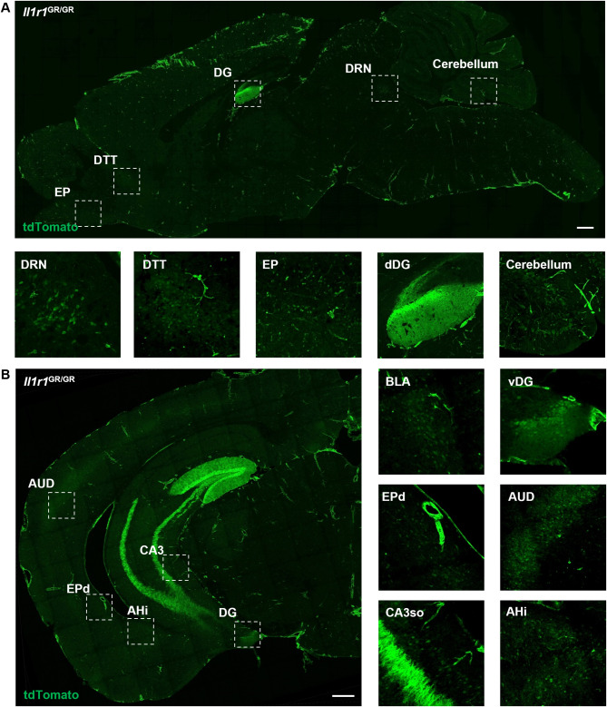
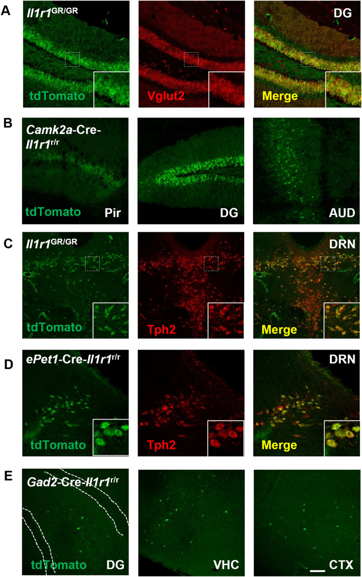
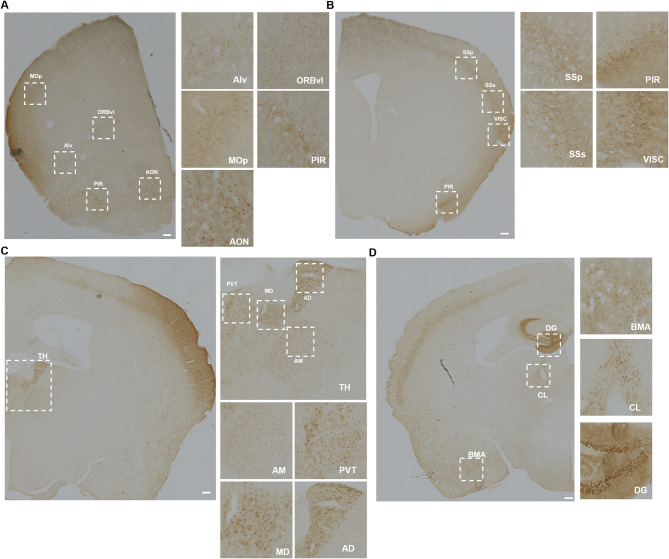
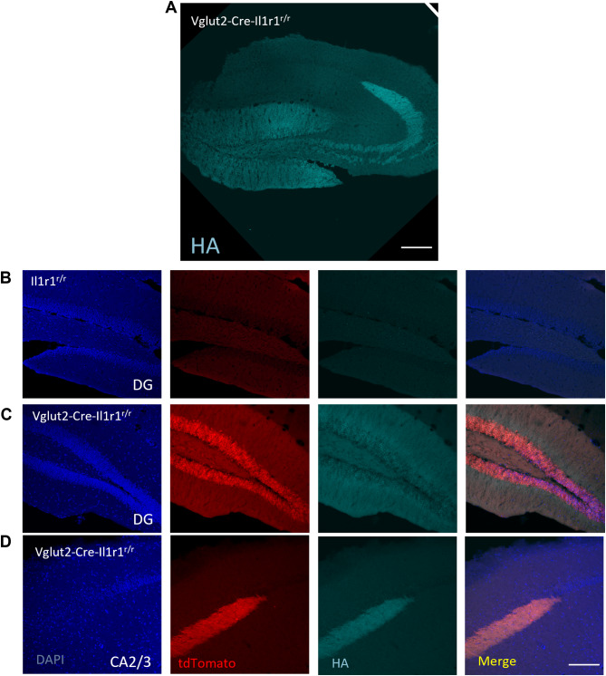
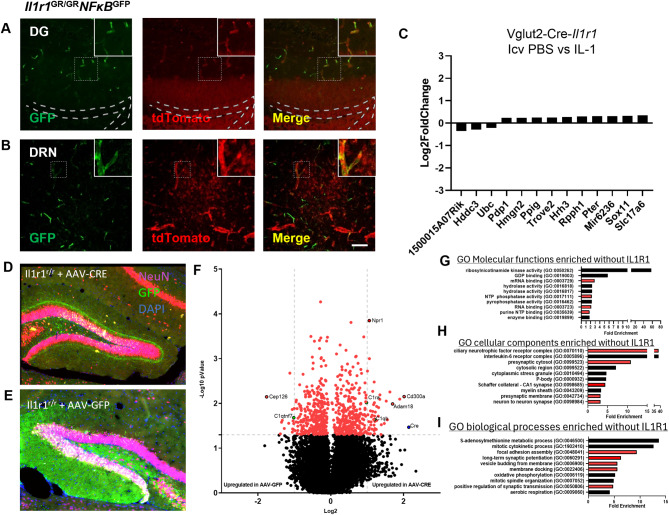
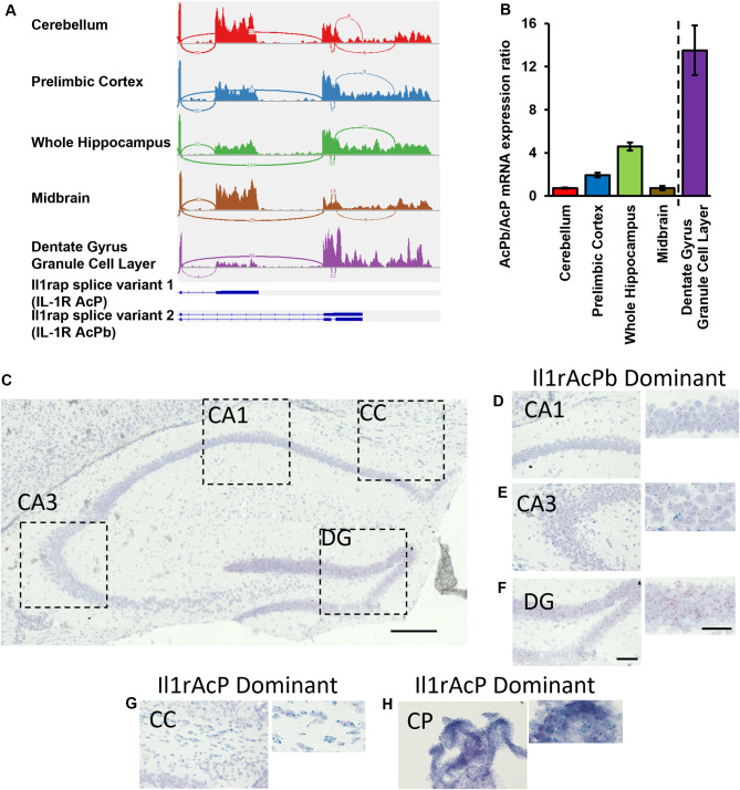
Interleukin-1 (IL-1) is a pro-inflammatory cytokine that exerts a wide range of neurological and immunological effects throughout the central nervous system (CNS) and is associated with the etiology of affective and cognitive disorders. The cognate receptor for IL-1, Interleukin-1 Receptor Type 1 (IL-1R1), is primarily expressed on non-neuronal cells (e.g., endothelial cells, choroidal cells, ventricular ependymal cells, astrocytes, etc.) throughout the brain. However, the presence and distribution of neuronal IL-1R1 (nIL-1R1) has been controversial. Here, for the first time, a novel genetic mouse line that allows for the visualization of IL-1R1 mRNA and protein expression (Il1r1GR/GR) was used to map all brain nuclei and determine the neurotransmitter systems which express nIL-1R1 in adult male mice. The direct responsiveness of nIL-1R1-expressing neurons to both inflammatory and physiological levels of IL-1β in vivo was tested. Neuronal IL-1R1 expression across the brain was found in discrete glutamatergic and serotonergic neuronal populations in the somatosensory cortex, piriform cortex, dentate gyrus, and dorsal raphe nucleus. Glutamatergic nIL-1R1 comprises most of the nIL-1R1 expression and, using Vglut2-Cre-Il1r1r/r mice, which restrict IL-1R1 expression to only glutamatergic neurons, an atlas of glutamatergic nIL-1R1 expression across the brain was generated. Analysis of functional outputs of these nIL-1R1-expressing nuclei, in both Il1r1GR/GR and Vglut2-Cre-Il1r1r/r mice, reveals IL-1R1+ nuclei primarily relate to sensory detection, processing, and relay pathways, mood regulation, and spatial/cognitive processing centers. Intracerebroventricular (i.c.v.) injections of IL-1 (20 ng) induces NFκB signaling in IL-1R1+ non-neuronal cells but not in IL-1R1+ neurons, and in Vglut2-Cre-Il1r1r/r mice IL-1 did not change gene expression in the dentate gyrus of the hippocampus (DG). GO pathway analysis of spatial RNA sequencing 1mo following restoration of nIL-1R1 in the DG neurons reveals IL-1R1 expression downregulates genes related to both synaptic function and mRNA binding while increasing select complement markers (C1ra, C1qb). Further, DG neurons exclusively express an alternatively spliced IL-1R Accessory protein isoform (IL-1RAcPb), a known synaptic adhesion molecule. Altogether, this study reveals a unique network of neurons that can respond directly to IL-1 via nIL-1R1 through non-autonomous transcriptional pathways; earmarking these circuits as potential neural substrates for immune signaling-triggered sensory, affective, and cognitive disorders.


Conflict of interest statement

Declarations Ethical approval All experiments were approved by the Florida Atlantic University Institutional Animal Care and Use Committee under protocol A22-39 and A22-14. Consent for publication Not applicable. Competing interests The authors declare no competing interests.

Figures

Fig. 1
Fig. 1
Representative images of neuronal IL-1R1 expression in the brain. Representative tilescan images of tdTomato labeling of a sagittal (A) and coronal (B) section from an Il1r1GR/GR mouse. Areas where high levels of neuronal IL-1R1 were found are marked with dashed squares and shown at higher magnification below (A) or to the right of the low-magnification image (B). Scale bar: 500 μm. DRN, dorsal raphe nucleus; DTT, dorsal tenia tecta; EP, endopiriform nucleus; dDG, dorsal dentate gyrus; vDG, ventral dentate gyrus; AUD, auditory cortex; CA3, Cornus ammonus 3; AHi, hippocampal-amygdalar transition area
Fig. 2
Fig. 2
Neuronal IL-1R1 is expressed in diverse types of neurons classified by different neurotransmitters. (A) Representative images of tdTomato and Vglut2 labeling in Il1r1GR/GR hippocampal sections. Dashed squares mark the area shown at higher magnification on the bottom right. (B) Representative images of tdTomato from different CamK2a-Cre-Il1r1r/r brain sections. Pir, Piriform cortex; DG, dentate gyrus; AUD, primary auditory cortex. (C) Representative images of tdTomato and Tph2 labeling in Il1r1GR/GR dorsal raphe sections. A dashed square marks the area shown at higher magnification on the bottom right. DRN, dorsal raphe nucleus. (D) Representative images of tdTomato and Tph2 labeling in ePet1-Cre-Il1r1r/r dorsal raphe sections. Dashed squares mark the area shown at higher magnification on the bottom right. (E) Representative images of tdTomato from different Gad2-Cre-Il1r1r/r brain sections, white dotted lines denote granule cell layer of the DG. DG, Dentate gyrus, VHC, ventral hippocampus, CTX, cortex. Scale bar: 200 μm
Fig. 3
Fig. 3
Representative images of IL-1R1 expression on glutamatergic neurons in different brain sections. (A) Representative images of tdTomato immunolabeling in Vglut2-Cre-Il1r1r/r mice, highlighting the cortex and olfactory areas. MOp, primary motor area; ORBvl, orbital area, ventrolateral part; Alv, agranular insular area, ventral part; PIR, piriform area; AON, anterior olfactory nucleus. The dashed squares indicate areas with high neuronal IL-1R1 expression and are magnified. Scale bar = 200 μm (B) Representative images of tdTomato immunolabeling in the Vglut2-Cre-Il1r1r/r somatosensory areas, visceral areas, and piriform area. SSp, primary somatosensory area; SSs, supplementary somatosensory area; VISC, visceral areas; PIR, piriform area. Scale bar: 200 μm (C) Representative images of DAB labeling in various thalamic nuclei of the Vglut2-Cre-Il1r1r/r mouse. AD, anterodorsal nucleus; MD, mediodorsal nucleus of thalamus; PVT, paraventricular nucleus of thalamus; AM, anteromedial nucleus. Scale bar: 200 μm (D) Representative images of tdTomato immunolabeling in Vglut2-Cre-Il1r1r/r mouse dentate gyrus (DG), central lateral nucleus of the thalamus (CL), and basomedial amygdalar nucleus (BMA). Scale bar = 200 μm
Fig. 4
Fig. 4
IL-1R1 protein expression is localized across the entire neuron of the dentate gyrus. (A) Representative tilescan image of immunofluorescent labeling of HA-tag in the hippocampus of glyoxal-fixed Vglut2-Cre-Il1r1r/r mice. (B-C) 20x micrograph of immunofluorescent labeling in the DG of DAPI, tdTomato, and HA-tag in Il1r1r/r (B) or Vglut2-Cre-Il1r1r/r (C) mice. (D) 20x micrograph of immunofluorescent labeling in the CA3 of DAPI, tdTomato, and HA-tag in Vglut2-Cre-Il1r1r/r (D) mice
Fig. 5
Fig. 5
Neuronal IL-1R1-mediated signaling is independent of NF-κB pathway and chronic restoration of DG neuronal IL-1R1 reveals negative regulation of synaptic-related pathways. (A-B) Representative images of GFP and tdTomato labeling in Il1r1GR/GRNFκBGFP DG (A) and DRN (B) sections. The dashed square marks the area shown at higher magnification on the top right. Dashed lines mark the dentate gyrus granule cell layer. DG, dentate gyrus; DRN, dorsal raphe nucleus. Scale bar = 200 μm. (C) Log2 fold change of top 13 differentially expressed genes of microdissected dentate gyrus following i.c.v. IL-1 (20 ng) compared to PBS. (D-E) Immunofluorescent labeling of NeuN (Red), GFP (Green), and DAPI (Blue) in Il1r1r/r mice injected with AAV-CMV-CRE-GFP into the right dentate (D) and AAV-CMV-GFP injected into the left dentate (E). (F) Volcano plot of differentially expressed genes between AAV-Cre versus AAV-GFP injected dentate gyrus neurons within Il1r1r/r mice (n = 3). Vertical dotted lines indicate a log2 value of 1 and horizontal dotted lines indicate a -Log10 p value of 1.3. Red data points were used in GO pathway enrichment analysis in G-I. (G-I) GO Molecular function (G), Cellular component (H), and Biological Process (I) pathway analysis of differentially expressed genes in the AAV-CMV-GFP injected Il1r1r/r DG neurons
Fig. 6
Fig. 6
IL-1R1 expressing neurons utilize alternatively spliced accessory protein, IL-1RAcPb. (A) Sashimi plots from RNAseq data analyses of different brain areas and schematics showing the last two exons of Il1rap. (D) Quantification of AcPb/AcP RNA transcripts ratio in different brain areas. Dashed line separates two different RNAseq experiments. Error bars represent the mean ± SEM. (C) Photomicrograph of the WT hippocampus following BaseScope in situ hybridization of exon junction regions for IL-1RAcP (Blue-Green) and IL-1RAcPb (Red). Scale bar = 300 μm Dotted lines indicate magnified regions presented in D-G. (D-F) Photomicrographs of predominantly IL-1RAcPb expression regions, such as the CA1 (D), CA3 (E), and DG (F), which are magnified images to the right of each corresponding image. (G-H) Photomicrographs of predominantly IL-1RAcP expressing regions of the corpus callosum (G) and the choroid plexus (H), which are magnified images to the right of each corresponding image. DG, dentate gyrus; CC, corpus callosum; CP, choroid plexus. Scale bar = 100 μm

References

    1. Avital A, Goshen I, Kamsler A, Segal M, Iverfeldt K, Richter-Levin G, Yirmiya R. Impaired interleukin-1 signaling is associated with deficits in hippocampal memory processes and neural plasticity. Hippocampus. 2003;13(7):826–34. 10.1002/hipo.10135. - PubMed
    1. Baganz NL, Lindler KM, Zhu CB, Smith JT, Robson MJ, Iwamoto H, Deneris ES, Hewlett WA, Blakely RD. A requirement of serotonergic p38α mitogen-activated protein kinase for peripheral immune system activation of CNS serotonin uptake and serotonin-linked behaviors. Translational Psychiatry. 2015;5(11):e671. 10.1038/tp.2015.168. - PMC - PubMed
    1. Bajo M, Patel RR, Hedges DM, Varodayan FP, Vlkolinsky R, Davis TD, Burkart MD, Blednov YA, Roberto M. Role of MyD88 in IL-1β and ethanol modulation of GABAergic Transmission in the Central Amygdala. Brain Sci. 2019;9(12). 10.3390/brainsci9120361. Article 12. - PMC - PubMed
    1. Bajo M, Varodayan FP, Madamba SG, Robert AJ, Casal LM, Oleata CS, Siggins GR, Roberto M. IL-1 interacts with ethanol effects on GABAergic transmission in the mouse central amygdala. Front Pharmacol. 2015;6:49. 10.3389/fphar.2015.00049. - PMC - PubMed
    1. Ban E, Milon G, Prudhomme N, Fillion G, Haour F. Receptors for interleukin-1 (α and β) in mouse brain: mapping and neuronal localization in hippocampus. Neuroscience. 1991;43(1):21–30. 10.1016/0306-4522(91)90412-h. - PubMed

Substances

LinkOut - more resources