. 2024 Dec 3;18(48):33058-33072.
doi: 10.1021/acsnano.4c08490. Epub 2024 Nov 20.

Distinct Inflammatory Programs Underlie the Intramuscular Lipid Nanoparticle Response

Affiliations

Distinct Inflammatory Programs Underlie the Intramuscular Lipid Nanoparticle Response

William Dowell et al. ACS Nano. .

Abstract

Developments in mRNA/lipid nanoparticle (LNP) technology have advanced the fields of vaccinology and gene therapy, raising questions about immunogenicity. While some mRNA/LNPs generate an adjuvant-like environment in muscle tissue, other mRNA/LNPs are distinct in their capacity for multiple rounds of therapeutic delivery. We evaluate the adjuvancy of components of mRNA/LNPs by phenotyping cellular infiltrate at injection sites, tracking uptake by immune cells, and assessing the inflammatory state. Delivery of 9 common, but chemically distinct, LNPs to muscle revealed two classes of inflammatory gene expression programs: inflammatory (Class A) and noninflammatory (Class B). We find that intramuscular injection with Class A, but not Class B, empty LNPs (eLNPs) induce robust neutrophil infiltration into muscle within 2 h and a diverse myeloid population within 24 h. Single-cell RNA sequencing revealed SM-102-mediated expression of inflammatory chemokines by myeloid infiltrates within muscle 1 day after injection. Surprisingly, we found direct transfection of muscle infiltrating myeloid cells and splenocytes 24 h after intramuscular mRNA/LNP administration. Transfected myeloid cells within the muscle exhibit an activated phenotype 24 h after injection. Similarly, directly transfected splenic lymphocytes and dendritic cells (DCs) are differentially activated by Class A or Class B containing mRNA/LNP. Within the splenic DC compartment, type II conventional DCs (cDC2s) are directly transfected and activated by Class A mRNA/LNP. Together, we show that mRNA and LNPs work synergistically to provide the necessary innate immune stimuli required for effective vaccination. Importantly, this work provides a design framework for vaccines and therapeutics alike.

Keywords: cancer vaccine; gene therapy; innate immunity; ionizable lipids; lipid nanoparticles; mRNA therapeutics; mRNA vaccines.


Conflict of interest statement

Notes

The authors declare no competing financial interest.

Figures

Figure 1.
Figure 1.
Distinct transcriptomic signatures induced by ionizable lipids. (A) Gene expression signature distinguishes inflammatory and noninflammatory ionizable lipids. Nine different eLNPs, differing only in the ionizable lipid, were made and injected intramuscularly into mice. 24 h after administration, muscle tissue was harvested, and RNA was collected for sequencing. Class A inflammatory and Class B noninflammatory ionizable lipids are distinguished by their ability to induce inflammatory gene expression within injected muscle tissue. (B) Gene expression signature induced by ionizable lipids. RNA was collected from injected muscle tissue at indicated time points and prepared for bulk RNA sequencing. Data shown are averaged Z-score values from biological duplicates, n = 2–4 mice. (C) Inflammatory Class A and noninflammatory Class B ionizable lipids. Ionizable lipids were classified based on the transcriptomic signature they induce within injected muscle tissue, as shown in (B). (D,E) Class A and Class B ionizable lipids differentially induce inflammatory genes by 24 h. RPKM values for indicated genes were calculated and grouped based on Class A or Class B ionizable lipid treatment. Data shown are average RPKM values from biological duplicates 24 h after LNP injection. Statistical significance was assessed by Students’ t test *P ≤ 0.05. (F) Gene ontology analysis reveals inflammatory gene expression programs induced by Class A and Class B ionizable lipids.
Figure 2.
Figure 2.
Myeloid infiltration into injection site within hours of injection. (A) Class A ionizable lipids induce robust neutrophil responses within hours of injection. Mice were intramuscularly injected with 2.5 μg eLNP formulated without an ionizable lipid (nonionizable) or with indicated ionizable lipids. Mice were intramuscularly injected with LNP, and single-cell suspensions were prepared for flow cytometric analysis at 2 h post-injection to characterize intramuscular immune cells. Representative neutrophil gating is shown, and summarized neutrophil frequencies of CD45+ cells are shown at right, n = 4–11 mice. Statistical significance was assessed by ANOVA. *P ≤ 0.05, **P ≤ 0.01, and ****P ≤ 0.0001. (B) Choice of ionizable lipid shapes eLNP capability to induce neutrophil infiltration into the site of injection within 24 h. Mice were injected intramuscularly with 2.5 μg of indicated eLNP, LNP lacking an ionizable lipid, or PBS. 24 h after injection, muscle tissue was harvested, and single-cell suspensions were generated for flow cytometry. Representative neutrophil gating is shown, and summarized neutrophil frequencies are shown at right, n = 4–14 mice. Statistical significance was assessed by ANOVA. *P ≤ 0.05 and **P ≤ 0.01. Mice were injected intramuscularly with 2.5 μg of indicated eLNP, LNP lacking an ionizable lipid, or PBS. 24 h after injection, muscle tissue was harvested, and single-cell suspensions were generated for flow cytometry. (C) Neutrophil infiltration into the site of injection not enhanced by mRNA in LNPs. Mice were intramuscularly injected with a total of 450 ng of mRNA encoding Luciferase encapsulated in 2.5 μg of LNP. Muscle tissue was harvested, and single-cell suspensions for flow cytometry were generated at 2 and 24 h post-injection, n = 5–12 mice. Statistical significance was assessed by ANOVA. (D) Neutrophil recruitment is confined to the site of injection. Neutrophil recruitment to the ipsilateral and contralateral limbs was assessed after SM-102 eLNP injection. Representative FACS plots showing neutrophil frequencies of CD45+ cells in indicated limbs after SM-102 eLNP treatment. Mean fold change in frequency is summarized at right, n = 3. (E) Neutrophil frequencies in muscle return to baseline by 3 days. Mice were intramuscularly injected with 2.5 μg of SM-102 eLNP, and neutrophil infiltration was assessed by flow cytometry 2, 3, or 4 days later.
Figure 3.
Figure 3.
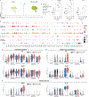
Single-cell RNA sequencing reveals inflammatory signature of myeloid infiltrates. (A) UMAP plot of single-cell RNA sequencing data of CD45+ immune cells isolated from SM-102 eLNP or PBS-injected muscle tissue. (B) SM-102 eLNP induces inflammatory gene expression within CD45+ cells in muscle. Dot plot showing fold change in expression of select genes in immune cells from SM-102 eLNP-treated mice relative to cells from PBS-treated mice. Relative levels of expression are stratified by cell type identity as depicted in (A) and determined in Supporting Information Figure S3B. (C) SM-102 eLNP induces ISG expression in neutrophils. Violin plots of select ISGs. Data shown are averaged expression of biological duplicates.(D) SM-102 eLNP induces inflammatory cytokine expression in distinct cell types within injected muscle tissue. Violin plots showing expression of select cytokines in immune cells within injected muscle tissue. Data shown are averaged expression values from biological duplicates. (E) Inflammasome priming in neutrophils within injected muscle. Violin plots showing Nlrp3 and Il1b expression values across cell types within injected muscle tissue. Data shown are averaged expression values from biological duplicates. (F) Inflammatory chemokine expression is dependent upon the ionizable lipid component of LNP. Serological analyses reveal that inflammatory chemokine expression is dependent upon ionizable lipid. Mice were injected intramuscularly with SM-102, ALC-0315, nonionizable eLNP or PBS. 24 h after injection, serum was collected and screened for soluble mediators using high-throughput ELISA. Data are displayed as means and standard deviations, n = 3. Statistical significance was assessed by ANOVA. *P ≤ 0.05, **P ≤ 0.01, and ***P ≤ 0.001.
Figure 4.
Figure 4.
Myeloid infiltrates are transfected and activated within the muscle with SM-102 mRNA/LNP. (A) Differential uptake and translation of mRNA payload between different muscle-infiltrating myeloid cells. 450 ng of Cre mRNA encapsulated in SM-102 containing LNPs was injected intramuscularly into Ai6 mice. 24 h after injection, muscles and spleens were harvested, and single-cell suspensions were generated for flow cytometry. Data are displayed as means with standard deviations, n = 8. (B–D) Directly transfected muscle-infiltrating myeloid cells are activated. (C) Directly transfected Ly6Chi monocytes within the muscle are activated. Mean fluorescence intensity plots normalized to the mode of indicated markers on monocytes. Data are summarized below. Contour plots showing differential expression of indicated markers between GFP- and GFP+ monocytes. Data are summarized below. (D) Directly transfected macrophages are activated by mRNA/LNPs. Mean fluorescence intensity plots normalized to the mode showing differential expression of indicated markers between GFP + and GFP-macrophages. Data are summarized at right. (E) Muscle-infiltrating DCs are activated by mRNA/LNPs. Mean fluorescence intensity plots normalized to the mode showing differential expression of indicated markers between GFP+ and GFP-DCs. Data are summarized at right. Data are presented as means and standard deviations, n = 8. Statistical significance was assessed by paired Students’ t test. *P ≤ 0.05, **P ≤ 0.01, ***P ≤ 0.001, and ****P ≤ 0.0001.
Figure 5.
Figure 5.
Splenic APCs are directly transfected and activated within 24 h of SM-102 mRNA/LNP IM administration. (A) Differential uptake and translation of mRNA payload between different immune cells. 450 ng of Cre mRNA encapsulated in SM-102 containing LNPs was injected intramuscularly into Ai6 mice. 24 h later, muscles and spleens were harvested, and single-cell suspensions were generated for flow cytometry. Data are displayed as means, n = 8. (B–F) Directly transfected splenocytes display an activated phenotype. MHCII and CD86 expression between indicated GFP+ and GFP- immune cells displayed as mean fluorescence intensity. Data are summarized at right. Data presented as means and standard deviations, n = 8. Statistical significance was assessed by paired Students’ t test. *P ≤ 0.05, **P ≤ 0.01, ***P ≤ 0.001, and ****P ≤ 0.0001.
Figure 6.
Figure 6.
Differential activation of splenic APCs by Class A and B mRNA/LNP. (A,B) Differential delivery of mRNA payload to splenic APCs by Class A and Class B containing mRNA/LNP. 5 μg Cre recombinase encapsulated in either SM-102 or TCL053 containing LNPs was intramuscularly injected into Ai6 mice. 24 h later, spleens were harvested, and single-cell suspensions were prepared for flow cytometry. Cre-mediated GFP expression was analyzed in splenic DCs or B cells 24 h after indicated treatment. Summary data are displayed as means and standard deviations, n = 4. Statistical significance was assessed by Students’ t test. ***P ≤ 0.001. (C,D) Differential activation of transfected APCs by Class A and Class B mRNA/LNP. Mean fluorescence intensity of CD86 and MHCII on GFP+ DC (C) or GFP+ B cells (D) from mice receiving indicated Cre mRNA/LNP. Data are displayed as means and standard deviations, n = 4. Statistical significance was assessed by paired Students’ t test. *P ≤ 0.05 and **P ≤ 0.01.

References

    1. Li Y; Tenchov R; Smoot J; Liu C; Watkins S; Zhou Q. A Comprehensive Review of the Global Efforts on COVID-19 Vaccine Development. ACS Cent. Sci. 2021, 7 (4), 512–533. - PMC - PubMed
    1. Baden LR; El Sahly HM; Essink B; Kotloff K; Frey S; Novak R; Diemert D; Spector SA; Rouphael N; Creech CB; McGettigan J; Khetan S; Segall N; Solis J; Brosz A; Fierro C; Schwartz H; Neuzil K; Corey L; Gilbert P; Janes H; Follmann D; Marovich M; Mascola J; Polakowski L; Ledgerwood J; Graham BS; Bennett H; Pajon R; Knightly C; Leav B; Deng W; Zhou H; Han S; Ivarsson M; Miller J; Zaks T. Efficacy and Safety of the mRNA-1273 SARS-CoV-2 Vaccine. N. Engl. J. Med. 2021, 384 (5), 403–416. - PMC - PubMed
    1. Polack FP; Thomas SJ; Kitchin N; Absalon J; Gurtman A; Lockhart S; Perez JL; Marc GP; Moreira ED; Zerbini C; Bailey R; Swanson KA; Roychoudhury S; Koury K; Li P; Kalina WV; Cooper D; Frenck RW; Hammitt LL; Türeci Ö; Nell H; Schaefer A; Ünal S; Tresnan DB; Mather S; Dormitzer PR; Şahin U; Jansen KU; Gruber WC. Safety and Efficacy of the BNT162b2 mRNA Covid-19 Vaccine. N. Engl. J. Med. 2020, 383 (27), 2603–2615. - PMC - PubMed
    1. Kim W; Zhou JQ; Horvath SC; Schmitz AJ; Sturtz AJ; Lei T; Liu Z; Kalaidina E; Thapa M; Alsoussi WB; Haile A; Klebert MK; Suessen T; Parra-Rodriguez L; Mudd PA; Whelan SPJ; Middleton WD; Teefey SA; Pusic I; O’Halloran JA; Presti RM; Turner JS; Ellebedy AH. Germinal Centre-Driven Maturation of B Cell Response to mRNA Vaccination. Nature 2022, 604 (7904), 141–145. - PMC - PubMed
    1. Turner JS; O’Halloran JA; Kalaidina E; Kim W; Schmitz AJ; Zhou JQ; Lei T; Thapa M; Chen RE; Case JB; Amanat F; Rauseo AM; Haile A; Xie X; Klebert MK; Suessen T; Middleton WD; Shi P-Y; Krammer F; Teefey SA; Diamond MS; Presti RM; Ellebedy AH. SARS-CoV-2 mRNA Vaccines Induce Persistent Human Germinal Centre Responses. Nature 2021, 596 (7870), 109–113. - PMC - PubMed

Publication types

LinkOut - more resources