

Esophageal melanosis: Two case reports and review of literature
- PMID: 39563752
- PMCID: PMC11572630
- DOI: 10.3748/wjg.v30.i42.4557
Esophageal melanosis: Two case reports and review of literature
Abstract
Background: Esophageal melanosis (EM) is a rare condition characterized by melanin pigmentation in the esophageal mucosa. It is not well understood and has been documented in less than 100 cases worldwide.
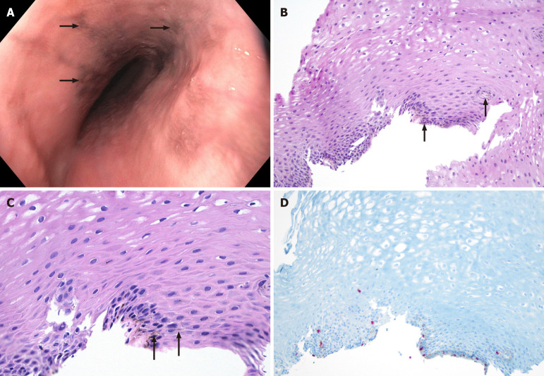
Case summary: We report two cases of African American patients who complained of significant weight loss (over 20 pounds in approximately six months) and abdominal pain during their first visit. The first case involves a 54-year female with a history of hepatic steatosis and polysubstance abuse, who also experiences nausea and vomiting. The second case is a 59-year-old male with hypertension and gastroesophageal reflux disease (GERD), who was diagnosed with esophageal squamous cell carcinoma. Both cases show benign melanocytes in the basal layer on the esophagus biopsy and are diagnosed as EM.
Conclusion: It is important to note that EM has been associated with malignancies such as carcinoma and melanoma. Therefore, accurate diagnosis and appropriate management are crucial. Patients with EM, especially those with concurrent risk factors (e.g., GERD, smoking), should be carefully monitored for any signs of malignancy.
Keywords: Case report; Esophageal melanosis; Esophagus; Gastroesophageal reflux disease; Melanoblasts.
©The Author(s) 2024. Published by Baishideng Publishing Group Inc. All rights reserved.
Conflict of interest statement
Conflict-of-interest statement: The authors declare that they have no conflict of interest to disclose.
Figures
References
-
- De La Pava S, Nigogosyan G, Pickren JW, Cabrera A. Melanosis of the esophagus. Cancer. 1963;16:48–50. - PubMed
-
- Özden A, Seven G, Savaş B, Üstün Y, Ensari A, Yusifova A. Özofageal melanositozis - Üç olgu ve literatürün gözden geçirilmesi. 2008. [cited 20 September 2024]. Available from: https://akademik.tgv.org.tr/index.asp?islem=makaledetay&sayfa=281 .
-
- Chang F, Deere H. Esophageal melanocytosis morphologic features and review of the literature. Arch Pathol Lab Med. 2006;130:552–557. - PubMed
-
- Yokoyama A, Omori T, Yokoyama T, Tanaka Y, Mizukami T, Matsushita S, Higuchi S, Takahashi H, Maruyama K, Ishii H, Hibi T. Esophageal melanosis, an endoscopic finding associated with squamous cell neoplasms of the upper aerodigestive tract, and inactive aldehyde dehydrogenase-2 in alcoholic Japanese men. J Gastroenterol. 2005;40:676–684. - PubMed
-
- Sharma SS, Venkateswaran S, Chacko A, Mathan M. Melanosis of the esophagus. An endoscopic, histochemical, and ultrastructural study. Gastroenterology. 1991;100:13–16. - PubMed
Publication types
MeSH terms
LinkOut - more resources
Full Text Sources
Medical
