

Resting-state functional magnetic resonance imaging and support vector machines for the diagnosis of major depressive disorder in adolescents
- PMID: 39564181
- PMCID: PMC11572682
- DOI: 10.5498/wjp.v14.i11.1696
Resting-state functional magnetic resonance imaging and support vector machines for the diagnosis of major depressive disorder in adolescents
Abstract
Background: Research has found that the amygdala plays a significant role in underlying pathology of major depressive disorder (MDD). However, few studies have explored machine learning-assisted diagnostic biomarkers based on amygdala functional connectivity (FC).
Aim: To investigate the analysis of neuroimaging biomarkers as a streamlined approach for the diagnosis of MDD in adolescents.
Methods: Forty-four adolescents diagnosed with MDD and 43 healthy controls were enrolled in the study. Using resting-state functional magnetic resonance imaging, the FC was compared between the adolescents with MDD and the healthy controls, with the bilateral amygdala serving as the seed point, followed by statistical analysis of the results. The support vector machine (SVM) method was then applied to classify functional connections in various brain regions and to evaluate the neurophysiological characteristics associated with MDD.
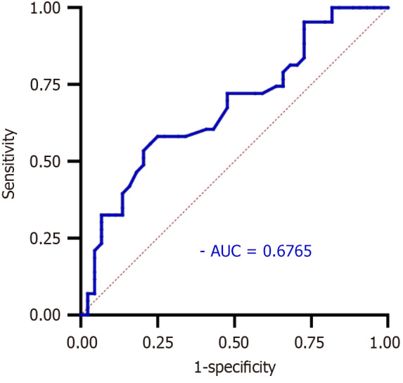
Results: Compared to the controls and using the bilateral amygdala as the region of interest, patients with MDD showed significantly lower FC values in the left inferior temporal gyrus, bilateral calcarine, right lingual gyrus, and left superior occipital gyrus. However, there was an increase in the FC value in Vermis-10. The SVM analysis revealed that the reduction in the FC value in the right lingual gyrus could effectively differentiate patients with MDD from healthy controls, achieving a diagnostic accuracy of 83.91%, sensitivity of 79.55%, specificity of 88.37%, and an area under the curve of 67.65%.
Conclusion: The results showed that an abnormal FC value in the right lingual gyrus was effective as a neuroimaging biomarker to distinguish patients with MDD from healthy controls.
Keywords: Adolescent; Biomarker; Machine learning; Major depressive disorder; Neuroimaging; Resting-state functional magnetic resonance imaging; Support vector machine.
©The Author(s) 2024. Published by Baishideng Publishing Group Inc. All rights reserved.
Conflict of interest statement
Conflict-of-interest statement: The authors declare that they have no conflict of interest.
Figures


Similar articles
-
Unveiling the invisible: How cutting-edge neuroimaging transforms adolescent depression diagnosis.World J Psychiatry. 2025 May 19;15(5):102953. doi: 10.5498/wjp.v15.i5.102953. eCollection 2025 May 19. World J Psychiatry. 2025. PMID: 40495844 Free PMC article.
-
Disruption of resting-state functional connectivity of right posterior insula in adolescents and young adults with major depressive disorder.J Affect Disord. 2019 Oct 1;257:23-30. doi: 10.1016/j.jad.2019.06.057. Epub 2019 Jul 2. J Affect Disord. 2019. PMID: 31299401
-
Reduction of Interhemispheric Homotopic Connectivity in Cognitive and Visual Information Processing Pathways in Patients With Thyroid-Associated Ophthalmopathy.Front Hum Neurosci. 2022 Jun 30;16:882114. doi: 10.3389/fnhum.2022.882114. eCollection 2022. Front Hum Neurosci. 2022. PMID: 35865354 Free PMC article.
-
Altered Resting-State Functional Activity in Medication-Naive Patients With First-Episode Major Depression Disorder vs. Healthy Control: A Quantitative Meta-Analysis.Front Behav Neurosci. 2019 May 7;13:89. doi: 10.3389/fnbeh.2019.00089. eCollection 2019. Front Behav Neurosci. 2019. PMID: 31133831 Free PMC article.
-
Machine learning approaches for classifying major depressive disorder using biological and neuropsychological markers: A meta-analysis.Neurosci Biobehav Rev. 2025 Jul;174:106201. doi: 10.1016/j.neubiorev.2025.106201. Epub 2025 May 10. Neurosci Biobehav Rev. 2025. PMID: 40354957 Review.
Cited by
-
Advancing the diagnosis of major depressive disorder: Integrating neuroimaging and machine learning.World J Psychiatry. 2025 Mar 19;15(3):103321. doi: 10.5498/wjp.v15.i3.103321. eCollection 2025 Mar 19. World J Psychiatry. 2025. PMID: 40109992 Free PMC article.
-
Unveiling the invisible: How cutting-edge neuroimaging transforms adolescent depression diagnosis.World J Psychiatry. 2025 May 19;15(5):102953. doi: 10.5498/wjp.v15.i5.102953. eCollection 2025 May 19. World J Psychiatry. 2025. PMID: 40495844 Free PMC article.
References
-
- Long X, Li L, Wang X, Cao Y, Wu B, Roberts N, Gong Q, Kemp GJ, Jia Z. Gray matter alterations in adolescent major depressive disorder and adolescent bipolar disorder. J Affect Disord. 2023;325:550–563. - PubMed
-
- Kang L, Wang W, Zhang N, Yao L, Tu N, Feng H, Zong X, Bai H, Li R, Wang G, Bu L, Wang F, Liu Z. Anhedonia and dysregulation of an angular gyrus-centred and dynamic functional network in adolescent-onset depression. J Affect Disord. 2023;324:82–91. - PubMed
-
- Dalal M, Holcomb JM, Sundaresan D, Dutta A, Riobueno-Naylor A, Peloquin GD, Benheim TS, Jellinek M, Murphy JM. Identifying and responding to depression in adolescents in primary care: A quality improvement response. Clin Child Psychol Psychiatry. 2023;28:623–636. - PubMed
Publication types
LinkOut - more resources
Full Text Sources
